J Clin Aesthet Dermatol. 2026;19(6):35–43.
by Haowei Han, DO; Valerie Foy, DO; Mairead Moloney, DO; Aleia Boccardi, DO; Graham H. Litchman, DO, MS; and Sourab Choudhury, DO
Drs. Han, Foy, Moloney, and Boccardi are with St. John’s Episcopal Hospital, Far Rockaway, New York. Dr. Litchman is with Touro University Nevada College of Osteopathic Medicine, Henderson, Nevada. Dr. Choudhury is with The Dermatology Specialists, New York, New York.
FUNDING: No funding was provided for this article.
DISCLOSURES: The authors have no relevant conflicts of interest.
OBJECTIVE: To describe the clinical characteristics, treatment responses, and outcomes of patients with Trichophyton indotineae infection in New York state and to contribute to the collective knowledge necessary for appropriate antifungal stewardship and management strategies. METHODS: We conducted a retrospective cohort study of 20 patients with culture-confirmed T. indotineae infection seen across New York City between June 2023 and April 2025. Demographic data, clinical features, medical history, and treatment outcomes were collected. All patient data were deidentified, and informed consent was waived. RESULTS: Patients were evenly distributed by gender, with a mean age of 43.8 years for men and 33.4 years for women. All patients reported pruritus and, notably, 90% had intact immune function. Common presentations included tinea corporis and tinea cruris. Itraconazole was the most effective first-line therapy; however, there was a high rate of recurrence after stopping the treatment. Griseofulvin, voriconazole, and fluconazole showed some success. LIMITATIONS: This study is limited by a small sample size and reliance on self-reported medical histories. Additionally, the study did not assess the impact of topical antifungal use or combination oral antifungal therapy on treatment efficacy. CONCLUSION: T. indotineae presents diagnostic and therapeutic challenges due to its atypical clinical features and antifungal resistance. Early recognition and appropriate systemic antifungal therapy are critical. Itraconazole remains the preferred first-line agent based on current clinical experience. Greater clinician awareness and further research are urgently needed to address the rising burden of this infection. KEYWORDS: Trichophyton indotineae, dermatophytes, antifungal resistant, antifungal stewardship, itraconazole, infectious disease
Introduction
Dermatophytosis is a superficial and inflammatory mycosis caused by keratinophilic fungi, including Trichophyton, Microsporum, and Epidermophyton species.1 Trichophyton rubrum, Trichophyton interdigitale, Trichophyton mentagrophytes, and Trichophyton tonsurans are the most common causes of tinea infections in humans. Most Trichophyton species are anthropophilic, meaning they are restricted to humans and typically cause mild inflammatory reactions. However, T. mentagrophytes is zoophilic, primarily affecting animals and triggering a strong inflammatory response in humans.1 It can cause kerion, tinea corporis, tinea barbae, and tinea pedis in the human body.1 A recently identified species, Trichophyton indotineae (previously known as Trichophyton mentagrophytes type VIII), has been seen in increasing numbers throughout the Indian subcontinent and has emerged as a public health concern.2 First described in 2019, its identification relied on sequencing the Internal Transcribed Spacer (ITS) region of ribosomal DNA (rDNA).3
Commonly used treatments for tinea include terbinafine, itraconazole, and fluconazole, which were approved by the US Food and Drug Administratin (FDA) in the 1990s, while griseofulvin was approved in the 1950s. Among these, only terbinafine is approved specifically for tinea corporis. In vitro studies have shown T. indotineae to be resistant to terbinafine, a commonly used first-line oral antifungal therapy targeting squalene epoxidase.4 This resistance is attributed to a point mutation in the squalene epoxidase gene (SQLE) phenylalanine-to-leucine amino acid change at codon 397 (F397L).3,5 Clinically, patients are typically immunocompetent and present with widespread, scaly, pruritic plaques on the trunk, groin, and extremities. Unlike classical tinea infections, T. indotineae commonly lacks central clearing.6 Many patients have a history of travel, particularly to the Indian subcontinent.7 Diagnosis of T. indotineae requires specialized laboratory testing using molecular methods, such as DNA sequencing, because culture alone may show T. mentagrophytes or T. interdigitale by most clinical laboratories.8
To date, T. indotineae has been reported in many distant continents, including North America, southern Africa, western and central Europe, and Latin America.5,9–12 The first 2 cases of T. indotineae in United States (US) were reported in May 2023 by dermatologists and public health officials in New York City.13,14 One immunocompetent patient reported her tinea infection had developed while she was in Bangladesh; the other patient developed the infection during her third trimester of pregnancy and denied any international travel history. These 2 cases underscored the potential transmission of T. indotineae within the US.13 However, due to diagnostic challenges, the prevalence and incidence of this infection in the US remain unclear. Furthermore, antifungal susceptibility testing (AFST) does not always correlate with clinical clearance, posing additional therapeutic obstacles for clinicians.14
To enhance antifungal stewardship and better understand the epidemiology of T. indotineae infections, our team conducted what we believe to be the largest case series to date, examining the treatments use and the clinical course.
Methods
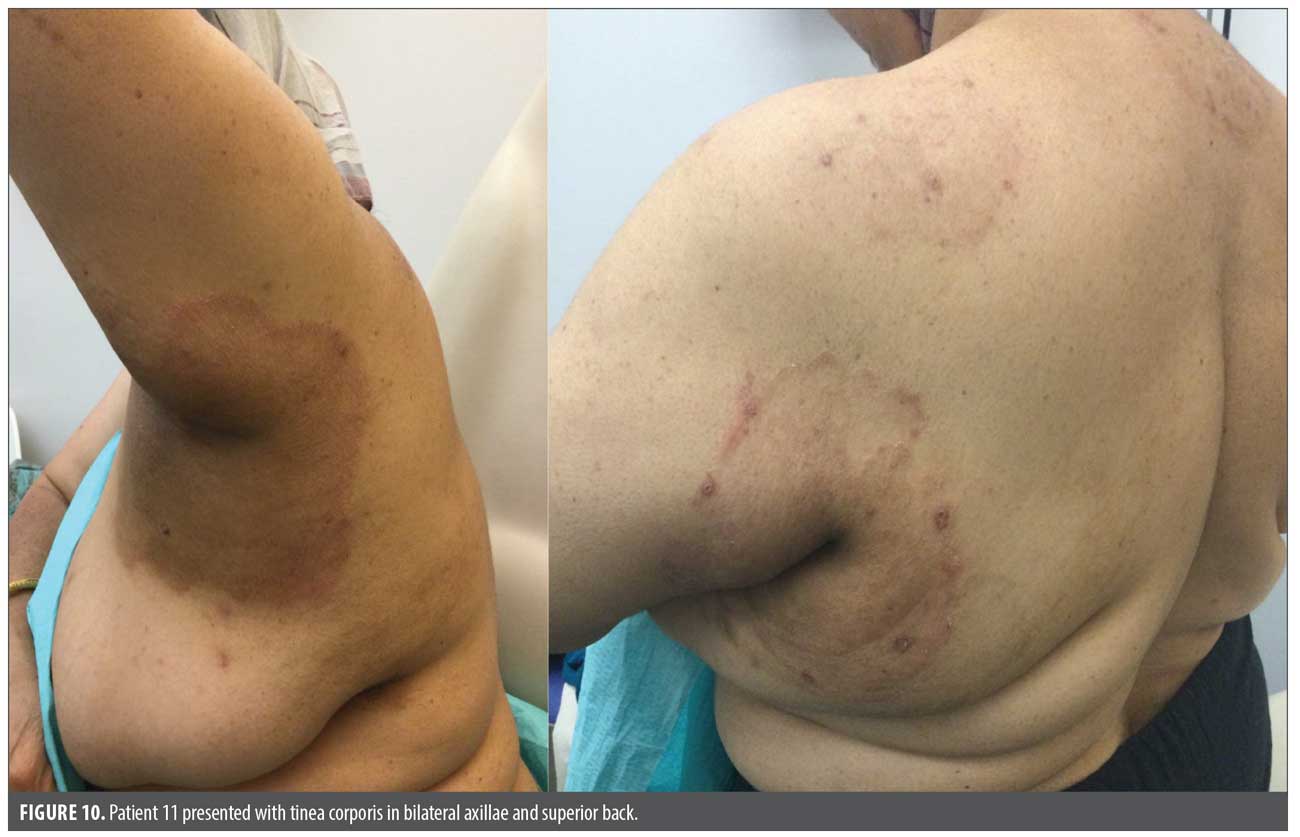
This retrospective cohort study included 20 cases of T. indotineae from private practice settings in New York City, New York, from June 2023 to April 2025. Information regarding patients’ demographics, clinical presentation, past medical history, and treatment outcomes was collected (Figures 1–10). Informed consent was waived because all data were deidentified.
Identification of T. indotineae. The diagnosis was reached by history (travel history, prior failure to oral antifungal therapy, pruritus), physical examination (widespread eczematous plaque ± excoriations), clinical suspicion, and confirmatory laboratory testing by the New York State Department of Health. Fungal cultures were performed at a community laboratory. Isolates identified as T. mentagrophytes complex were referred to the New York State Department of Health Wadsworth Center for species-level confirmation using ITS rDNA sequencing. Of the 20 isolates, all 20 were confirmed as T. indotineae, and all had been preliminarily identified as T. mentagrophytes.

Results
In terms of patient demographics, 50% were men, 50% were women, and ethnically, all were from the Indian subcontinent. The mean age of male patients was 43.8 years (SD: 14.1), while the mean age of female patients was 33.4 years (SD: 22.0) (Table 1). Eight patients (40%) reported a travel history to or immigration from Bangladesh; 1 patient (5%) immigrated from India and another patient (n=1, 5%) immigrated from Nepal. The detailed demographic information, clinical findings, treatments, and outcomes are listed in Supplemental Table 1.
Access Supplemental Table 1 Supplemental Table 1.
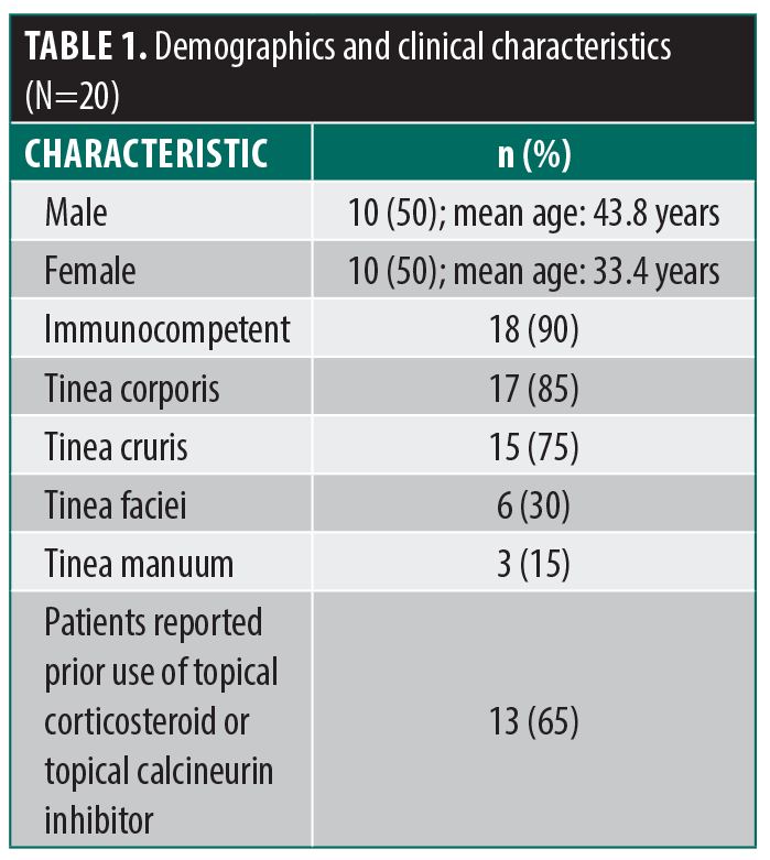
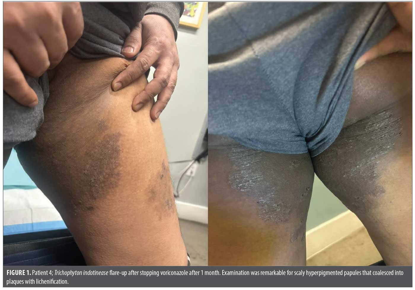
Clinically, all patients reported pruritus. Sixty-five percent (13/20) reported prior use of topical corticosteroids or topical calcineurin inhibitors. Most patients (18/20, 90%) were immunocompetent, and 2 patients had diabetes. Notably, patients 4, 5, 6, and 14 were related and lived in the same household. The wife of patient 16 also had active disease; however, culture was not performed, as the diagnosis was made based on clinical examination and history. She was subsequently treated with antifungal therapy and demonstrated a good clinical response.
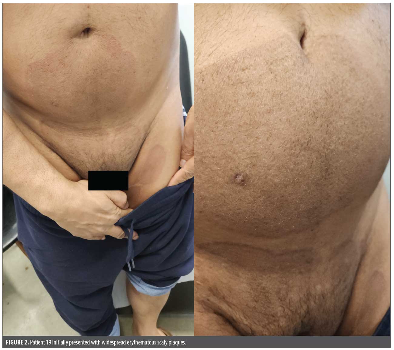
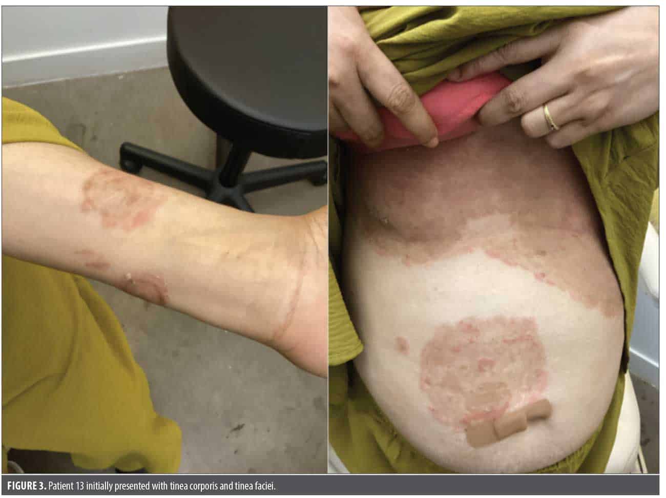
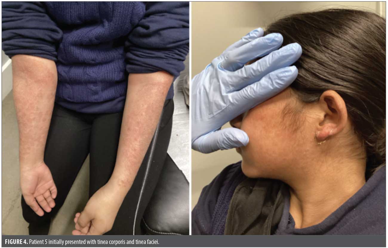
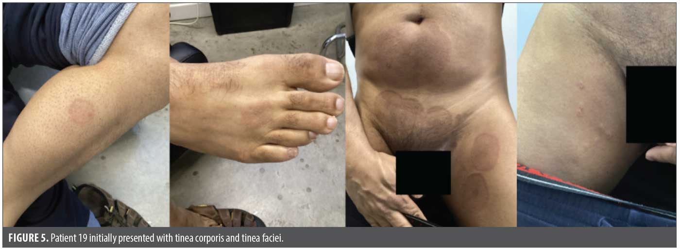
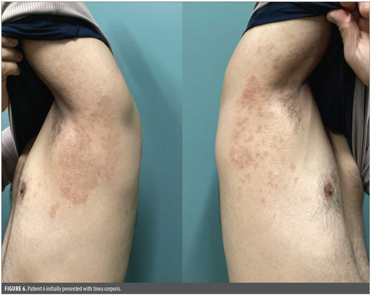
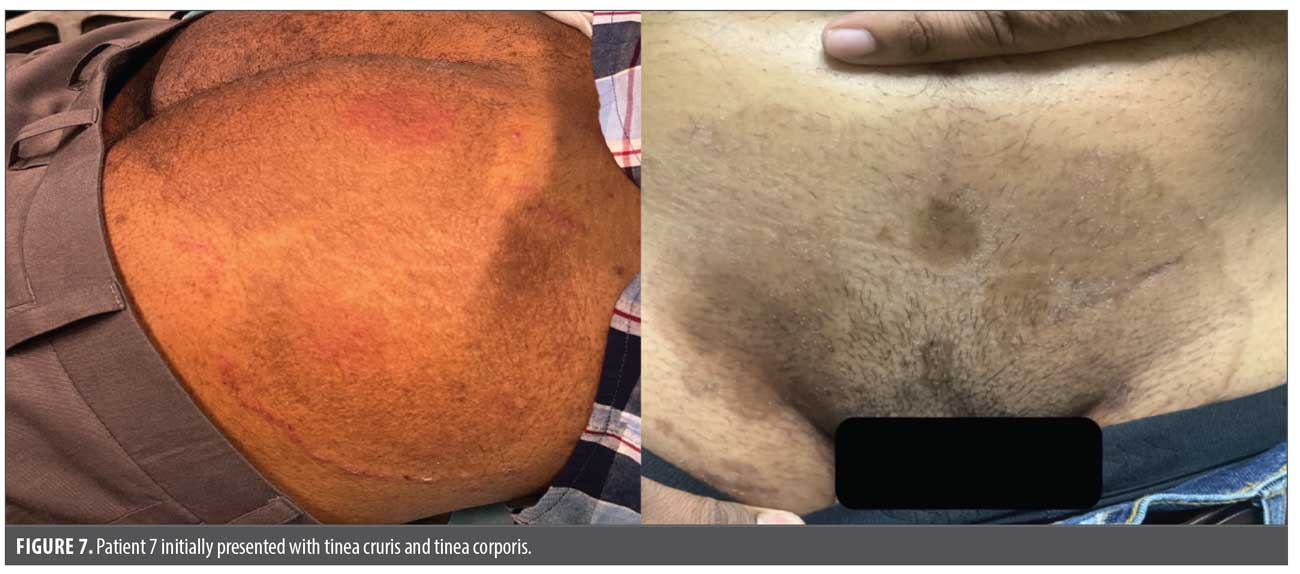
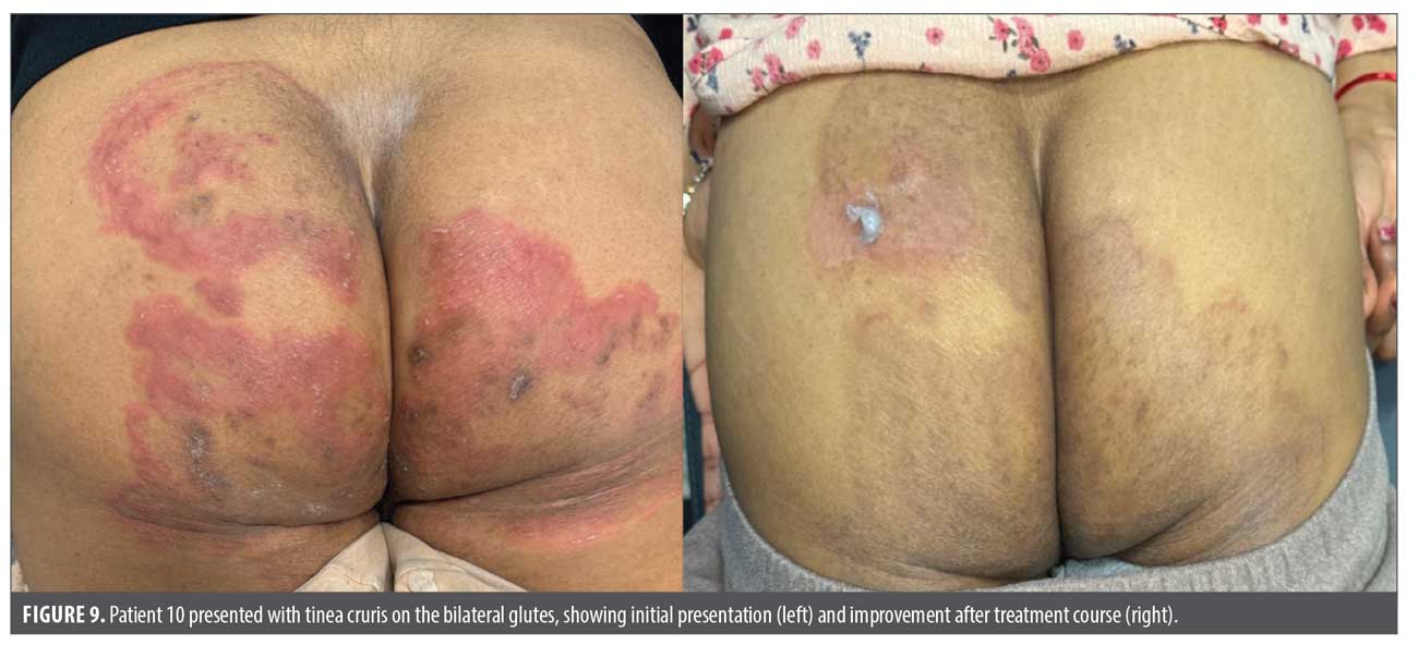
There were a variety of clinical presentations: 17 patients (85%) had tinea corporis, 15 patients (75%) had tinea cruris, 6 patients (30%) had tinea faciei, and 3 patients (15%) had tinea manuum. None of the patients developed tinea capitis or onychomycosis, which aligns with prior knowledge that T. indotineae is a rare cause of these conditions. In contrast to adult counterparts, tinea faciei was more common in adolescent and pediatric patients.
Regarding diagnostic workup, skin biopsies were performed on 9 patients, with 4 (44%) demonstrating spongiotic dermatitis. One additional patient reported a prior biopsy by an outside dermatologist that also showed spongiotic dermatitis. Among patients with spongiotic dermatitis on biopsy, all had either positive cultures or strong clinical suspicion of tinea and were subsequently treated with appropriate antifungal therapy with or without adjunctive topical steroids.
The mean duration of systemic antifungal therapy was 4.3 months (median: 4.1 months). The mean follow-up period was 6.9 months (median: 5.75 months). Among patients who experienced recurrence, the mean time to recurrence was 10.3 weeks (median: 6 weeks). Patients 8 and 9 each experienced 2 recurrences, and both events for each patient were included in the recurrence analysis. Notably, 1 recurrence in patient 9 occurred during antifungal taper. Patient 17 also experienced a recurrence during taper, therefore time to recurrence after complete discontinuation of therapy was not included in the analysis.
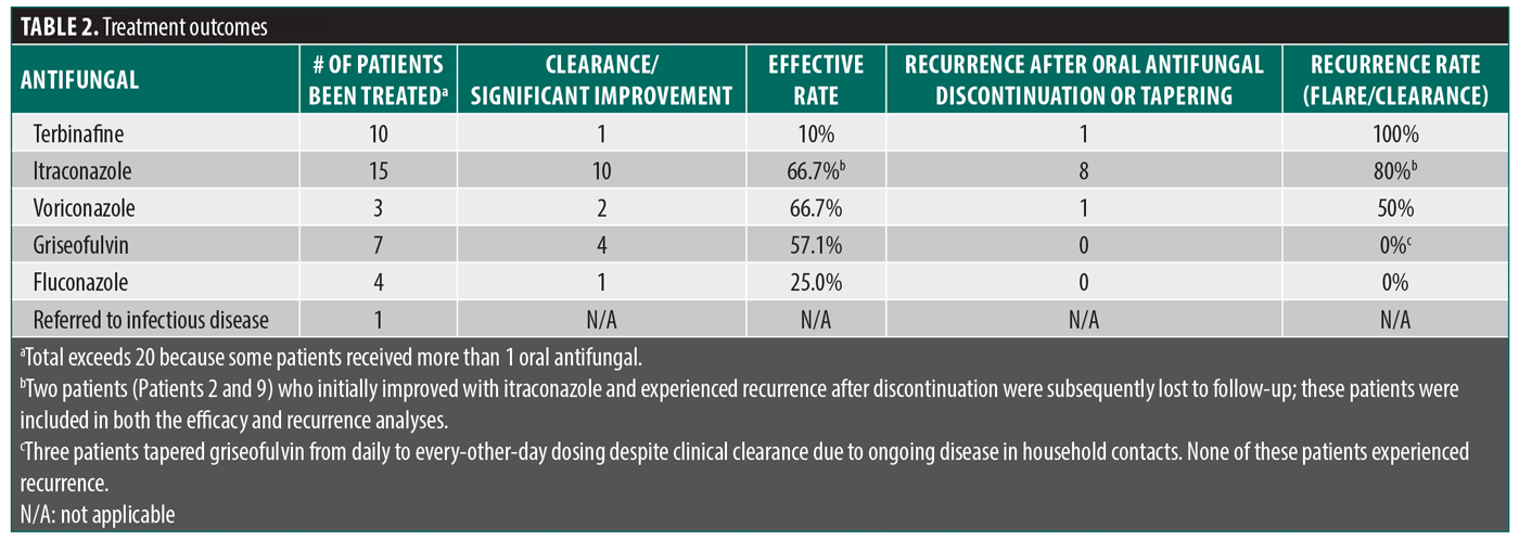
Among the 20 patients, 6 (30%) were lost to follow-up. Ten patients (50%) achieved clearance or improvement with the initial course of itraconazole; however, 8 of these 10 patients (80%) experienced recurrence after discontinuation or tapering of the medication. One patient (5%) achieved clearance with voriconazole but experienced a flare 4 weeks after discontinuation. Five patients (25%) achieved clearance with griseofulvin; however, 3 required continued therapy due to ongoing household transmission. One patient (5%) demonstrated improvement with fluconazole. Additionally,1 patient was referred to an infectious disease specialist. Overall, 10 patients (50%) experienced flaring or recurrence after cessation of oral antifungal therapy (Table 2).
Discussion
In July 2023, a reference laboratory analyzed the genetic profile and AFST of 21 strains of T. indotineae from North America, collected between 2021 and 2022. 5 A further database search identified 3 additional T. indotineae isolates, the earliest of which dated back to 2017. Interestingly, the earliest strain did not harbor an F397L codon change but instead exhibited a leucine-to-serine amino acid substitution at codon 393 on the SQLE.5 The majority of T. indotineae strains had a terbinafine minimal inhibitory concentration (MIC) >2 μg/mL and remained susceptible to itraconazole, with MIC values ranging from ≤0.03 to 0.125 μg/mL. Regarding the source of T. indotineae isolates, all were cultured from skin samples.
To the best of the authors’ knowledge, this is the first and largest case series conducted in New York City. Caplan et al13,14 identified 11 patients with T. indotineae. Our observations are consistent with those reports, as T. indotineae typically causes widespread inflammatory lesions in patients with intact immune systems. Moreover, based on the author’s (S.C.) clinical experience, many additional patients are treated empirically for T. indotineae based on history and physical examination without confirmatory culture, such as those with a history of travel to the Indian subcontinent, failure of terbinafine treatment, and extensive pruritic rashes affecting the body and groin. Itraconazole was the most commonly used agent, but griseofulvin demonstrated a higher clearance rate in this small cohort. This may be due to longer duration, higher dosages, different SQLE mutation profiles, or the overall smaller sample size. Of the 6 patients lost to follow-up, 15 were receiving itraconazole and 7 were receiving griseofulvin.14 According to a literature review conducted by Sonego et al,15 which included 58 published cases of T. indotineae, the most effective treatment leading to disease resolution was itraconazole at a dosage of 200 mg/day for a variable duration of 1 to 12 weeks, with 15 cases reported. Interestingly, Sonego et al15 also noted improvement with topical or oral terbinafine, although recurrences were observed later in the disease course.¹⁵ Compared to the report by Sonego et al,15 griseofulvin was ineffective in 5 patients but appeared to be effective in our study.
T. indotineae poses a significant public health concern due to challenges in diagnosis and management. Diagnosis is often complicated by the absence of central clearing and the frequent finding of spongiotic dermatitis in histopathology. However, a helpful diagnostic clue is a history of travel to the Indian subcontinent (particularly India or Bangladesh) or contact with someone who has, as cases have been reported in the US even without a significant travel history. Widespread use of antifungal and topical corticosteroids; multiple familial contacts; unhygienic practices; treatment by nondermatologists with inappropriate drugs, doses, and duration; and poor compliance with treatment have contributed to the emergence of antifungal-resistant species.15,16 This resistance complicates treatment, leading to prolonged infections and increased risk of transmission. In particular, misuse or overuse—such as using topical corticosteroids without confirming a fungal diagnosis—can mask symptoms, delay proper therapy, and promote the growth of resistant strains. Consequently, infections may even masquerade as spongiotic dermatitis, likely due to prior corticosteroid use or tinea incognito. A case study demonstrated that tinea incognito can present with spongiotic changes and a negative periodic acid-Schiff stain, further obscuring diagnosis.17 The treatment course for T. indotineae is often prolonged and may require extended maintenance therapy, even after clinical resolution, to prevent relapse and ensure complete mycologic cure.
Clinically, T. indotineae tends to be chronic, involving large body surface areas with frequent recurrences, often due to household transmission. The disease is often mismanaged with oral terbinafine or other ineffective treatments without proper fungal culture, leading to increased resistance. Past management often includes multiple courses of topical and systemic antifungal therapies, topical corticosteroids, and repeated biopsies, increasing the risk of adverse effects. A diagnostic challenge for community clinicians is that routine fungal culture cannot distinguish T. indotineae from other members of the T. mentagrophytes/T. interdigitale complex. Species-level confirmation requires molecular methods such as ITS sequencing, which are not readily available at most commercial laboratories. In New York, isolates can be referred to the New York State Department of Health Wadsworth Center for identification. Clinicians should suspect T. indotineae in patients presenting with widespread, pruritic dermatophytosis, particularly in those with a travel history to the Indian subcontinent, failure of terbinafine therapy, or household contacts with similar infections. In such cases, empiric initiation of itraconazole while awaiting confirmatory testing is reasonable, given the known inefficacy of terbinafine against most T. indotineae strains. Emerging polymerase chain reaction–based assays may eventually allow for a more rapid identification process. Prompt initiation of effective antifungal treatment, such as itraconazole, should be considered and continued until complete clearance of infection. In fact, treatment often becomes chronic, and continued antifungal therapy 2 to 3 times per week for several months is often necessary even after clinical clearance, especially if household contacts remain infected. In fact, the author’s (SC) clinical experience demonstrates that most patients needed 12 weeks to achieve clearance and several took up to 24 weeks to completely clear. Loss to follow-up was common, potentially related to the prolonged treatment course and persistent pruritus, leading to patient dissatisfaction. These data highlight the importance of examining and treating all household members and close contacts. Prompt identification and treatment are critical to preventing the continued spread. As with scabies and certain sexually transmitted infections, it is recommended that when a patient is diagnosed with T. indotineae, close contacts and household members should be examined and treated as necessary to prevent ongoing transmission.
Future directions may include the development of newer antifungal therapies, such as oral formulations of efinaconazole and tavaborole, both of which are approved for the treatment of onychomycosis caused by T. rubrum and T. mentagrophytes.
This study’s limitations include reliance on patients’ self-reported medical histories, a small sample size, and, most importantly, the lack of AFST reports, which prevents correlation between AFST results and clinical response. Differences in observed efficacy between agents in this series should be interpreted cautiously given the small sample size and may not fully reflect broader treatment patterns. Additionally, none of the patients in this study received combination oral antifungal therapy, and topical antifungal use was documented but not considered in the analysis.
Conclusion
Our case series highlights the clinical challenges associated with diagnosing and managing T. indotineae infections. Given its chronicity, frequent recurrence, and potential for household transmission, early recognition and appropriate systemic antifungal therapy are critical for successful outcomes. Itraconazole remains the most effective first-line treatment based on current evidence, although voriconazole, fluconazole, and griseofulvin may offer an alternative option in select cases. Greater awareness among clinicians and improved antifungal stewardship are essential to address the growing public health impact of this emerging pathogen.
References
- Bolognia JL, Schaffer JV, Duncan KO, Ko C. Dermatology Essentials. 1st ed. Elsevier Health Sciences; 2014.
- Ebert A, Monod M, Salamin K, et al. Alarming India-wide phenomenon of antifungal resistance in dermatophytes: a multicentre study. Mycoses. 2020;63(7):717–728.
- Uhrlaß S, Verma SB, Gräser Y, et al. Trichophyton indotineae-an emerging pathogen causing recalcitrant dermatophytoses in India and worldwide-a multidimensional perspective. J Fungi (Basel). 2022;8(7):757.
- Wolverton SE, Wu JJ. Comprehensive Dermatologic Drug Therapy. 4th ed. Elsevier Health Sciences; 2019.
- Cañete-Gibas CF, Mele J, Patterson HP, et al. Terbinafine-resistant dermatophytes and the presence of Trichophyton indotineae in North America. J Clin Microbiol. 2023;61(8):e0056223.
- De Marco A, Liguori G, Cafarchia C, et al. Cutaneous infections caused by Trichophyton indotineae: case series and systematic review. J Clin Med. 2025;14(4):1280.
- Xu Z, Caplan AS. Extensive tinea corporis and tinea cruris from Trichophyton indotineae. N Engl J Med. 2024;391(19):1837.
- Recognizing Trichophyton indotineae. Accessed 23 Dec 2024. https://www.aad.org/member/clinical-quality/clinical-care/emerging-diseases/dermatophytes/recognizing-trichophyton-indotineae
- Messina F, Santiso G, Romero M, Bonifaz A, Fernandez M, Marin E. First case report of tinea corporis caused by Trichophyton indotineae in Latin America. Med Mycol Case Rep. 2023;41:48–51.
- Mosam A, Shuping L, Naicker S, et al. A case of antifungal-resistant ringworm infection in KwaZulu-Natal Province, South Africa, caused by Trichophyton indotineae. Public Health Bulletin South Africa. 2023.
- Dellière S, Joannard B, Benderdouche M, et al. Emergence of difficult-to-treat tinea corporis caused by Trichophyton mentagrophytes complex isolates, Paris, France. Emerg Infect Dis. 2022;28(1):224–228.
- Süß A, Uhrlaß S, Ludes A, et al. Ausgeprägte tinea corporis durch ein terbinafin-resistentes Trichophyton-mentagrophytes-isolat vom Indischen genotyp bei einem säugling aus Bahrain in Deutschland. Extensive tinea corporis due to a terbinafine-resistant Trichophyton mentagrophytes isolate of the Indian genotype in a young infant from Bahrain in Germany. Hautarzt. 2019;70(11):888–896.
- Caplan AS, Chaturvedi S, Zhu Y, et al. Notes from the field: first reported US cases of tinea caused by Trichophyton indotineae – New York City, December 2021-March 2023. MMWR Morb Mortal Wkly Rep. 2023;72(19):536–537.
- Caplan AS, Todd GC, Zhu Y, et al. Clinical course, antifungal susceptibility, and genomic sequencing of Trichophyton indotineae. JAMA Dermatol. 2024;160(7):701–709.
- Sonego B, Corio A, Mazzoletti V, et al. Trichophyton indotineae, an emerging drug-resistant dermatophyte: a review of the treatment options. J Clin Med. 2024;13(12):3558.
- Appannavar S, Kiran, Pise G, et al. Recurrent and chronic dermatophytosis: culprit- not just antifungal resistance. Int J Res Dermatol. 2021;7(2):213.
- Diep D, Calame A, Cohen PR. Tinea corporis masquerading as a diffuse gyrate erythema: case report and a review of annular lesions mimicking a dermatophyte skin infection. Cureus. 2020;12(6):e8935.
OX40/OX40L Costimulatory Pathway: A Potential Therapeutic Target for Allergic Contact Dermatitis?