
J Clin Aesthet Dermatol. 2026;19(10):24–29.
Tebyan Khalfalla, BS; David Cotter, MD, PhD; and Aamir Hussain, MD
Ms. Khalfalla is with the University of Rochester School of Medicine and Dentistry, Rochester, New York. Dr. Cotter is with Las Vegas Dermatology, Las Vegas, Nevada. Dr. Hussain is with the Donald and Barbara Zucker School of Medicine at Hofstra/Northwell, Hempstead, New York.
FUNDING: No funding was provided for this article.
DISCLOSURES: The authors have no relevant conflicts of interest.
Abstract: BACKGROUND: Surgical intervention is rarely used in managing cutaneous mast cell disorders. OBJECTIVE: To analyze studies on surgical management of cutaneous mast cell disorders in humans and review practical considerations for these surgeries. METHODS: We conducted a literature search of major medical databases using terms related to mast cell disease and surgical treatment in human patients from inception of the databases to April 20, 2026. RESULTS: Nineteen of 981 screened articles met inclusion criteria. There were 13 cases of solitary mastocytomas: 3 mast cell sarcomas (MCS), 1 multilocalized mastocytomas, 1 solitary mastocytoma that transformed into MCS, and 1 atypical mastocytoma to borderline MCS. For localized mastocytomas, wide local excision achieved adequate symptom control in most cases. Systemic disease carried a poor prognosis overall despite both surgical and adjuvant therapies. Tryptase was more likely to be elevated in more severe systemic, recurrent, or malignant disease. CONCLUSION: Surgical intervention has both diagnostic and therapeutic value for isolated solitary mastocytomas and in certain cases of MCS. Careful perioperative management is crucial. Further research is necessary to determine the value of premedication. Keywords: Mastocytosis, mastocytoma, tryptase, mast cell disease, mast cell sarcoma
Introduction
Mastocytosis is characterized by an inappropriate proliferation of mast cells in the skin, gastrointestinal tract, kidneys, bone marrow, or other organs. The most current diagnostic guidelines adopted by the World Health Organization in 2021 divide this disease into overarching subtypes: cutaneous mastocytosis (CM) and systemic mastocytosis (SM), each of which is further stratified by prognostic and clinical criteria.1 CM, the focus of this review, is characterized by neoplastic mast cells limited to the skin and is the more common presenting subtype in pediatric patients, carrying a good prognosis with possible resolution at puberty. The cutaneous lesions of CM are categorized by their morphology, and the commonly described variants include monomorphic and polymorphic maculopapular CM, also known as urticaria pigmentosa, as well as cutaneous mastocytoma and diffuse CM.2 Adult patients are more likely to experience SM, which frequently presents with cutaneous lesions. Cutaneous lesions can be the first sign of SM and carry a poor prognosis, specifically for nonindolent SM, due to internal organ involvement.1 Both forms can exhibit Darier’s sign and may appear similar on histology with increased numbers of mast cells in the dermis and perivascularly.
Mast cell degranulation can be triggered by various stimuli, including crosslinking of immunoglobulin E, via toll-like receptors, stem cell factor receptors, specific ligand-mediated G protein-coupled receptor activation, the KIT receptor, and more.3 Subsequent degranulation then causes the release of preformed mediators including histamine and tryptase, which is followed within a few minutes by the synthesis of prostaglandin D2, leukotrienes, and platelet activating factor, and finally by the generation of numerous cytokines with both pro- and anti-inflammatory effects.4 Symptoms of mast cell activation are heterogenous in both CM and SM and can range vastly from bothersome pruritus or flushing to life-threatening anaphylaxis.5–7
Surgical procedures, specifically to alleviate the cutaneous manifestations for isolated mastocytoma or mast cell sarcoma (MCS) via surgical excision, are rarely performed in humans with mastocytosis. This is possibly due to concerns about perioperative complications such as mast cell degranulation or anaphylaxis, though surgical recommendations are largely derived from case reports and expert opinions rather than controlled trials.8 However, there exists abundant veterinary literature describing surgical management of CM in animals. By extrapolation, surgery may provide a therapeutic option in certain human populations.
The objectives of this review are to identify appropriate patient types for surgical management of cutaneous manifestations of mastocytosis and MCS and define the best practices to minimize perioperative complications.
Methods
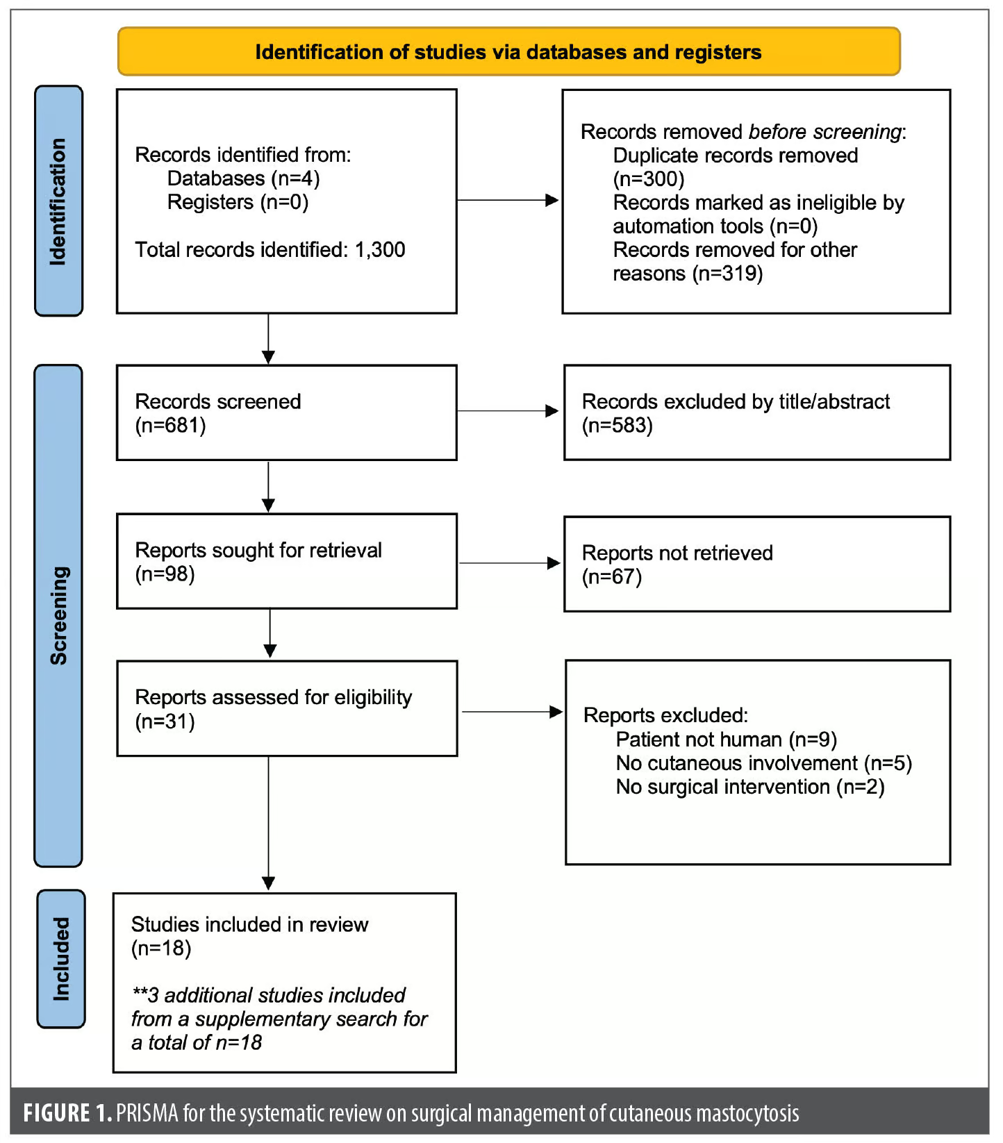
A literature search using PubMed, PubMed Central, Google Scholar, and the Cochrane Library was performed for the terms “mast cell disease,” “mastocytosis,” or “mastocytoma,” in association with the terms “surgical,” “surgery,” “excision,” or “surgical treatment” from database inception to April 20, 2026. The search was conducted adhering to the updated 2020 standards of the Preferred Reporting Items for Systematic Reviews and Meta-analyses (PRISMA) statement (Figure 1). Cases were included if they involved human patients, cutaneous forms of mast cell disease were diagnosed, and surgical intervention was performed for isolated CM or cutaneous MCS. During the review process, articles describing animal cases and cases of SM without cutaneous involvement were excluded. Two reviewers (author AH and author TK) independently reviewed the studies, and discrepancies were resolved through consensus.
Results
A total of 981 unique articles were screened, and 31 met criteria to have the full text screened. Of these, 16 met eligibility criteria for inclusion. Supplementary searching identified 3 additional eligible case reports with the addition of the search terms “surgical intervention,” for a final total of 19 included cases. There were 13 cases of CMs, 3 MCSs, 1 multilocalized mastocytoma, 1 CM that transformed into MCS, and 1 atypical mastocytoma to borderline MCS. Eight of the patients were women, 10 were men, and 1 case did not specify a gender. The cases exhibited a broad age distribution ranging from 13 days to 71 years. Tryptase levels were available in 2 MCS cases (range: 18–28 ng/mL in 1 case and >122 ng/mL in the other) and 3 CM cases (5.3–14.0 ng/mL); there was also a case of an atypical CM to borderline MCS (2.9 ng/mL). Each case is detailed in Table 1.
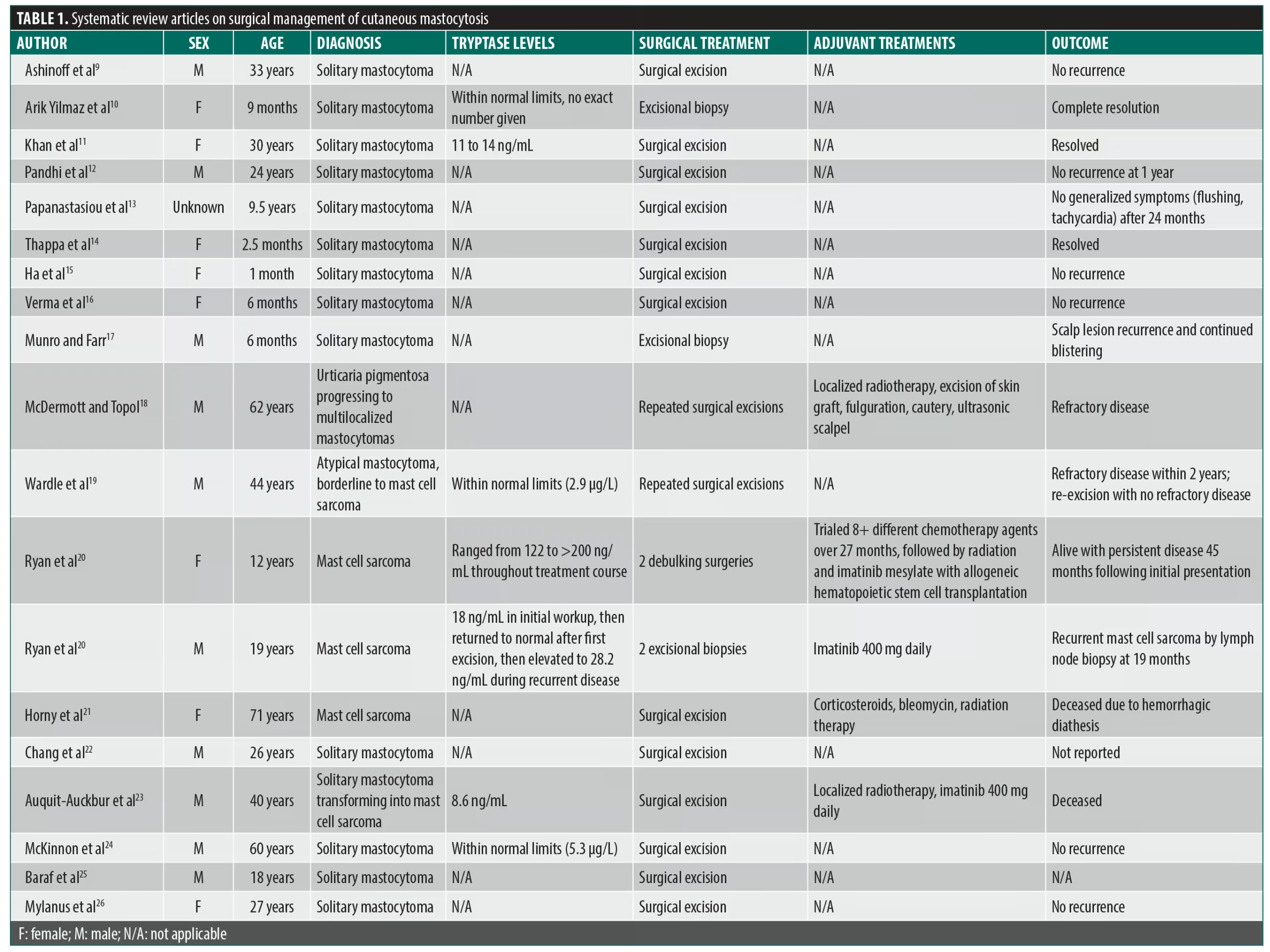
Patients experienced a full resolution of symptoms in 10 of the 13 cases of solitary mastocytoma treated with surgical resection or excisional biopsy. One patient had resolution of systemic symptoms but mild localized recurrent blistering after excision, and 2 patients did not have follow-up information available. Surgery was performed to confirm the diagnosis (excisional biopsy) or for debulking in patients with an MCS and solitary mastocytoma transforming into MCS. These aggressive tumors were treated with multimodal therapy including imatinib (3 cases), systemic corticosteroids (1 case), radiation therapy (3 cases), and stem cell transplant (1 case). Two cases of MCS presented in childhood (ages 12 and 19) and 1 presented in adulthood (age 71), with the additional case of malignant transformation into MCS occurring at age 40; the pediatric cases were both alive at follow up while both adults were deceased. Of note, none of the cases in this review were associated with SM.
Discussion
For solitary mastocytomas, simple wide local excision appeared effective, with the majority of cases showing complete resolution of symptoms without recurrence. Of the 13 mastocytoma cases in this review, 10 achieved full symptom resolution following surgical resection or excisional biopsy.9–17 Adjuvant medical therapy was rarely required.
Two cases underscore the indications for surgical intervention. Ha et al15 described a 13-day-old neonate with a rapidly growing facial mastocytoma successfully treated with excisional biopsy and no postoperative complications or recurrence. Verma et al16 reported a 2-month-old infant in whom a solitary mastocytoma caused life-threatening recurrent apnea, respiratory distress, diarrhea, and limpness—all of which resolved completely following surgical excision at 6 months of age, representing the first reported case of surgical excision resolving such acute multisystem manifestations in a solitary cutaneous mastocytoma.
In contrast, outcomes were generally worse for more disseminated or aggressive subtypes, even those considered nonmalignant. Munro and Farr17 reported ongoing blistering at the surgical site after excising a solitary scalp lesion. Meanwhile, McDermott and Topol18 found some success in debulking several refractory tumors with an ultrasonic scalpel after localized radiotherapy, excision, and cautery failed.
The cases of transforming tumors may lower the threshold for excising even benign-behaving mastocytomas due to the exceedingly rare possibility of malignant degeneration to MCS. Poor surgical control and recurrence were seen for transforming tumors and primary MCSs. For example, Wardle et al19 described a man with an atypical, morphologically high-grade cutaneous mast cell tumor (borderline between mastocytoma and MCS) managed by serial excision following recurrence more than 6 years after diagnosis, with persistently normal serum tryptase throughout. Additionally, 2 cases of MCS described by Ryan et al20 demonstrated failed surgical control even with multi-agent chemotherapy regimens after initial mass debulking. Moreover, Horny et al21 described patient death secondary to intractable bleeding after resection of cutaneous metastases from an aggressive subglottic mass. It is important to note that these cases represent rare and severe manifestations of the disease, and the outcomes may not be generalizable to all patients with cutaneous mastocytomas.
In 1 interesting case, initial excision of a benign primary MCS preceded the appearance of a more aggressive sarcoma at the surgical site by 4 years, despite clear margins at the time of the initial excision.27 Unfortunately, following re-excision of the sarcoma with clear margins and adjuvant radiotherapy, the patient still developed lymph node and bone metastases within a few months and ultimately succumbed to lethal hemorrhage of the metastases. While this description of malignant transformation in this disease process is exceptionally rare, further studies may be necessary to understand the epidemiology of and molecular events underpinning malignant degeneration of presumed-benign mast cell tumors.
The role of tryptase
Tryptase levels were reported infrequently but provided some insight into disease extent when available. For the 2 mastocytoma cases with levels, Arik Yilmaz et al10 and Khan et al11 described normal preoperative serum tryptase, suggestive of isolated solitary cutaneous disease. Both were effectively treated with surgical excision. Meanwhile, tryptase was elevated intermittently throughout the treatment course of 2 MCSs, with levels fluctuating from 122 ng/mL to >200 ng/mL (normal range: 1–15 ng/mL), in the cases reported by Ryan et al.20 Persistently high tryptase mirrored aggressive biology and heralded subsequent relapse and mortality. Normal levels may indicate truly localized pathology where surgery may succeed as monotherapy for primary cutaneous disease.
Tryptase has a low intra-individual variability and can be helpful for risk stratifying for anaphylactic reactions.28 Additionally, tryptase levels can help differentiate CM from SM. World Health Organization guidelines include tryptase levels >20 ng/mL as a minor criterion for SM; however, this is nuanced, as pediatric patients with diffuse disease may exhibit serum tryptase levels >100 ng/mL without having SM.1 Furthermore, trending tryptase levels following the excision of lesions with a history or examination concerning for more aggressive disease may be prudent.
The role of DDPCR for the KIT D816V mutation
Droplet digital polymerase chain reaction (ddPCR) is a highly sensitive molecular diagnostic tool that can detect low levels of the KIT D816V mutation in peripheral blood.29 Given the strong association between KIT D816V and SM, ddPCR may be helpful in the workup for patients with CM to rule out SM, potentially reducing the need for more invasive diagnostic testing in selected cases.28 Collins et al30 reported a case of MCS with a KIT p.D816V, detected with ddPCR, with the individual consequently found to have underlying SM. As such, ddPCR has emerged as a useful adjunct in distinguishing isolated cutaneous disease from SM.
Considerations for surgery
Surgical treatment may be reserved for patients who fail medical therapies including antihistamines, antileukotrienes, topical corticosteroids, or targeted KIT inhibitors such as avapritinib or for patients with localized skin disease that causes them significant distress.4 Importantly, before surgical management, symptomatic CM should be worked up to exclude SM. Not every patient may be appropriate for operative management, especially asymptomatic patients or pediatric patients for whom surgical treatment may require sedation or restraint.31 Indications to operate include the presence of a lesion on an area that experiences friction or pressure potentially leading to mast cell degranulation and systemic symptoms, such as flexural areas, palms, soles, and scalp.4 Some tips for procedural management are included in Figure 2.32
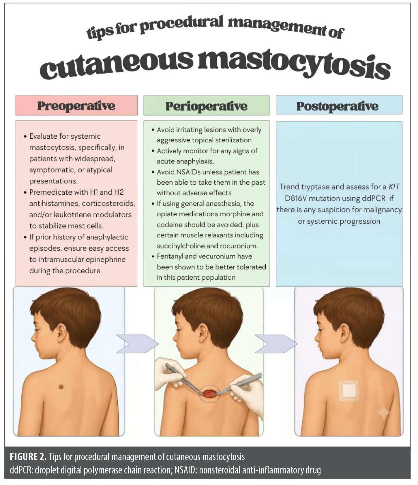
Of the 19 cases evaluated in our review, none reported perioperative complications, such as hypotension or anaphylactic shock, even when multiple large tumors were surgically operated on such as in the case described by McDermott and Topol.18 Notably, of the 19 cases, none of the patients received special preoperative treatment. However, given the paucity of literature describing surgical treatment of these lesions, a thorough history, physical examination, and laboratory markers such as tryptase may still be warranted to gauge the risk of perioperative complications. Even in the absence of positive findings, it is prudent to consider preoperative mast cell–inhibiting medications, as there is the rare possibility of anaphylaxis even in patients with CM.7 Further research may evaluate what indications might exist for triaging patients to higher-acuity surgical centers as opposed to excision in the dermatology office.
Patients with both CM and SM may be at an increased risk of anaphylaxis when undergoing certain general anesthesia, and it is recommended that surgically managed patients be postoperatively monitored for at least 24 hours since delayed anaphylaxis has occurred hours after surgery in some patients.7,33–35 Additionally, friction and pressure are known causes of irritation and degranulation of cutaneous mastocytomas and may precipitate anaphylaxis. In vitro studies have shown stabilization of rat mast cells with direct injection of lidocaine, and local anesthesia has been shown to be safe overall in mastocytosis; however, the safety of injecting directly into mast cell lesions in humans is uncertain.36,37 Systemic lidocaine is not recommended in patients with mastocytosis, though lidocaine injections can be safely used in these patients. Regarding general anesthesia, in a cohort of 219 procedures, 91.8% of known mastocytosis cases used chronic antimediator therapy and/or preoperative prophylaxis and 10 required intraoperative treatment for potential mast cell activation.38 This suggests that this protocol may be effective with a relatively low-risk profile if general anesthesia is necessary, such as in pediatric patients.
As mentioned in the previous section, tryptase levels have an undetermined significance in benign solitary lesions; however, they may have some utility in gauging the progression of malignant lesions. In patients with excised masses demonstrating mast cell infiltration or myeloid features on histopathology, trending serum tryptase levels postexcision can help identify residual systemic disease, progression to SM, or an associated neoplasm, though the likelihood of an event is low. Additionally, ddPCR of KIT D816V offers a promising new molecular tool to evaluate for the presence or development of SM.
This study was limited by the small sample size of case reports and lack of outcome data in some cases. Tryptase levels were unavailable for many patients, which limited the ability to analyze the utility of this biomarker across different presentations. Cutaneous mast cell tumors, especially malignant forms, are quite rare, which makes the generalizability of the findings difficult. However, this review aimed to synthesize the available evidence on surgical management of these lesions and spark a multidisciplinary conversation that could potentially lead to better guidelines and protocols for surgical treatment in the future. Further large-scale studies with robust data collection on outcomes are needed to develop higher-quality evidence-based recommendations.
Conclusion
Although rarely performed, surgical intervention has both diagnostic and therapeutic value for isolated mastocytomas. Careful triaging of procedures in addition to preoperative medication and perioperative management will help to minimize possible adverse outcomes. Transformation from mastocytoma to MCS was reported in 1 case, suggesting that surgical removal may be warranted for persistent isolated mastocytomas. The prognosis for MCS remains poor, and debulking surgery may be used among other multimodal approaches. The degree of tryptase level spike or persistence of high levels on serial measurements may directly relate to prognosis. Further study should examine tryptase trends as a way to help tailor and monitor treatment across the spectrum of presentations. This study was limited by the small sample size of case reports and lack of outcome data in some cases. Further studies and multidisciplinary input from numerous specialties are needed to devise surgical management guidelines.
References
- Valent P, Akin C, Hartmann K, et al. Updated diagnostic criteria and classification of mast cell disorders: a consensus proposal. Hemasphere. 2021;5(11):e646.
- Hartmann K, Escribano L, Grattan C, et al. Cutaneous manifestations in patients with mastocytosis: consensus report of the European Competence Network on Mastocytosis; the American Academy of Allergy, Asthma & Immunology; and the European Academy of Allergology and Clinical Immunology. J Allergy Clin Immunol. 2016;137(1):35–45.
- Gülen T. A puzzling mast cell trilogy: anaphylaxis, MCAS, and mastocytosis. Diagnostics. 2023;13(21):3307.
- Castells M, Butterfield J. Mast cell activation syndrome and mastocytosis: initial treatment options and long-term management. J Allergy Clin Immunol Pract. 2019;7(4):1097–1106.
- Ertugrul A, Baskaya N, Cetin S, Bostanci I. Anaphylaxis and epinephrine autoinjector use in pediatric patients with cutaneous mastocytosis. Pediatr Dermatol. 2021;38(5):1080–1085.
- González de Olano D, de la Hoz Caballer B, Núñez López R, et al. Prevalence of allergy and anaphylactic symptoms in 210 adult and pediatric patients with mastocytosis in Spain: a study of the Spanish network on mastocytosis (REMA). Clin Exp Allergy. 2007;37(10):1547–1555.
- Rama TA, Castells M. Triggers of anaphylaxis in mastocytosis patients: evidence of the current drug-avoidance recommendation. Curr Treat Options Allergy. 2023;10:442–457.
- Bernstein JA, Storey ES, Bauer RW. Moh’s micrographic surgery for the management of a periocular mast cell tumor in a dog. Vet Ophthalmol. 2013;16(3):234–239.
- Ashinoff R, Soter NA, Freedberg IM. Solitary mastocytoma in an adult. Treatment by excision. J Dermatol Surg Oncol. 1993;19(5):487–488.
- Arik Yilmaz E, Orhan D, Andiran F, Soyer O. Rare cause of recurrent urticaria in childhood and its unusual presentation: solitary mastocytoma on vulva. J Dermatol. 2017;44(2):213–214.
- Khan K, Kupferman ME, Gardner JM, Ivan D. Solitary mastocytoma in an adult with an unusual clinical presentation. J Am Acad Dermatol. 2011;65(3):683–684.
- Pandhi D, Singal A, Aggarwal S. Adult onset, hypopigmented solitary mastocytoma: report of two cases. Indian J Dermatol Venereol Leprol. 2008;74(1):41–43.
- Papanastasiou D, Magiakou MA, Giannopoulou E, Kaklamanis L. Solitary mastocytoma with generalized symptoms. Article in German. Monatsschr Kinderheilkd. 1988;136(4):203–205.
- Thappa DM, Jeevankumar B. Solitary mastocytoma. Indian Pediatr. 2005;42(4):390.
- Ha NH, Lee YJ, Park MC, et al. Solitary mastocytoma presenting at birth. Arch Craniofac Surg. 2018;19(2):127–130.
- Verma R, Verma RP. Surgical resection of a solitary cutaneous mastocytoma with resolution of apnea and multi-system symptoms. J Pediatr Surg Case Rep. 2017;22:39–41.
- Munro CS, Farr PM. Solitary mastocytoma causing recurrent blistering in infancy. Arch Dis Child. 1992;67(8):1038–1039.
- McDermott WV, Topol BM. Systemic mastocytosis with extensive large cutaneous mastocytomas: surgical management. J Surg Oncol. 1985;30(4):221–225.
- Wardle CLW, Oldhoff JM, Diepstra A, et al. Case report of a clinically indolent but morphologically high-grade cutaneous mast cell tumor in an adult: atypical cutaneous mastocytoma or mast cell sarcoma? J Cutan Pathol. 2021;48(11):1404–1409.
- Ryan RJH, Akin C, Castells M, et al. Mast cell sarcoma: a rare and potentially under-recognized diagnostic entity with specific therapeutic implications. Mod Pathol. 2013;26(4):533–543.
- Horny HP, Parwaresch MR, Kaiserling E, et al. Mast cell sarcoma of the larynx. J Clin Pathol. 1986;39(6):596–602.
- Chang IJ, Yang CY, Sung FY, Ng KY. A red-brown plaque on the nape. Solitary mastocytoma. Arch Dermatol. 2004;140(10):1275–1280.
- Auquit-Auckbur I, Lazar C, Deneuve S, et al. Malignant transformation of mastocytoma developed on skin mastocytosis into cutaneous mast cell sarcoma. Am J Surg Pathol. 2012;36(5):779–782.
- McKinnon EL, Rand AJ, Proia AD. Solitary mastocytoma in the eyelid of an adult. Am J Ophthalmol Case Rep. 2018;9:103–105.
- Baraf CS, Shapiro L. Solitary mastocytoma: case report in an adult. Arch Dermatol. 1969;99:589–590.
- Mylanus EA, Wielinga EW, van de Nes JA. A solitary manifestation of mastocytosis in the head and neck. Eur Arch Otorhinolaryngol. 2000;257(5):270–272.
- Sverrild A, van der Sluis S, Kyvik KO, et al. Genetic factors account for most of the variation in serum tryptase–a twin study. Ann Allergy Asthma Immunol. 2013;111(4):286–289.
- Valent P, Akin C, Metcalfe DD. Mastocytosis: 2016 updated WHO classification and novel emerging treatment concepts. Blood. 2017;129(11):1420–1427.
- Greiner G, Gurbisz M, Ratzinger F, et al. Digital PCR: a sensitive and precise method for KIT D816V quantification in mastocytosis. Clin Chem. 2018;64(3):547–555.
- Collins N, Willard N, Pan Z. Mast cell sarcoma with KIT p.D816V mutation and concurrent systemic mastocytosis. J Hematop. 2024;17(4):281–287.
- Skaljic M, McGinnis A, Streicher JL. Comfort positioning during procedures in pediatric dermatology. Pediatr Dermatol. 2020;37(2):396–398.
- Bocca-Tjeertes IFA, van de Ven AAJM, Koppelman GH, et al. Medical algorithm: peri-operative management of mastocytosis patients. Allergy. 2021;76(10):3233–3235.
- Matsumoto NP, Yuan J, Wang J, et al. Mast cell sarcoma: clinicopathologic and molecular analysis of 10 new cases and review of literature. Mod Pathol. 2022;35(7):865–874.
- Brockow K, Bonadonna P. Drug allergy in mast cell disease. Curr Opin Allergy Clin Immunol. 2012;12(4):354–360.
- Carter MC, Uzzaman A, Scott LM, et al. Pediatric mastocytosis: routine anesthetic management for a complex disease. Anesth Analg. 2008;107(2):422–427.
- Tanasi I, Olivieri E, Oberti M, et al. Safety of local anesthesia and prevalence of hypersensitivity reactions in adult patients with clonal mast cell diseases: a retrospective single-center study. J Allergy Clin Immunol Pract. 2021;9(8):3224–3226.
- Yanagi H, Sankawa H, Saito H, Iikura Y. Effect of lidocaine on histamine release and Ca2+ mobilization from mast cells and basophils. Acta Anaesthesiol Scand. 1996;40(9):1138–1144.
- Lau S, Sprung J, Volcheck GW, et al. Perioperative management of mastocytosis. J Anesth. 2023;37(5):741–748.
From the 2nd Annual RAPIDS Immuno-Dermatology Conference: From Approval to Adoption—Translating Innovation Into Clinical Practice