
J Clin Aesthet Dermatol. 2026;19(10):50–53
Sabine Obagi, MD, and Clay J. Cockerell, MD, MBA, JD
Dr. Obagi is with the Department of Medicine, Harbor-UCLA Medical Center, Torrance, California. Dr. Cockerell is with Cockerell Dermatopathology, Dallas, Texas.
FUNDING: No funding was provided for this article.
DISCLOSURES: The authors have no relevant conflicts of interest.
Abstract: OBJECTIVE: To evaluate the clinical utility and diagnostic concordance of the designation “rule out melanoma” (ROM) in dermatopathology requisitions and to assess its potential contribution to diagnostic inflation. METHODS: An observational analysis was conducted of 13,000 dermatopathology requisition forms submitted between January 1 and July 1, 2025, in which ROM was listed as the primary or sole clinical diagnosis. Concordance between clinical designation and histopathologic diagnosis was assessed. RESULTS: Of 13,000 cases, 1,196 (9.2%) were diagnosed as melanoma (invasive or melanoma in situ). The remaining 11,804 cases (90.8%) represented nonmelanoma diagnoses, most commonly solar lentigo, seborrheic keratosis, melanocytic nevi (including dysplastic nevi), pigmented actinic keratosis, and pigmented basal cell carcinoma. LIMITATIONS: This analysis represents a single-institution observational dataset. CONCLUSION: The ROM designation demonstrates low positive predictive value and provides limited actionable diagnostic information. These findings suggest that current clinical labeling practices may contribute to reduced diagnostic specificity and broader patterns of diagnostic inflation. Keywords: Dermatopathology, clinical correlation, rule out melanoma, melanoma in situ, overdiagnosis, electronic medical records, diagnostic amplification
Introduction
Clinicopathologic correlation represents a central principle of dermatopathology, particularly in the evaluation of pigmented lesions, where clinical morphology, lesion evolution, and risk stratification directly inform histopathologic interpretation.1 In contemporary practice, this collaborative exchange has been increasingly constrained by electronic medical record (EMR) workflows that prioritize efficiency and structured inputs over descriptive clinical reasoning. As a result, detailed morphologic descriptions are frequently replaced by abbreviated diagnostic labels. Furthermore, “canned” clinical descriptions are often proffered, such as “irregular, multi-brown lesion” for virtually all lesions without further description.
Among these, the phrase “rule out melanoma” (ROM) has become ubiquitous. Although originally intended to communicate clinical concern, its widespread application has rendered it a nonspecific designation that often fails to convey meaningful diagnostic information.2
In this study, we sought to determine the clinical utility of ROM and its implications for diagnostic accuracy.
Methods
An internal observational analysis was conducted of 13,000 dermatopathology requisition forms submitted between January 1 and July 1, 2025. Of these, virtually all were submitted by either board-certified dermatologists or dermatology-specific providers (physician assistants or nurse practitioners).
Inclusion criteria required that ROM be listed as either the primary or sole clinical diagnosis. Histopathologic outcomes were reviewed, and concordance between clinical designation and final diagnosis was assessed.
Results
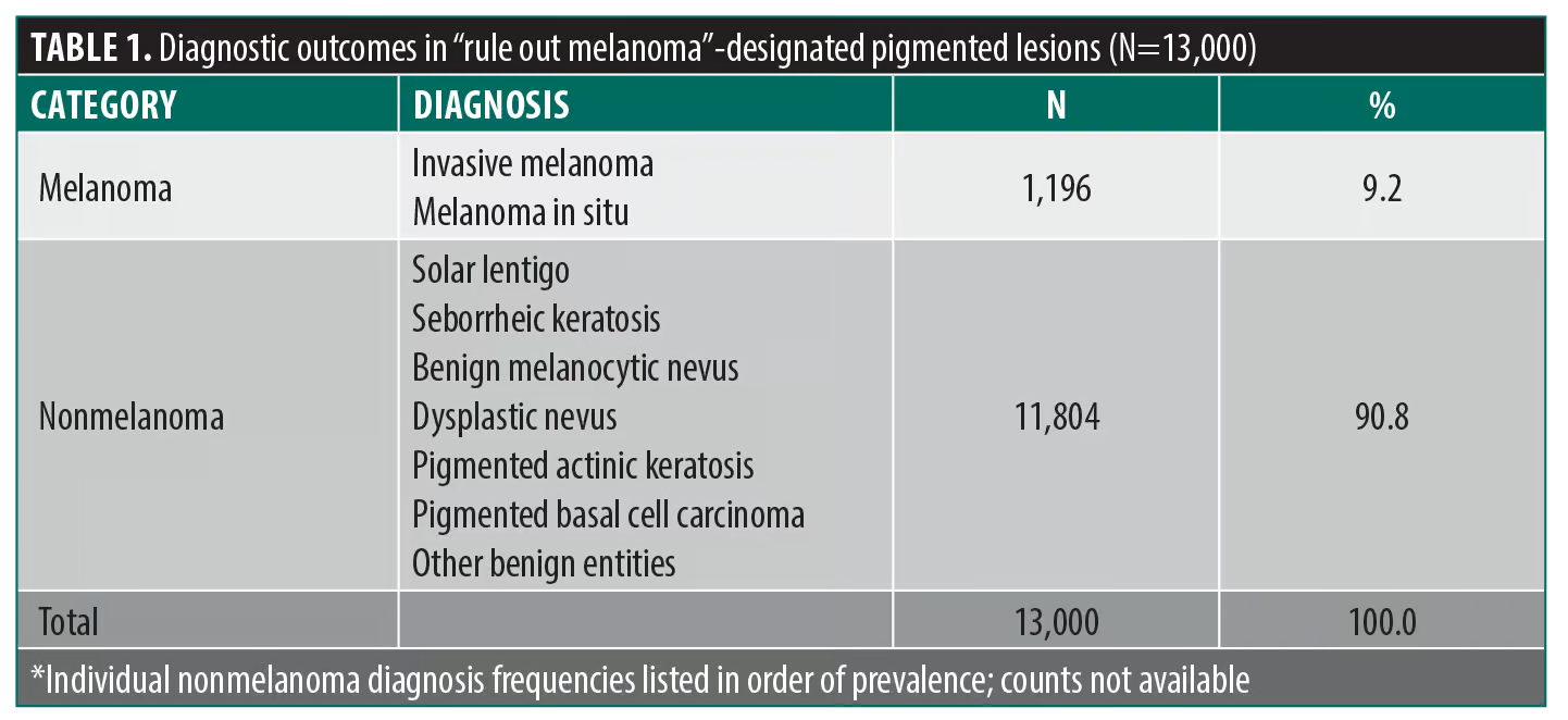
Of the 13,000 specimens that met the inclusion criteria, 1,196 (9.2%) were diagnosed as melanoma (invasive or melanoma in situ). The remaining 11,804 cases (90.8%) represented nonmelanoma diagnoses. These included solar lentigo, seborrheic keratosis, benign melanocytic nevi, dysplastic nevi, pigmented actinic keratosis, and pigmented basal cell carcinoma (Table 1).
Contextual comparison with population-level incidence. Population-based data indicate that invasive melanoma occurs at a rate of approximately 25 to 30 cases per 100,000 individuals annually, with total melanoma incidence (including melanoma in situ) estimated at approximately 60 to 70 cases per 100,000.3 In contrast, the present observational cohort, representing a highly selected population of biopsied lesions, demonstrated a melanoma yield of 9.2%.
If all lesions designated as ROM in this cohort had represented true melanoma, the implied incidence would approach several thousand cases per 100,000 individuals annually, orders of magnitude higher than established population-based estimates. This discrepancy highlights the extent to which ROM labeling is applied beyond cases of meaningful clinical suspicion.
Discussion
The conceptual limitations of “rule out” diagnosis. The phrase “rule out melanoma” is neither a diagnosis nor a structured differential diagnosis. It does not communicate probability, morphologic reasoning, or prioritization of diagnostic possibilities. Instead, it represents a binary acknowledgment that melanoma has been considered.4
Effective clinicopathologic correlation requires transmission of pretest probability, which allows the dermatopathologist to interpret histopathologic findings within a clinically meaningful framework. Because the clinical information that is proffered in this context is generally formulaic, generated by an EMR without specific information and description of the lesion in question, the ROM designation fails to provide sufficient pretest probability. A more informative approach would include descriptive morphology, lesion size, and a prioritized differential diagnosis, such as “asymmetric 7-mm pigmented macule on the upper back with interval change; moderate concern for melanoma vs dysplastic nevus.”
Additional information, including dermoscopic features and clinical photographs, should be incorporated in appropriate circumstances and when available.
Beyond lesion-level morphology, several additional elements can meaningfully raise or lower pretest probability and should be conveyed when relevant. These include a personal or family history of melanoma, a history of dysplastic (atypical) nevi, and the patient’s Fitzpatrick skin type and degree of cumulative photodamage. Equally important, the differential diagnosis should be framed to include the benign entities that most commonly mimic melanoma clinically, such as dermatofibroma, dermal (intradermal) melanocytic nevus, and seborrheic keratosis, rather than defaulting to melanoma alone. Explicitly naming these alternatives conveys genuine diagnostic reasoning and helps calibrate, rather than inflate, the dermatopathologist’s index of suspicion.
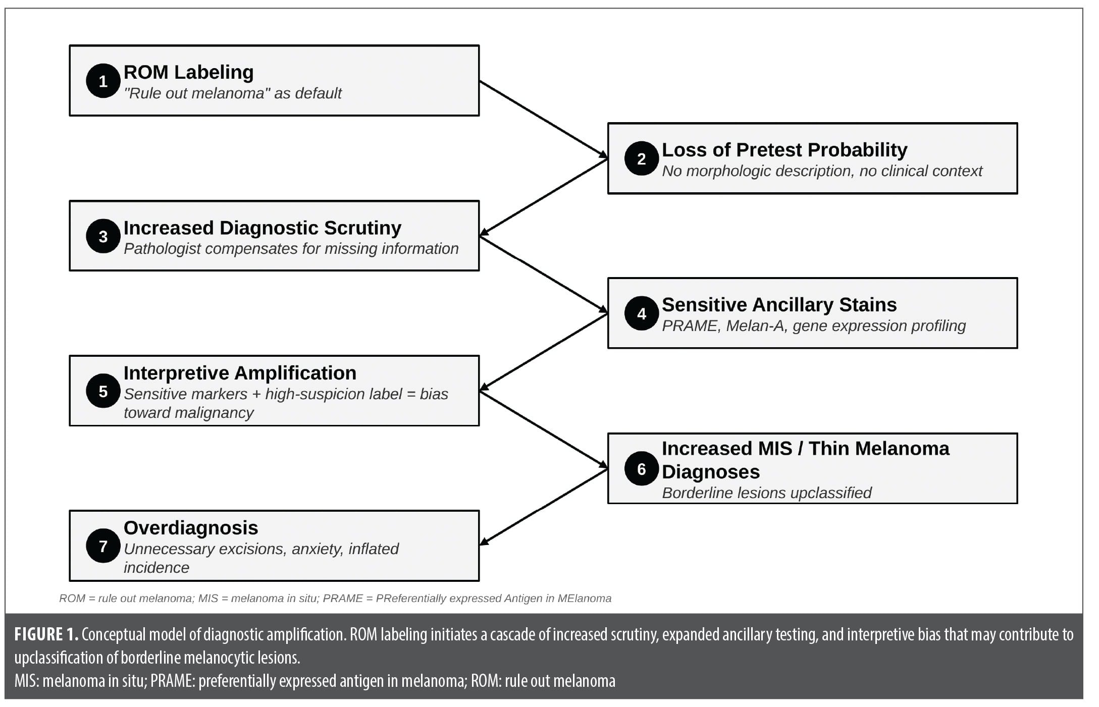
Diagnostic amplification and ancillary testing. ROM labeling influences the behavior of the dermatopathologist confronted with a pigmented lesion and this limited information. When melanoma is explicitly raised, dermatopathologists often increase diagnostic scrutiny and use ancillary studies.
Immunohistochemical markers such as preferentially expressed antigen in melanoma (PRAME) and Melan-A are frequently employed in this setting. While useful, these markers are highly sensitive and may lack specificity in certain contexts. Increased staining can occur in benign or mildly atypical lesions, particularly in sun-damaged skin.5 This introduces a risk of overinterpretation, particularly when combined with high-suspicion clinical labeling. The interaction between nonspecific clinical input raising suspicion of melanoma and sensitive diagnostic tools may amplify diagnostic uncertainty rather than resolve it.
These ancillary studies also carry meaningful and frequently underappreciated costs. Each immunohistochemical stain adds incremental technical and professional charges; single-antibody immunohistochemistry, such as PRAME or Melan-A, is commonly reimbursed on the order of $90 to $120 per stain by commercial payers, and several stains are often ordered on a single specimen. Molecular assays are considerably more expensive, with gene expression profiling for melanocytic lesions reimbursed in the range of several thousand dollars per test.6 When applied broadly to lesions with low pretest probability, more than 90% of which proved benign in the present cohort, these incremental expenditures aggregate into a substantial and potentially avoidable burden on healthcare spending.7 Critically, this cost is not borne by payers alone. Patients are frequently responsible for a portion through deductibles, coinsurance, and out-of-network or balance billing, such that low-yield testing prompted by nonspecific clinical labeling can translate into direct and unexpected financial consequences for individuals.5
This process may be conceptualized as diagnostic amplification (Figure 1).
Melanoma in situ; Surveillance, Epidemiology, and End Results (SEER) limitations; and overdiagnosis. Melanoma incidence has risen substantially over recent decades, particularly for melanoma in situ, while mortality has remained relatively stable.8–10
SEER datasets have historically focused on invasive melanoma and incompletely captured melanoma in situ.3 However, current estimates suggest that melanoma in situ incidence approaches that of invasive melanoma, with the American Cancer Society projecting over 107,000 new cases in 2025 alone.11 Published analyses estimate that 29% to 60% of melanoma diagnoses, and more than 80% of melanoma in situ, may represent overdiagnosis.10,12
The Melanocytic Pathology Assessment Tool and Hierarchy for Diagnosis (MPATH-Dx) framework reflects ongoing efforts to align diagnostic terminology with biological behavior and acknowledges uncertainty in the classification of early melanocytic lesions.13 Because of the overdiagnosis of melanoma in situ noted above, discussion has been ongoing to reclassify borderline lesions into risk-stratified categories that may behave less like true melanoma.
Cognitive bias and diagnostic threshold drift. Clinical labeling influences diagnostic interpretation through anchoring and framing effects.14,15 When lesions are labeled as suspicious for melanoma, borderline histopathologic features may be interpreted more aggressively.
Increased surveillance has also been shown to increase biopsy rates and melanoma diagnoses independent of underlying biological risk.16
Implications for Clinical Practice
The findings of this study suggest that the ROM designation provides limited diagnostic value and may contribute to reduced specificity in clinical communication.
Replacing nonspecific “rule out” terminology with structured clinical descriptions, including lesion size, asymmetry, dermoscopic features, and prioritized differential diagnoses, represents an important opportunity to improve diagnostic accuracy and restore clinicopathologic correlation.17 Incorporating relevant clinical history, including any personal or family history of melanoma or dysplastic nevi and the patient’s skin phototype, further strengthens this communication.
Limitations
This analysis represents a single-institution observational dataset and may not fully generalize across all practice settings. However, the large sample size supports broader relevance.
Conclusion
In the evaluation of pigmented skin lesions, the ROM designation demonstrates low positive predictive value and limited diagnostic utility.
When considered alongside population-level melanoma incidence, rising melanoma in situ diagnoses, and evolving classification frameworks, these findings suggest that current clinical labeling practices contribute to reduced diagnostic specificity and may play a role in diagnostic inflation.
References
- Nguyen CV, Miller DD. The role of dermatopathology in inpatient care. Semin Cutan Med Surg. 2017;36(1):17–22.
- Arimany Manso J, Martin Fumadó C, Mascaró Ballester JM. Medical malpractice issues in dermatology: clinical safety and the dermatologist. Actas Dermosifiliogr (Engl Ed). 2019;110(1):20–27.
- American Cancer Society. Cancer Facts & Figures 2025. Atlanta, GA: American Cancer Society; 2025. Accessed 6 Sep 2026. https://www.cancer.org/research/cancer-facts-statistics/all-cancer-facts-figures/2025-cancer-facts-figures.html
- Davis LE, Shalin SC, Tackett AJ. Current state of melanoma diagnosis and treatment. Cancer Biol Ther. 2019;20(11):1366–1379.
- Marks E, Jarell A, Ludzik J, et al. A physician’s guide to the use of gene expression profile ancillary diagnostic testing for cutaneous melanocytic neoplasms. J Clin Aesthet Dermatol. 2023;16(4):12–20.
- Cassarino DS, Lewine N, Cole D, et al. Budget impact analysis of a novel gene expression assay for the diagnosis of malignant melanoma. J Med Econ. 2014;17(11):782–791.
- Sheerin J, Collgros H, Chamberlain A, et al. A clinical perspective on melanoma overdiagnosis. Australas J Dermatol. 2025;66(7):388–395.
- Kurtansky NR, Dusza SW, Halpern AC, et al. An epidemiologic analysis of melanoma overdiagnosis in the United States, 1975–2017. J Invest Dermatol. 2022;142(7):1804–1811.
- Karponis D, van Bodegraven B, Mistry K, et al. Incidence and mortality of melanoma in situ and malignant melanoma in England between 2001 and 2020. Br J Dermatol. 2025;193(4):687–695.
- Bjørch MF, Gram EG, Brodersen JB. Overdiagnosis in malignant melanoma: a scoping review. BMJ Evid Based Med. 2024;29(1):17–28.
- Galambus J, Smythe C, Cockerell CJ. Pathology of melanoma: new concepts (2025 update). Dermatol Clin. 2025;43(3):409–418.
- Adamson AS, Naik G, Jones MA, Bell KJ. Ecological study estimating melanoma overdiagnosis in the USA using the lifetime risk method. BMJ Evid Based Med. 2024;29(3):156–161.
- Lott JP, Elmore JG, Zhao GA, et al; International Melanoma Pathology Study Group. Evaluation of the Melanocytic Pathology Assessment Tool and Hierarchy for Diagnosis (MPATH-Dx) classification scheme for diagnosis of cutaneous melanocytic neoplasms: results from the International Melanoma Pathology Study Group. J Am Acad Dermatol. 2016;75(2):356–363.
- Bucchi L, Mancini S, Zamagni F, et al; AIRTUM Working Group. Patient presentation, skin biopsy utilization and cutaneous malignant melanoma incidence and mortality in northern Italy: trends and correlations. J Eur Acad Dermatol Venereol. 2023;37(2):293–302.
- Bucchi L, Mancini S, Zamagni F, et al. North-south differences in incidence and surveillance of cutaneous malignant melanoma in Italy. Tumori. 2024;110(4):264–272.
- Whiteman DC, Olsen CM, MacGregor S, et al; QSkin Study. The effect of screening on melanoma incidence and biopsy rates. Br J Dermatol. 2022;187(4):515–522.
- Cockerell CJ. Real-world medicolegal issues in dermatology and dermatopathology. J Clin Aesthet Dermatol. 2026;19(1):27–29.
From the 2nd Annual RAPIDS Immuno-Dermatology Conference: From Approval to Adoption—Translating Innovation Into Clinical Practice