
J Clin Aesthet Dermatol. 2026;19(8):41–49.
Potluri Sri Harshitha, B.Tech; Anupama Damarla, PhD; Vangala Bhargavi, B.Tech; Tedla Sai Charan, B.Tech; Batchu Sai Karthik, B.Tech; Karthika, M.Tech; Sumathi D, PhD; Gouranga Mandal, PhD
Ms. Harshitha, Ms. Bhargavi, Mr. Charan, and Mr. Karthik are with the School of Computer Science and Engineering, Vellore Institute of Technology, Andhra Pradesh University, Amaravati, India. Dr. Damarla is with the Department of Data Science, Malla Reddy University, Hyderabad, India. Ms. Karthika is with the School of Computer Science and Engineering, Vellore Institute of Technology, Andhra Pradesh University, Amaravati, India. Dr. Sumathi D is with the Department of Cybersecurity, Dayananda Sagar University, Bangalore, India. Dr. Mandal is with Manipal Institute of Technology, Manipal Academy of Higher Education, Manipal, India.
FUNDING: No funding was provided for this article.
DISCLOSURES: The authors have no relevant conflicts of interest.
Skin segmentation of lesions can be challenging because it relies on numerous factors such as size, shape, and position. Convolutional neural networks (CNN) have shown exceptional efficacy in automating image segmentation. The inability of current CNNs to maintain local features and emphasize pertinent information restricts decision-making ability. To achieve more precise medical image segmentation, this research suggests a CNN with a spatial feature mechanism (UCMNet). The UNet architecture serves as the foundation for UCMNet. To retain an image’s local features during downsampling and increase the relevance of the feature maps supplied to the upsampling module relative to the ground truth, a spatial multiple-scale characteristic (SA) module is included in the decoder. Comprehensive tests were conducted on the 3D-TBP dataset. The suggested UCMNet outperformed UNet in skin lesion tissue segmentation, with an average Dice score improvement of 0.82% and 0.85%, respectively. The suggested UCMNet outperformed UNet in skin lesion tissue segmentation, with an average Dice score improvement of 0.03 and the Intersection over Union also rose by 0.08. The pixel error fell from 0.7132 to 0.6083 pixels, indicating better segmentation performance. When the segmentation area contains interference factors and the initial image of skin lesions has an indistinct boundary, UCMNet can achieve better results. It demonstrated strong segmentation ability by incorporating attention mechanisms into the encoder and applying comprehensive attention methods. Keywords: Dermoscopic images, skin lesion segmentation, deep learning, UCM-Net, CNN
Introduction
According to the World Health Organization, approximately 10 million people worldwide died from cancer in 2022, with approximately 1.2% due to skin cancer. The International Agency for Research on Cancer (IARC) estimated there were 332,000 new cases of melanoma and over 1.2 million new cases of nonmelanoma skin cancer cases worldwide in 2022.1 Skin cancer symptoms include asymmetric moles, borders with irregular or blurred edges, and large patches in various shades of brown, red, black, white. Dermatologists have emphasized the early detection of skin cancer, especially melanoma, as critical and potentially lifesaving. Early detection can aid in higher survival rates, allow use of less aggressive treatment, lower healthcare costs, and improve quality of life.2 Thus, an automated system for the detection of skin cancer, which includes data acquisition, data preprocessing, segmentation, and classification, can play a significant role in early diagnosis.
The 3-dimensional (3D) total body photography (3D-TBP) dataset is a medical image dataset used primarily in skin cancer research and detection, especially focusing on melanoma. It consists of high-resolution, 3D body surface scans of real patients to study skin lesion distribution patterns on the human body and to improve early detection of melanoma through full-body analysis. The Unique Features of 3D-TBP Include 3D modeling, images of multiple lesions per patient, and the ability to be combined with dermoscopic and clinical imaging. Data Cleaning, or Preprocessing, is an important step, as it can result in more accurate Results, Better Model Performance, and reduced Inconsistencies, making Analysis Easier.
To perform this step, we will collect images from the 3D-TBP, which contains real-time patient data. These images may present challenges such as blurriness or hair around the lesion area, which can hinder accurate analysis and detection of the lesion. The result of Data Cleaning can be a reliable, accurate, and usable dataset.
After Data Processing, the advanced processing, segmentation, and classification techniques come into picture. The segmentation includes UNet algorithm, which takes an input image and outputs a segmentation mask where each pixel is classified into a category, such as lesion or nonlesion. So, a possible output could be a green color lesion around the contour. After this, we perform the classification, where we classify the Images as either Class 1, malignant (positive), and Class 0, benign (negative).
Machine learning, ensemble learning, and deep learning approaches. Machine learning, ensemble learning, and deep learning models are used after classification. The machine learning models include support vector machine, logistic regression, and linear regression; Ensemble Learning Methods Include Random Forest and Boosting, and the Deep Learning Models include MobileNet architecture. The result of applying these approaches include accuracy, precision, and recall.
In this paper, we will:
- Address the challenge of accurately segmenting skin lesions, which is influenced by lesion size, shape, and location
- Propose a new model, UCMNet, to enhance segmentation accuracy in medical imaging. It is robust in handling indistinct lesion boundaries and interference in segmentation regions
- Demonstrate that attention mechanisms can significantly boost convolutional neural network (CNN) performance in clinical image segmentation
- Introduce a Spatial Attention (SA) module in the decoder that preserves local image features during downsampling
- Enhance feature relevance in upsampling, improving alignment with the ground truth
- Evaluate the International Skin Imaging Collaboration (ISIC) 2024 dataset3 for skin lesion segmentation and compare the performance against the baseline UNet
Literature Review
In recent years, skin cancer, especially malignant melanoma, has been on the rise, notably among individuals with lighter skin tones who spend more time in the sun. Diagnosing skin cancer accurately is still a major challenge, largely due to the fact that melanoma lesions often come in different shapes, sizes, and colors. Additionally, their borders are usually blurry and there is little contrast between the lesion and the surrounding healthy skin, making it tough for even advanced systems to correctly detect them. These factors often result in segmentation challenges, such as missing parts of the lesion or overestimating its boundaries. Although CNNs have brought improvements in image segmentation, many encoder-decoder frameworks, such as ResNet, and tightly connected networks depend heavily on datasets unrelated to medical imaging. Furthermore, limitations such as poorly tuned parameters, limited insights from pretrained features, and a lack of multiscale context awareness, reduce their success in real-world melanoma segmentation tasks.4
To deal with these challenges, researchers developed a modified version of the UNet model by using DenseNet77 as the encoder. This change allowed the model to notice finer details in the skin images, which helped the decoder create clearer and more accurate outlines of the lesion areas. By addressing the wide-ranging visual differences in skin lesions, this updated model delivered improved segmentation results compared to earlier approaches.5
Apart from segmentation, identifying and classifying skin lesions accurately and at the right time are equally important, as these can greatly influence the effectiveness of treatment. To support early diagnosis, researchers built a model called InSiNet using a CNN designed to identify whether a skin lesion is benign or malignant. It was evaluated on well-known datasets, including HAM10000 (2018), ISIC 2019, and ISIC 2020. During testing, InSiNet consistently delivered better results than many popular models, not only in terms of accuracy but also in processing speed. Compared to models such as GoogleNet, DenseNet-201, ResNet152V2, and EfficientNetB0 and even classic methods such as random forests and logistic regression, InSiNet proved to be faster and more reliable, making it a strong candidate for real-world clinical use.6
For prognostic evaluation, especially in older patients diagnosed with ulcerative melanoma, survival prediction tools are crucial. A robust nomogram was constructed using SEER data (2004–2015), incorporating prognostic variables such as age, tumor size, cancer stage, and treatment details. Cox regression models were used to construct and validate the tool, with receiver operating characteristic curves, calibration plots, and decision curve analyses confirming its high predictive accuracy.7
While radiology adopted artificial intelligence (AI) early on,8 dermatology has taken a more gradual approach. Recently, however, doctors have started using deep learning methods, particularly CNNs, to examine skin images. This shift is helping spot signs of skin cancer, like malignant lesions, sooner and with fewer diagnostic mistakes.9 Especially in underserved regions, these AI systems offer a scalable means of enhancing access to care and reducing the psychological and economic burden of late-stage diagnoses.10 Automated diagnostic systems are also being explored to overcome challenges in hospitals dealing with large volumes of skin cancer cases and a shortage of dermatologists.10 Hybrid models that combine deep neural networks for feature extraction with machine learning classifiers have shown promise.11 Feature extraction techniques such as the Contourlet Transform and Local Binary Pattern Histograms have enhanced the precision of classification tasks, offering reliable alternatives to traditional diagnostic methods.11
More than 14,000 studies were reviewed to explore how tools like AI and machine learning are being applied to detect skin cancer. Among them, 272 studies were found to be directly relevant,12 with many reporting encouraging results, particularly in general healthcare and local clinic settings. However, there is still not enough evidence on how well these tools work in places where skin cancer cases are rare. Skin lesions and tumors are more difficult for less experienced dermatologists to diagnose accurately. Doctors’ methods for interpreting and analyzing lesion imaging is labor intensive and frequently erroneous due to subjective evaluation, primarily due to the difficulty of correctly portraying skin flaws on camera.13
For AI to become a normal part of medical routines, more testing in real-world settings is needed, along with a better grasp of how affordable, useful, and accepted these systems are among both doctors and patients. The fact that machine learning techniques necessitate human-engineered features is a disadvantage. This problem was partially resolved by deep learning, which offered the ability to automatically extract features. To produce a better choice, the collaborative learning technique takes advantage of the diversity of learners.14 For the purpose of training the model, machine learning with supervision needs data, which presents significant system requirement issues for mobile applications in terms of processing and storage. Consequently, numerous studies concentrate on the implementation of cloud-based learning algorithms,15 which use a network connection for outsourcing computations requiring a high amount of data. However, there are several disadvantages to this strategy, including latency and privacy issues, that should be taken into account when dealing with sensitive material.15 These data augmentation methods like rotation, flipping, scaling, and color transformation have been proven to enhance the robustness and generalization ability of the CNN’s in skin lesion classification.16
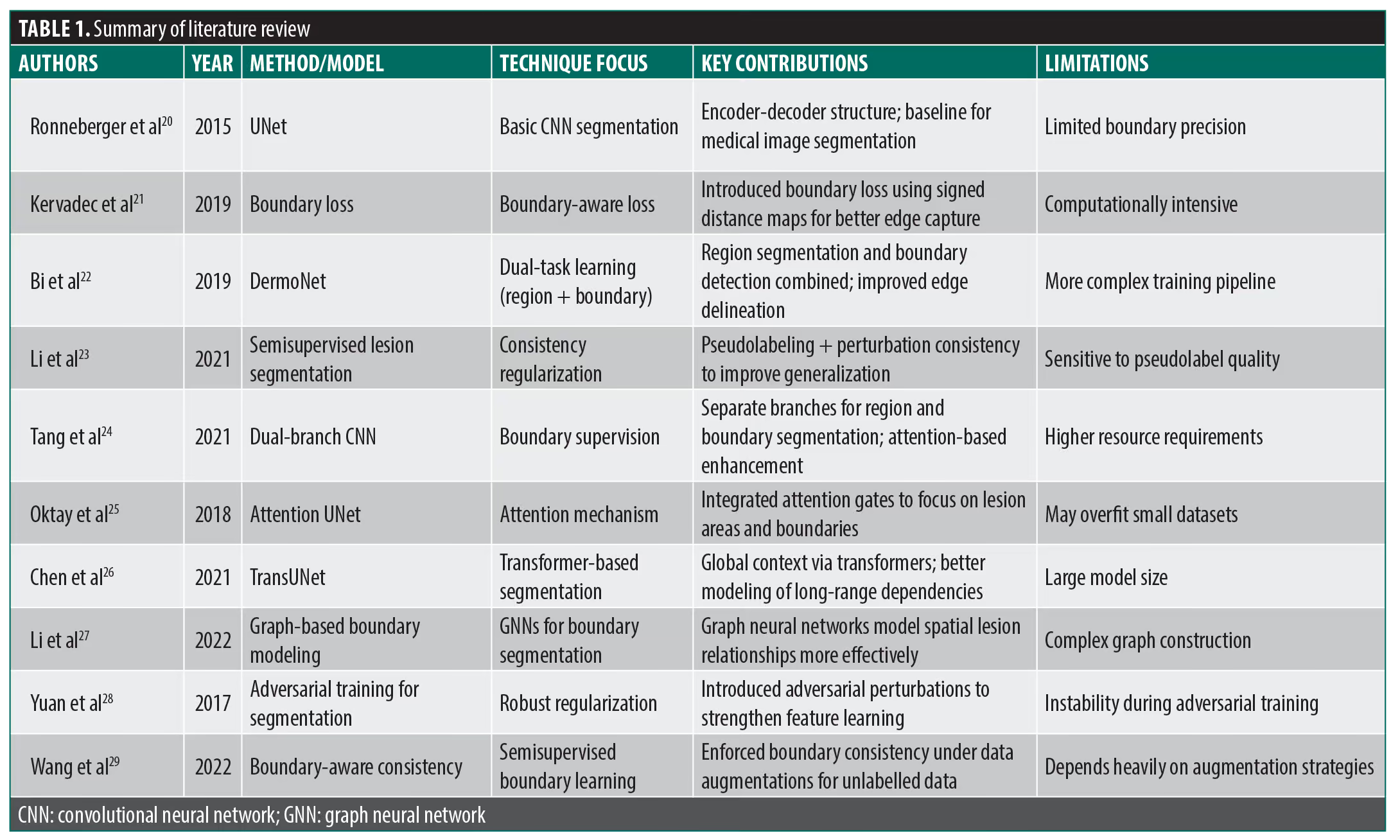
Given the severity of the conditions that lead to skin cancer, scientists have created a number of early detection methods. In order to make skin cancer more treatable in its early stages, lesion criteria, including symmetry, color, size, and shape, are employed to detect and differentiate it from melanoma.17 Mahum and Aladhadh18 employed a CNN with ReLU as an activation function to identify lesions using a generalized Gaussian distribution approach for image segmentation. The model’s performance was measured in terms of sensitivity, specificity, and accuracy. The suggested method’s accuracy was 98.32% on the ISIC 2016 dataset. As evidence of the influence of architectural depth, deep neural networks with consistently good performance included ResNet-50, ResNet34, Inception V3, and ResNet 152. Additionally, the study investigated the relationship between accuracy and dataset size, exposing a range of model responses. Outliers, anomalies, and the impact of certain datasets (such as unbalanced classes) were revealed by temporal trends and model-specific studies, offering important new information for further study and model development.19 Other works were summarized in Table 1.
Methodology
Dataset description. This study is based on data from the ISIC 2024 Challenge,3 a leading initiative focused on improving skin cancer detection using 3D-TBP. The dataset includes high-resolution JPEG images, with each image showing a single skin lesion. To maintain consistency and keep attention on the lesion area, the images were cropped to 15 mm×15 mm; this standard size helps improve performance in image segmentation tasks. In addition to the images, the dataset offers detailed metadata, including patient age, gender, lesion identification, biopsy date, and histopathology results. These details are useful for classifying lesions into benign, malignant, or intermediate categories, improving the accuracy of classification models. To ensure a high-quality analysis, only lesions confirmed through biopsy were included. Their related information was carefully documented in metadata tables. The lesion images were extracted from the 3D-TBP dataset using the official Canfield ISIC 2024 Tile Export Tool, which ensures accuracy and consistency. The metadata were further refined to suit machine learning needs, making the dataset reliable and well prepared for research in medical image analysis.
Data processing. The initial vital step after acquiring the large ISIC 2024 dataset from Kaggle required data preprocessing of its more than 400,000 skin cancer photographs. The models used for segmentation and classification depend on preprocessing quality because it influences the accuracy and reliability output of these models. Once the Kaggle environment received the imported dataset, preprocessing was started to transform it into an analyzable state.
The presence of hair follicles on skin sections became a major technical obstacle during the preprocessing operations. The lesion detection task became complicated because the hair follicles covered many of the skin areas that needed evaluation. The lack of resolution in fixing this issue could lead the models to mistake visual data, thus compromising their detection capabilities for skin cancer lesions. The objective of this subsection is to remove follicles that appeared in the output image of the preceding module.
Hair removal involved 3 steps: contouring the hairs, creating a mask, and employing in-painting to eliminate hairs in the image. This process began with generating a grayscale image from an RGB image to identify hair contours. The outlines of the hairs in the grayscale image were identified by subtracting the original image from its closing version using:
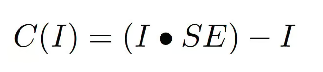
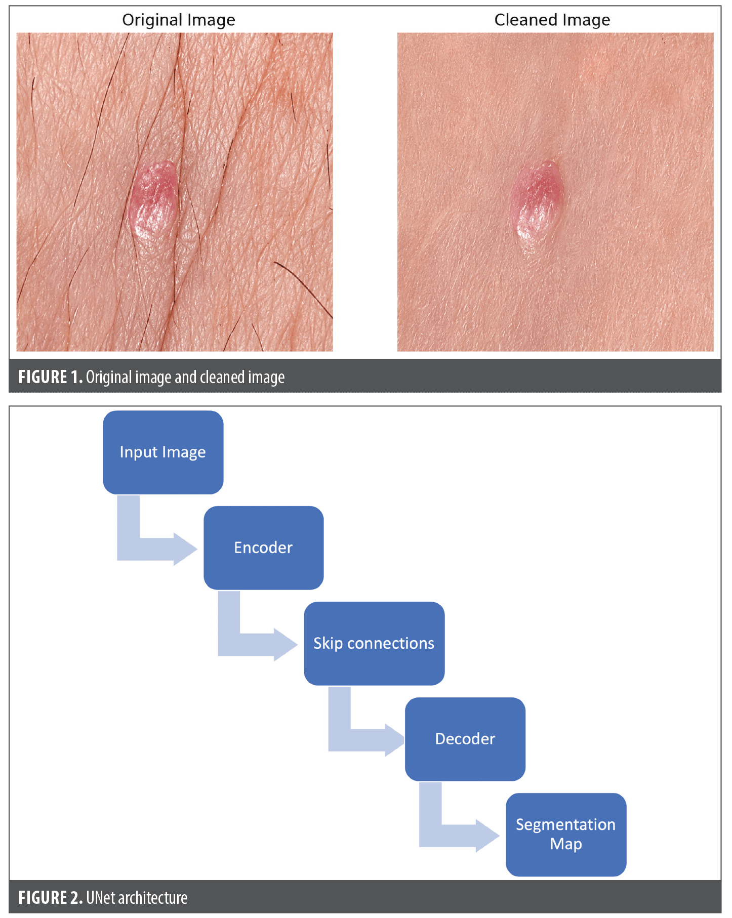
where I denotes the image, SE denotes the structure element (component), • is the closing operation, and C(I) is the outline image. A mask was then created using gray-level slicing to define the in-painting region. The mask contains zeroes or nonzero pixels, indicating the area for in-painting. The mask was then used in the third phase to eliminate hair from the RGB image. This also continued adjusting the image size and normalization of the pixel values and setting appropriate brightness and contrast levels to maintain consistent data integrity. The developed images then were saved as “Cleaned Images” following their improvement stage.
The best quality input data were given to the models since this folder acted as the basis for all the segmentation and classification operations. The difference between the original and preprocessed images are depicted in Figure 1.
Segmentation and classification using UNet. After preprocessing raw images to obtain clean images, the UNet architecture was implemented for segmentation in the initial phase of the process.The UNet architecture is a symmetric encoder-decoder framework used in biomedical image analysis. It extracts spatial features using sequential convolutional layers, introducing nonlinearity and reducing spatial dimensions while retaining essential features. The architecture also employs max-pooling layers to reduce computational costs and improve the hierarchical representation of image features. The feature reduction process occurs through dense network convolutional layers, generating a compact yet descriptive feature map at the bottleneck region.
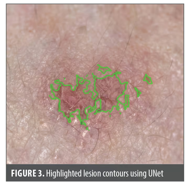
The model’s reconstruction of spatial resolution occurs through upsampling operations. UNet produces pixel-wise classification probabilities through convolutional operations, delivering segmented high-definition masks targeting medical lesions (Figure 2). Its flexible design makes it an essential framework for medical image analysis, providing stable and accurate segmentation for clinical use. The processed cleaned images entered the UNet model, which then performed an accurate identification of needed regions of interest. The segmentation process designated the identified lesion areas through the addition of green borders. The identified boundary represented the affected regions with clarity, which proved that the segmentation process successfully distinguished the lesion areas from other image elements. The UNet Architecture of this project generated the Highlighted Lesion Contours Image depicted in Figure 3. An explanation of learning and inference using the UNet architecture is provided by the following mathematical equation:
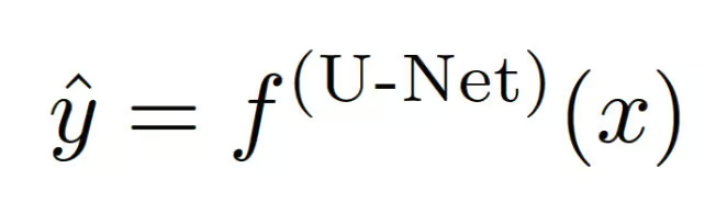
where input x represents a multiscale feature and U-Net f(U-Net) yields the logits ŷ with respect to x, where ŷ indicates the appropriate pixel-wise probability S. The feature map of layer l is computed to produce characteristics (features) at multiple levels within f(U-Net) of N layers of convolutional operation, as represented in the following equation:
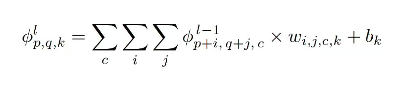
Subsequent convolution and Max-Pooling layers were employed to augment the receptive field, facilitating the acquisition of higher levels information completely in the equation below:
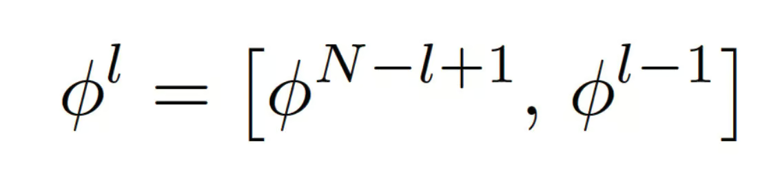
Finally, given the softmax function by the following equation:
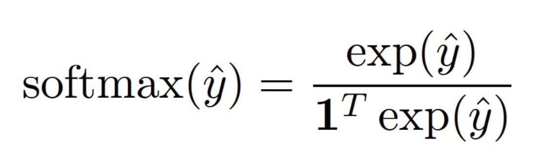
with regard to input x, ŷ is the prediction of f(U-Net), and for every category, α is the flexible weights vector. The following equation dynamically calculates each component of α:
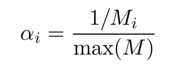
where Mi is the total count of category i and αi is the adaptive weight of category i.
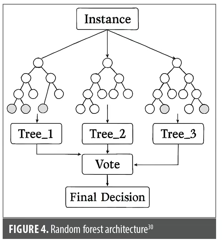
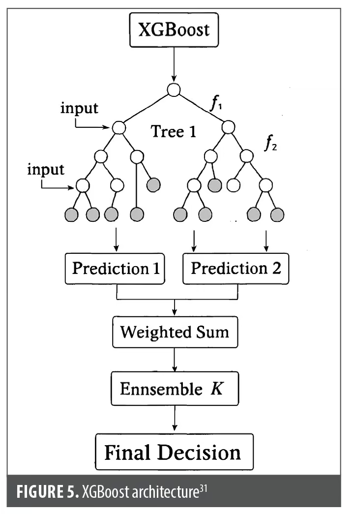
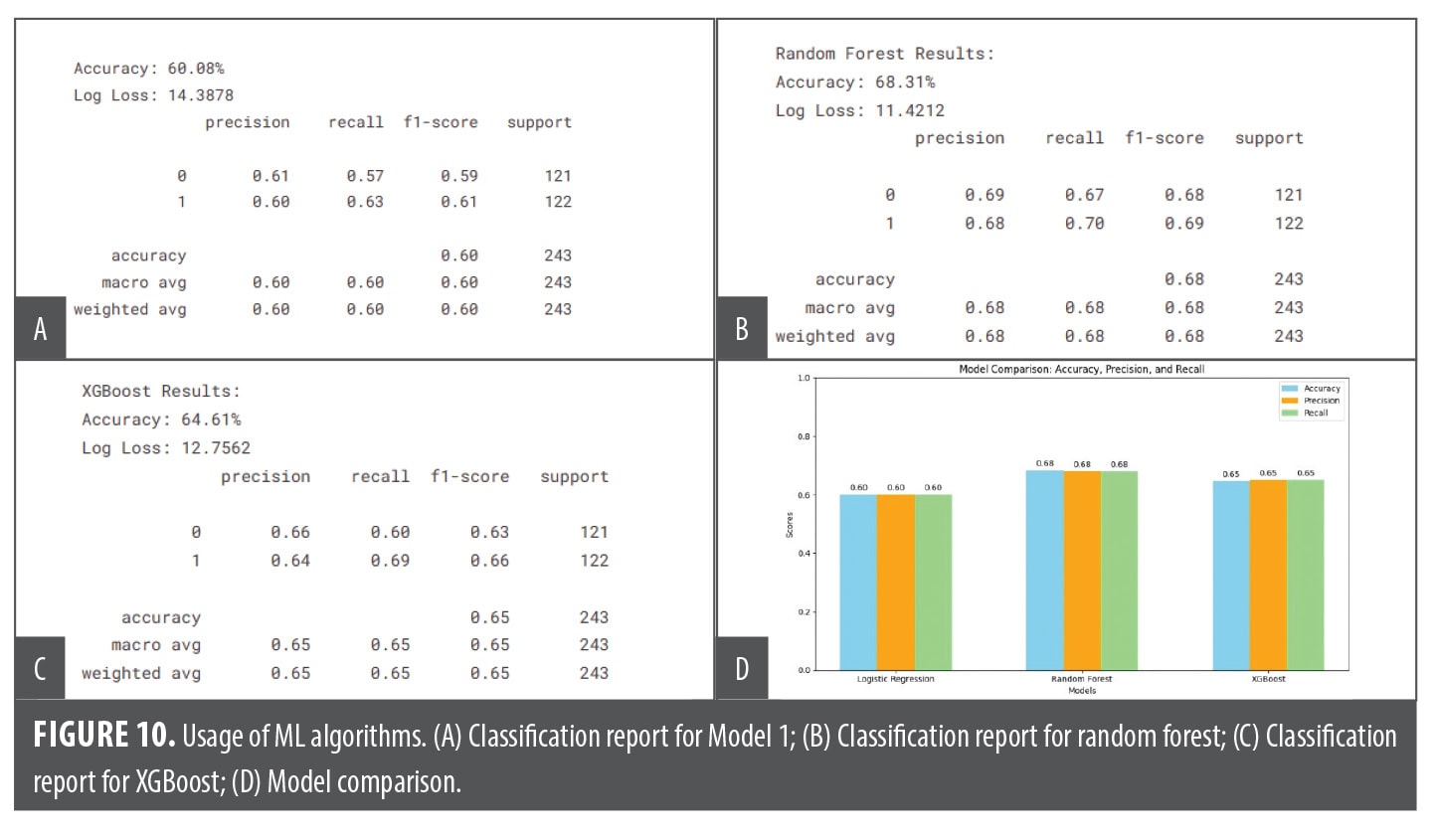
Traditional machine learning models including Support Vector Machines as well as Random Forest models and XGBoost were executed during the classification process. Random Forest and XGBoost possess specific abilities through which they become suitable tools for postsegmentation skin lesion diagnosis. Bootstrap sampling enables Random Forest to function as an ensemble learning method by creating various decision trees through sampled data subsets that include replacement elements. Individual tree learning progresses on sample subsets through variable grouping processes to achieve unpredictable outcomes and minimize overfitting risks during the training session. The ensemble prediction from decision trees generates the final output, because this method operates effectively against noise while handling high-dimensional data. Sequential tree development allows XGBoost to function as an ensemble method with weak learners, which produces a final analysis. The sequence of trees performs loss function minimization to correct previous errors. The optimization methods within XGBoost depend on parallel processing together with tree pruning methods and L1 and L2 regularization to achieve better performance accuracy at faster computational speeds. The training process of XGBoost shows variable significance through its feature importance methodology. Our approach used segmented images as input for these models to reach classification results, which showed the success of our method in differentiating between benign and malignant cases. You can view the model architectural diagrams for Random Forest in Figure 4 and XGBoost in Figure 5.
Segmentation using UCMNet. In the second part of the procedure, we implemented the UCMNet architecture for segmentation. UCMNet builds upon the foundational UNet architecture by addressing its limitations, particularly in segmenting irregular and ambiguous boundaries. The encoder in UCMNet follows a structure similar to UNet, using convolutional and pooling layers to capture spatial features at multiple scales. However, UCMNet introduces multiscale feature extraction modules, which enhance the model’s ability to capture detailed information from both small and large regions of the image. These modules employ dilated convolutions to expand the receptive field without adding computational overhead, enabling the detection of fine details alongside broader contextual patterns.
The architecture of UCMNet is shown in Figure 6. One contribution of UCMNet is the incorporation of boundary-aware modules. The specialized modules are meant to detect and refine lesion edges from high-resolution features of previous encoder layers. The architecture overcomes the problems associated with complex biomedical images by enhancing the representation of irregular shapes and weak boundaries. These modules refine the features and yield a much-improved segmentation in case of the intricate structure or a poorly defined one. The decoder of UCMNet is fed with the upsampling process of UNet, which can reconstruct the spatial resolution using transposed convolutional layers as shown in Figure 7. Unlike UNet, the decoder of UCMNet has boundary-aware modules with enriched features and skip connections for transferring fine-grained details from the encoder. The SA module is designed to learn the positional intensities (spatial weights) of the feature maps at every decoder stage in an adaptable manner as well as carry out fusion procedures. All other feature map levels are reshaped to resemble the feature map exhibiting the greatest correlation to ground truth in the SA module and then fused spatially using the learnt weights, as illustrated in Figure 7. Prior to using interpolation to boost the resolution, we initially implemented a 1×1 convolutional layer to reduce the number of feature channels to match that of the previous level. To adjust the channel number and resolution for downsampling, we employed a 3×3 convolutional layer with a stride of 2. Let f’i be a feature map that has been modified at the i-th level to match the fourth level in terms of channels and resolution. Lastly, we combined the fourth-level feature map with the modified 3-feature maps using the spatial feature fusion module shown below:
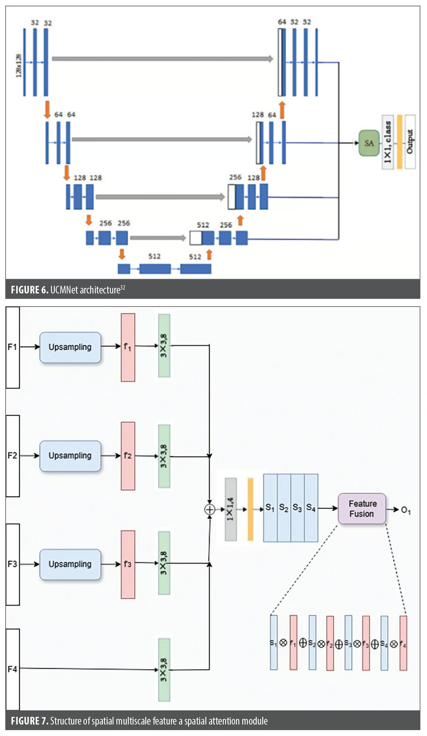

where f’1, f’2, f’3 are feature maps to be adjusted to the same resolution; S1, S2, S3, and S4 are positional intensities (spatial weights) of the 4 different level feature maps; and O1 is the final output of the segmented image. Here, S1, S2, S3, and S4 equaled 0 or 1, which specifies the pixel values calculated with the below equation.13
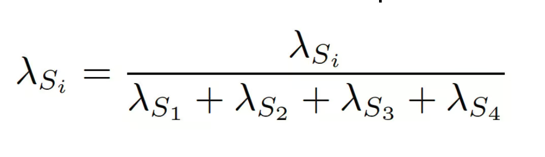
Using 1×1 convolutional layers, which can be trained using conventional backpropagation, we computed the scalar weight maps for λS1, λS2, λS3, and λS4 from S1, S2, S3, S4 intensities (spatial weights). Validated through advanced metrics such as Intersection over Union (IoU) and Dice coefficient, the segmentation masks were outputted by the output layer of UCMNet using pixel-wise classification. These metrics quantify the overlap between predicted and ground truth masks, showing that UCMNet has better capability to segment the complex lesion boundaries. In addition, it was found that the architecture is robust with respect to noise and image artifacts,thus making it appropriate for medical imaging tasks requiring a high level of precision and reliability.
The same workflow was applied to the UNet-based approach. The dataset was taken initially and then thoroughly preprocessed, ensuring the images were cleaned and enhanced and hair follicles were removed and, most importantly, improving the visibility of the lesion. Then, the preprocessed images were saved to a dedicated folder.
Instead of UNet, UCMNet architecture was used for segmentation. Using this advanced model, we achieved such a precision segmentation of the lesion regions that we were also able to evaluate them with metrics (IoU, Precision, Recall, and Dice coefficient) to see if the segmentation process was correct and robust. In the classification stage, we used a simple yet efficient deep learning algorithm (MobileNet) to classify images into 2 classes: malignant or benign.
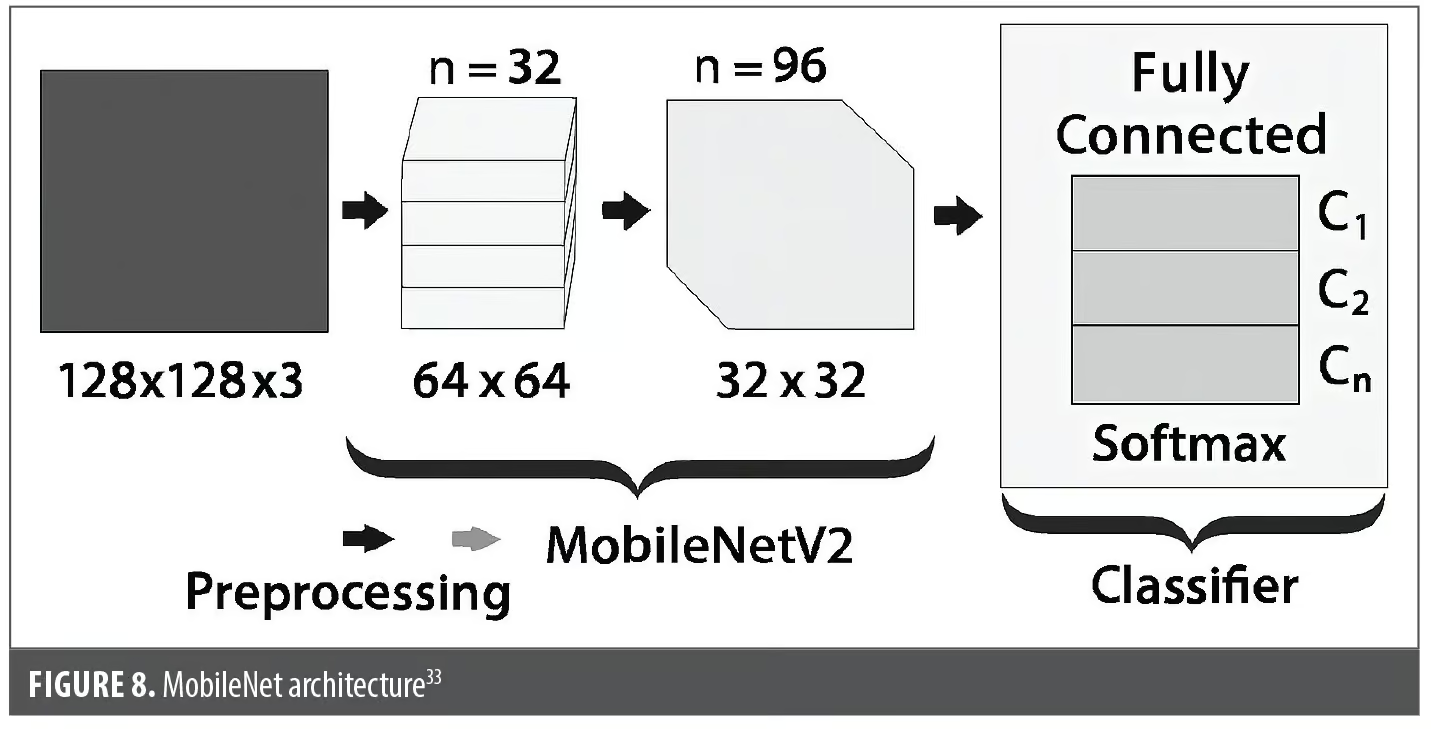
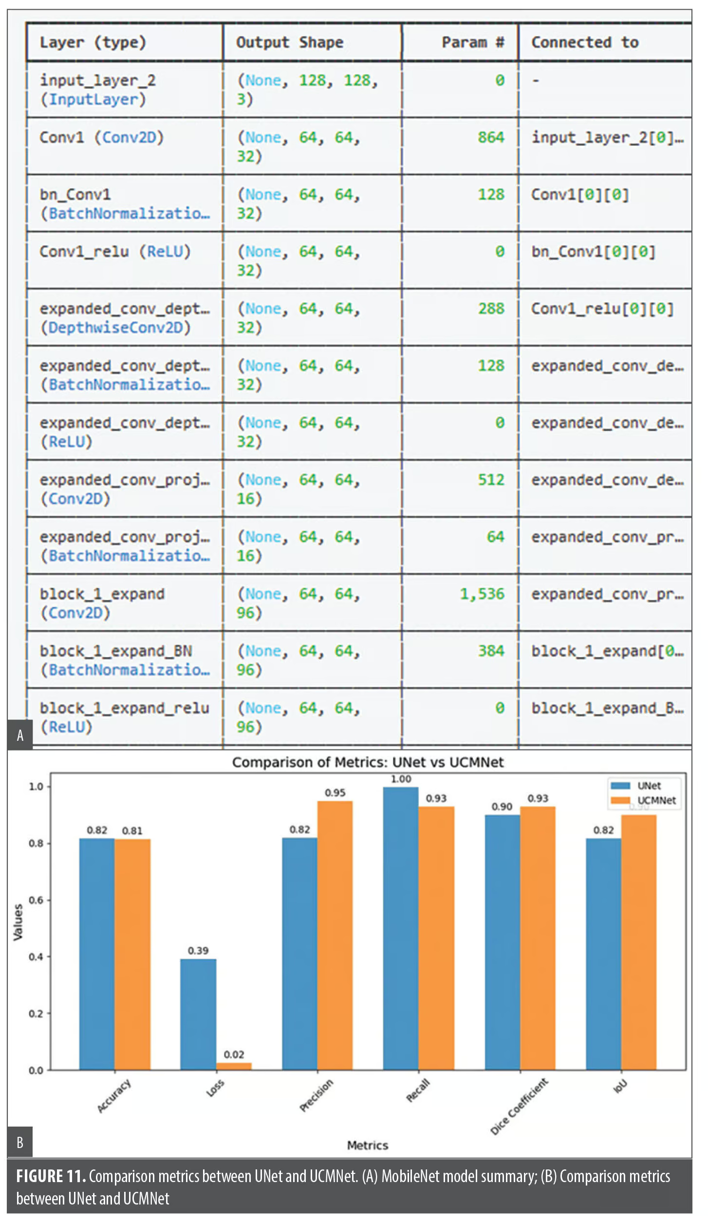
MobileNet is a lightweight CNN model that is efficient at computation, which includes being able to run the same tests with high performance, especially on resource-constrained devices. With depth-wise separable convolutions as the core building block, the architecture was simpler: it used orders of magnitude and fewer parameters, and it drastically reduced the computational overhead compared to traditional CNNs. The resulting structure splits each standard convolution into 2 operations: (1) depth-wise convolutions, which apply 1 convolutional filter per input channel to find spatial features, and (2) pointwise convolutions that aggregate depth-wise convolutions in channels together using 1×1 filters. Techniques such as batch normalization and ReLU activation following each of the convolutional layers are also included in the architecture to improve training stability and feature extraction. The hyperparameters for MobileNet are width multiplier (α), modifying the number of filters,and resolution multiplier (ρ), shrinking the spatial dimensions of the input image. Finally, the MobileNet has fully connected layers as its final layers, followed by a softmax classifier for multiclass classification. These design principles make MobileNet highly efficient, adaptable,and capable of handling high-dimensional data without requiring significant computational resources. We obtained the model summary, analyzed its performance, and evaluated the classification accuracies, which demonstrated the effectiveness of this approach in distinguishing between benign and malignant lesions. Figure 8 shows the MobileNet Architecture.
Results and Discussion
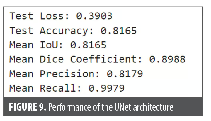
Figure 9 metrics highlight the performance of the UNet architecture, demonstrating the effectiveness of the segmentation process. A high Dice coefficient and recall emphasize the model’s precision and reliability in accurately detecting lesion boundaries. Figure 10 depicts the usage of machine learning Algorithms and their respective performance metrics. Figure 11A depicts the usage of Mobile Net and the respective Model Summary; Figure 11B illustrates comparison Metrics between UNet and UCMNet. Boundary-aware models are designed to enhance sensitivity around lesion edges, improving diagnostic accuracy and supporting clinical decision-making. Finally, the following project demonstrates the functionality indicating that the lesion belongs to the class with a match value of 0.0, identifying it as benign.
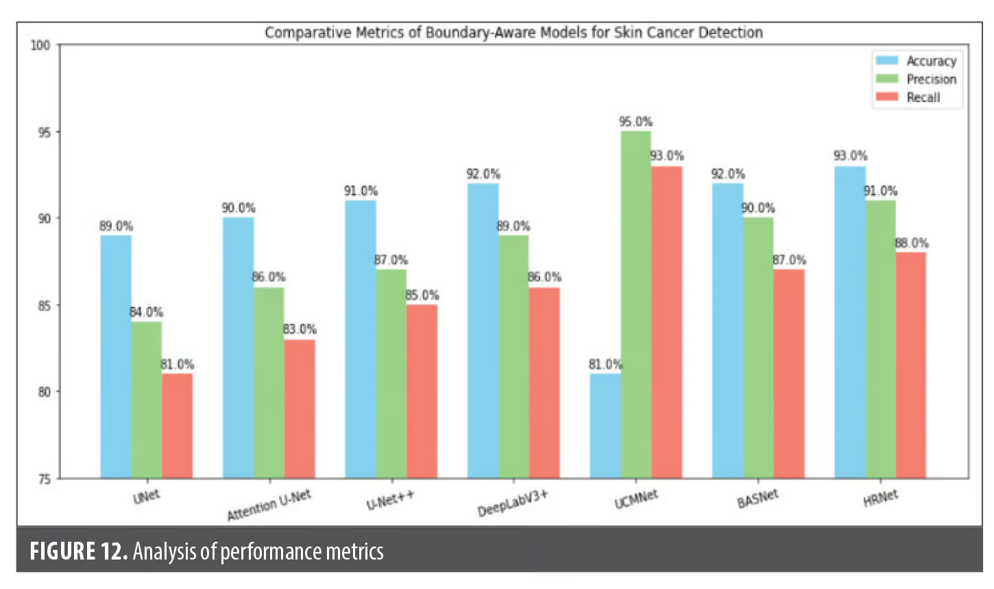
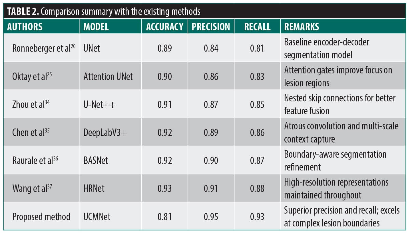
By emphasizing edge sharpness, small lesion visibility, and contextual refinement, boundary-aware models suh as UCMNet, BASNet, and HRNet provide improved precision and recall for skin cancer detection. As a result, they are well suited for difficult medical datasets like 3D-TBP and ISIC. Table 2 and Figure 12 denote the comparative summary of the existing methods with the proposed method. UCMNet excels in precision and recall, indicating highly accurate lesion boundary detection.
 Conclusion
The proposed system effectively combines UNet and UCMNet architectures for accurate skin lesion segmentation and machine learning algorithms and MobileNet for classification, achieving robust performance on various metrics such as Accuraccy, IoU, Dice coefficient, Precision, and recall using ISIC 2024 dataset. Advanced preprocessing techniques, including hair follicle removal, ensured high-quality input data, resulting in reliable differentiation between cancerous and noncancerous lesions. This solution highlights the potential of integrating deep learning models with preprocessing pipelines to address medical imaging challenges. Future work can focus on exploring advanced segmentation models like Transformer-based architectures, extending classification to additional skin cancer types, and incorporating explainable AI for interpretable predictions. Efforts can also include optimizing models for real-time mobile applications, integrating larger and diverse datasets to enhance generalizability, and deploying the system on cloud platforms for scalable, accessible diagnostic services while collaborating with medical professionals to validate and integrate into clinical workflows.
References
- Cancer. World Health Organization; 2026. Accessed 5 Jul 2026. https://www.who.int/news-room/fact-sheets/detail/cancer
- Garbe C, Amaral T, Peris K, et al. European consensus-based interdisciplinary guideline for melanoma. Part 1: Diagnostics – Update 2024. Eur J Cancer. 2025;215:115152.
- ISIC 2024 – skin cancer detection with 3D-TBP. Kaggle. Accessed 5 Jul 2026. https://www.kaggle.com/competitions/isic-2024-challenge/data
- Miradwal S, Mohammad W, Jain A, Khilji F. Lesion segmentation in skin cancer detection using UNet architecture. In: Mercier-Laurent E, Jayaraman B, Ravisankar P, et al, eds. Computational Intelligence and Data Analytics. Springer Nature Singapore; 2022:329–340.
- Nawaz M, Nazir T, Masood M, et al. Melanoma segmentation: a framework of improved DenseNet77 and UNET convolutional neural network. Int J Imaging Syst Technol. 2022;32(6): 2137–2153.
- Reis HC, Turk V, Khoshelham K, Kaya S. InSiNet: a deep convolutional approach to skin cancer detection and segmentation. Med Biol Eng Comput. 2022;60(3):643–662.
- Yan J, Wang H, Lu X, Li F. Development and validation of a nomogram for elderly patients with ulcerative melanoma. Melanoma Res. 2024;34(3):207–214.
- Esteva A, Kuprel B, Novoa RA, et al. Dermatologist-level classification of skin cancer with deep neural networks. Nature. 2017;542(7639):115–118.
- Brinker TJ, Hekler A, Enk AH, et al. Deep learning outperformed 136 of 157 dermatologists in a head-to-head dermoscopic melanoma image classification task. Eur J Cancer. 2019;113:47–54.
- Das K, Cockerell CJ, Patil A, et al. Machine learning and its application in skin cancer. Int J Environ Res Public Health. 2021;18(24):13409.
- Tembhurne JV, Hebbar N, Patil HY, Diwan T. Skin cancer detection using ensemble of machine learning and deep learning techniques. Multimedia Tool Applications 2023;82(18):27501–27524.
- Jones OT, Matin RN, van der Schaar M, et al. Artificial intelligence and machine learning algorithms for early detection of skin cancer in community and primary care settings: a systematic review. Lancet Digit Health. 2022;4(6):e466–e476.
- Mazhar T, Haq I, Ditta A, et al. The role of machine learning and deep learning approaches for the detection of skin cancer. Healthcare. 2023;11(3):415.
- Imran A, Nasir A, Bilal M, et al. Skin cancer detection using combined decision of deep learners. IEEE Access. 2022;10:118198–118212.
- Dai X, Spasic I, Meyer B, et al. Machine learning on mobile: an on-device inference app for skin cancer detection. In: 2019 Fourth International Conference on Fog and Mobile Edge Computing (FMEC). IEEE; 2019:301–305.
- Shorten C, Khoshgoftaar TM. A survey on image data augmentation for deep learning. J Big Data. 2019;6(1):1–48.
- Dildar M, Akram S, Irfan M, et al. Skin cancer detection: a review using deep learning techniques. Int J Environ Res Public Health. 2021;8(10):5479.
- Mahum R, Aladhadh S. Skin lesion detection using hand-crafted and DL-based features fusion and LSTM. Diagnostics (Basel). 2022;12(12):2974.
- Adamu S, Alhussian H, Aziz N, et al. The future of skin cancer diagnosis: a comprehensive systematic literature review of machine learning and deep learning models. Cogent Eng. 2024;11:2395425.
- Ronneberger O, Fischer P, Brox T. U-Net: convolutional networks for biomedical image segmentation. In: Navab N, Hornegger J, Wells WM, Frangi AF, eds. Medical Image Computing and Computer-Assisted Intervention – MICCAI 2015; Springer; 2015: 234–241.
- Kervadec H, Bouchtiba J, Desrosiers C, et al. Boundary loss for highly unbalanced segmentation. In: Proceedings of the 2nd International Conference on Medical Imaging with Deep Learning. Proceedings of Machine Learning Research; 2019:285–296.
- Bi L, Kim J, Ahn E, Kumar A, Fulham M, Feng D. Dermoscopic image segmentation via multistage fully convolutional networks. IEEE Trans Biomed Eng. 2017;64(9):2065–2074.
- Li XM, Yu LQ, Chen H, Fu CW, Heng PA. Semi-supervised skin lesion segmentation via transofrmation consistent self-ensembling model. arXiv preprint arXiv:1808.03887.
- Wang J, Wei L, Wang L,et al. Boundary-aware transformers for skin lesion segmentation. In International conference on medical image computing and computer-assisted intervention (pp. 206–216). Cham: Springer International Publishing. 2021.
- Oktay O, Schelmper J, Le Folgoc L, et al. Attention U-Net: learning where to look for the pancreas. . arXiv. Preprint posted online 20 May 2018. Accessed 5 Jul 2026.
- Chen J, Lu Y, Yu Q, et al. TransUNet: transformers make strong encoders for medical image segmentation. arXiv. Preprint posted online 8 Feb 2021. Accessed 5 Jul 2026.
- Li X, Yu L, Chen H, et al. Gated fully fusion for skin lesion segmentation with graph-based boundary modeling. Med Image Anal. 2022;76:102305.
- Yuan Y. Hierarchical convolutional-deconvolutional neural networks for automatic liver and tumor segmentation. arXiv. Preprint posted online 12 Oct 2017. Accessed 5 Jul 2026.
- Wang Y, Xiao B, Bi X, Li W, Gao X. Boundary-aware prototype in semi-supervised medical image segmentation. IEEE Transact Image Processing. 2024;33:5456–5467.
- Shafi ASM, Imran Molla MM, Jui JJ, Rahman MM. Detection of colon cancer based on microarray dataset using machine learning as a feature selection and classification techniques. SN Appl Sci. 2020;2:1243.
- Chen S. Predicting Lung Cancer Stage by Expressions of Protein-Encoding Genes. Adv Biosci Biotechnol. 2023;14(8):368–377.
- Kim MJ, Chae SG, Bae SJ, Hwang KG. Unsupervised few shot learning architecture for diagnosis of periodontal disease in dental panoramic radiographs. Sci Rep. 2024;14(1):23237.
- Ghosh N, Jana B, Jana S, Sao NK. Face Mask Detection Exploiting CNN and MobileNetV2. In: Mandal JK, Jana B, Lu TC, De D, eds. Proceedings of International Conference on Network Security and Blockchain Technology. Springer; 2024:67–84.
- Zhou, Z., Rahman Siddiquee, M. M., Tajbakhsh, N., & Liang, J. (2018). Unet++:A nested U-Net architecture for medical image segmentation. Deep Learn Med Image Anal Multimodal Learn Clin Decis Support. 2018;11045:3–11.
- Chen LC, Zhu Y, Papandreou G, et al. Encoder-decoder with atrous separable convolution for semantic image segmentation. arXiv. Preprint posted online 22 Aug 2018. Accessed 5 Jul 2026.
- Raurale S, McAllister J, del Rincon JM. EMG wrist-hand motion recognition system for real-time embedded platform. In: Proceedings of the IEEE International Conference on Acoustics, Speech and Signal Processing (ICASSP). IEEE; 2019. p. 1523–1527.
- Wang J, Sun K, Cheng T, et al. Deep high-resolution representation learning for visual recognition. IEEE Trans Pattern Analysis Machine Intelligence. 2021;43(10):3349–3364.
From the 2nd Annual RAPIDS Immuno-Dermatology Conference: From Approval to Adoption—Translating Innovation Into Clinical Practice