
J Clin Aesthet Dermatol. 2026;19(8):34–40.
Amber Khalil, MBChB; Hiva Fassihi, FRCP, PhD; Mark A. Birch-Machin, PhD; Jonathan Brookes, BSc; Amy Bowman, PhD; Gewei Zhu, PhD; Megan Hughes; Florence McDermott; and Emma Craythorne, FRCP, MBChB
Dr. Khalil is with Ninewells Hospital, NHS Tayside, Scotland, United Kingdom. Prof. Fassihi is with St John’s Institute of Dermatology, Guy’s and St Thomas’ NHS Foundation Trust, London, United Kingdom, and King’s College London, London, United Kingdom. Prof. Birch-Machin is with Dermatological Sciences, Translational and Clinical Research Institute, Newcastle University and Skin Life Analytics Ltd, Newcastle upon Tyne, United Kingdom. Mr. Brookes, Dr. Bowman, and Dr. Zhu are with Skin Life Analytics Ltd, Newcastle upon Tyne, United Kingdom. Ms. Hughes is with Klira Ltd, London, United Kingdom. Ms. McDermott is with Francis Holland School, London, United Kingdom. Dr. Craythorne is with St John’s Institute of Dermatology, Guy’s and St Thomas’ NHS Foundation Trust and Klira Ltd, London, United Kingdom.
FUNDING: Product supply for the trial was provided by Klira Ltd. Sampling materials and laboratory processing were provided by Skin Life Analytics Ltd, and paid for by Klira Ltd.
DISCLOSURES: Dr. Craythorne reports being the founder of Klira Ltd. Dr. Birch-Machin reports being the founder and codirector of Skin Life Analytics Ltd. Mr. Brookes reports being the codirector of Skin Life Analytics Ltd. Drs. Zhu and Bowman report that they are employees of Skin Life Analytics Ltd. All other authors report no conflicts of interest.
Abstract: OBJECTIVE: To evaluate whether a daily antioxidant-enriched sunscreen preserves mitochondrial DNA (mtDNA) integrity vs vehicle, whether habitual sunscreen users gain additional protection from antioxidants, and whether mtDNA swabs can detect individual-level responses. METHODS: Adults completed a randomized, double-blind, vehicle-controlled 12-week trial. Cheek swabs collected at baseline and Week 12 underwent blinded quantitative polymerase chain reaction assays. mtDNA damage was expressed as ΔCt, where higher values indicate greater damage and a negative change reflects improved integrity. The primary outcome was change in ΔCt. RESULTS: Forty-one participants provided valid samples (antioxidant sunscreen: n=19; vehicle: n=18). Mean ΔCt change favored the antioxidant sunscreen (–0.48 vs 0.35; P=0.06; Mann-Whitney U, P=0.07). Responder rates were higher with antioxidant sunscreen (73.7% vs 38.9%; Fisher exact, P=0.05). Among habitual sunscreen users, mtDNA integrity improved with antioxidant enrichment (median ΔCt: −0.67; P=0.04) and worsened with vehicle (median ΔCt: 0.58; P=0.03). In this subgroup, responder rates were also higher with active treatment (75% vs 25%; P=0.03). mtDNA integrity demonstrated a clear individual-level sensitivity to change. LIMITATIONS: This was a single-site pilot study and lacked a no sunscreen-only comparator arm. CONCLUSION: Daily use of an antioxidant-enriched sunscreen produced consistent molecular evidence of reduced mtDNA damage, including statistically significant within-participant improvement among habitual sunscreen users. Keywords: Mitochondrial DNA damage, oxidative stress, antioxidant sunscreen, in vivo biomarker, randomized controlled trial, quantitative PCR, photoprotection
Introduction
Chronic sun exposure accelerates skin photoaging, leading to wrinkles, dyspigmentation, and dermal laxity. UV-A (320–400 nm) penetrates the dermis and generates reactive oxygen species (ROS).1–4 Through activator protein-1 activation and matrix metalloproteinase upregulation, UV-A–induced ROS promote collagen and elastin degradation as well as oxidative mitochondrial DNA (mtDNA) mutations.5 These mutations impair electron transport chain function and increase ROS production, creating a self-amplifying cycle of dysfunction.6 As a direct target of UV-A–mediated oxidative injury, mitochondrial integrity is a key mechanistic target for photoprotection.
mtDNA is more vulnerable than nuclear DNA due to its lack of histones and limited repair capacity, leading to accumulation of characteristic lesions such as the 4,977-bp “common deletion” in sun-exposed skin.6 Cumulative mtDNA mutations are hallmarks of photoaging,7 and mtDNA integrity represents a biologically grounded biomarker for in vivo photoprotection assessment.8
Skin is also exposed to oxidative stress from visible light, particularly the blue light or high-energy visible (HEV) range (approximately 380–500 nm), which can generate ROS in skin,9 as well as from infrared radiation and atmospheric pollutants such as ozone, particulate matter, heavy metals, and tobacco smoke.10
Fibroblasts harboring mtDNA deletions demonstrate altered contractility.11,12 Smokers accumulate mtDNA deletions in lung tissue.13 The skin’s antioxidant network, including superoxide dismutases, catalase, glutathione peroxidases, peroxiredoxins, glutathione, ubiquinol, α-tocopherol, and ascorbate, mitigates ROS under normal conditions.14,15 However, environmental exposures can overwhelm these systems, leading to increased mitochondrial ROS production, oxidative damage, inflammatory signaling (nuclear factor-κB, aryl hydrocarbon receptor), and cellular senescence.16–20 Combining UV filters with antioxidants may address broader oxidative stress.
Daily sunscreen use prevents photoaging and photocarcinogenesis.21–23 However, even formulations with high SPF permit residual UV-A,24 visible, and infrared radiation.25 Topical antioxidants, therefore, complement UV filters across these wavelengths.25,26 Antioxidants reduce UV-induced erythema, sunburn cells, nuclear DNA damage, and the mtDNA common deletion.27–30 However, most evidence derives from ex vivo or reconstructed skin models that lack full physiologic context.31,32 mtDNA integrity offers a noninvasive biomarker of photodamage in vivo.7,8,33
To our knowledge, no previous study has evaluated sunscreen or antioxidant photoprotection on mtDNA integrity in vivo in human skin. Prior mtDNA photodamage studies have been performed across a range of experimental in vitro systems, including cultured keratinocytes, cultured dermal fibroblasts, and reconstructed human skin models containing both epidermal and dermal components.7,8,33 In contrast, the present noninvasive swab-based assay is expected to sample predominantly superficial epidermal keratinocyte-rich material in vivo. Residual oxidative stress persists with high-factor sunscreens,34,35 highlighting opportunities for additive antioxidant benefit.36–41
Methodological advances have also validated the long/short amplicon quantitative polymerase chain reaction (qPCR) assay for assessing mtDNA integrity in cell-based or ex vivo systems rather than randomized human trials. 7,8,33 This trial provides the first in vivo evidence that a combined sunscreen-antioxidant formulation can preserve mtDNA integrity in human facial skin under ambient environmental exposure.
Accordingly, we conducted a randomized, double-blind, vehicle-controlled clinical trial to evaluate whether daily use of an encapsulated sunscreen (SPF 50) containing a multi-antioxidant complex reduces mtDNA damage in vivo more effectively than a vehicle-only cream. We hypothesized that antioxidant-enriched sunscreen would preserve mtDNA integrity more effectively than the vehicle during daily use. The study also assessed, within the prespecified subgroup of habitual daily sunscreen users, whether the antioxidant-enriched formulation provided greater mitochondrial protection than participants’ own real-life sunscreen routines. Finally, the trial evaluated mtDNA integrity as a scalable biomarker for biological effects of photoprotection, including detection of individual-level mitochondrial improvement.
Methods
A 12-week, randomized, double-blind, vehicle-controlled clinical trial was conducted during the spring (March–May) in the United Kingdom to minimize variability in UV exposure, temperature, and humidity. Adults aged 18 years and older were recruited from community and clinic sources. Eligibility was independent of prior sunscreen use. Baseline questionnaires documented sunscreen habits (daily, intermittent, none), and habitual daily use defined a prespecified subgroup for analysis.
Participants were randomized 1:1 to receive either an encapsulated sunscreen (SPF 50) containing a multi-antioxidant complex or a matched vehicle-only cream without sunscreen or antioxidants. Encapsulation prevents systemic absorption and improves antioxidant stability and delivery.42 The formulation contained encapsulated chemical filters (octinoxate, avobenzone, octocrylene) encapsulated in lecithin,43 titanium dioxide,44 resveratrol,45 glabridin,46 astaxanthin,47 and ubiquinone.48
Exclusion criteria were active facial dermatologic disease; laser, peel, or other energy-based procedures within the past 3 months; use of topical or systemic retinoids, antioxidants, or anti-inflammatory agents within 4 weeks; recent acute illness; pregnancy or breastfeeding; smoking or regular e-cigarette use; or planned high-UV travel during the study period. Participants taking medications known to influence mitochondrial function (antibiotics,49 statins,50 metformin,51 hormonal therapy,52 etc) were eligible only if doses had been stable for >3 months; those with unstable dosing were excluded from efficacy analyses. Similarly, background medications, supplements, and skincare were permitted if stable for >3 months and unchanged during the study; deviations led to exclusion from efficacy analyses.
Randomization used computer-generated permuted blocks with allocation concealment. The products were indistinguishable in appearance and texture, and participants, investigators, and laboratory personnel were blinded to treatment assignment. Samples were processed in coded form. The vehicle formulation was identical to the active formulation except for the absence of UV filters and antioxidants. A small nonrandomized comparator group continued its usual skincare routine to contextualize environmental background effects; this group was unblinded by design and excluded from randomized comparisons. No interim analyses or protocol changes occurred.
Participants applied the assigned product once daily in the morning to the entire face, following standardized instructions corresponding to a target application dose of 2 mg/cm2, the standard quantity used in sunscreen testing.53 The encapsulated delivery system was designed to support stability and uniform cutaneous distribution of filters and antioxidants.42 Participants were asked not to use additional daytime facial products, including other sunscreens, antioxidant serums, moisturizers, or makeup during the study. Adherence was assessed at Week 12 by self-report and by inspection of returned products.
Skin sampling used a validated noninvasive protocol.7,8,33,54,55 At baseline and Week 12, a synthetic-tipped swab was rubbed across a defined 2×2-cm cheek area using standardized strokes. Swabs were placed in coded vials and couriered to a central laboratory. Total DNA was extracted, and mtDNA integrity was quantified using qPCR assays amplifying a 1-kb lesion-sensitive fragment and an 83-bp control fragment for mtDNA content. mtDNA damage was expressed as ΔCt (higher = greater damage; negative change = improvement). Because cheek swabbing is a superficial sampling method, the recovered DNA is expected to derive predominantly from epidermal keratinocyte-rich material rather than dermal fibroblasts.
The primary outcome was the between-arm difference in change in mtDNA damage (ΔCt) from baseline to Week 12. The secondary outcome was the within-participant change in ΔCt among habitual daily sunscreen users, using each participant’s baseline value as the biological benchmark of their real-life sunscreen-only routine. Exploratory outcomes included comparison with the nonrandomized group and assessment of mtDNA integrity as a biomarker of photoprotection under routine use. Responders were defined a priori as ΔCt<0. As a pilot study, no formal sample size calculation was performed.
All analyses used a modified intention-to-treat (mITT) population including all randomized participants with valid paired mtDNA data. Between-arm comparisons used Welch t tests with confirmatory Mann–Whitney U tests; effect sizes were summarized using Hedges g. Within-participant changes in habitual daily sunscreen users were evaluated using paired tests as appropriate. Responder rates were compared using Fisher exact tests with Wilson 95% CIs. All statistical tests were 2-sided with α=0.05, and analyses were performed using GraphPad Prism version 10.6.1.
The study was registered on the Integrated Research Application System (IRAS; Project ID 364490) and assessed by the UK Health Research Authority (HRA), which determined that Research Ethics Committee (REC) review was not required. The trial was registered on ClinicalTrials.gov (Identifier: NCT07301515; Available at: https://clinicaltrials.gov/study/NCT07301515), and the ethics exemption documentation was supplied. The study was conducted under institutional governance as minimal-risk cosmetic research and was performed in accordance with the principles of the Declaration of Helsinki (2013 revision). Written informed consent was obtained from all participants.
Results
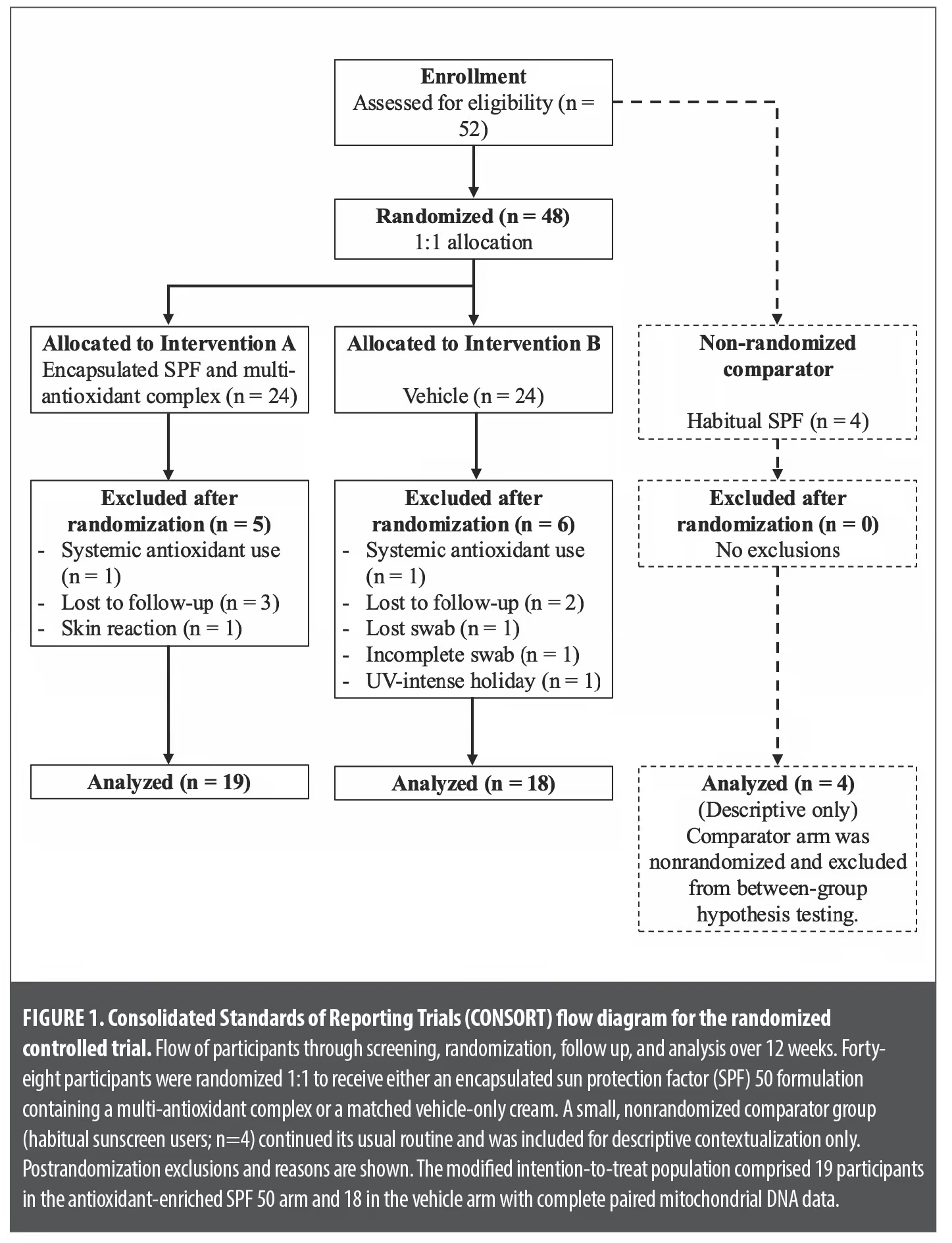
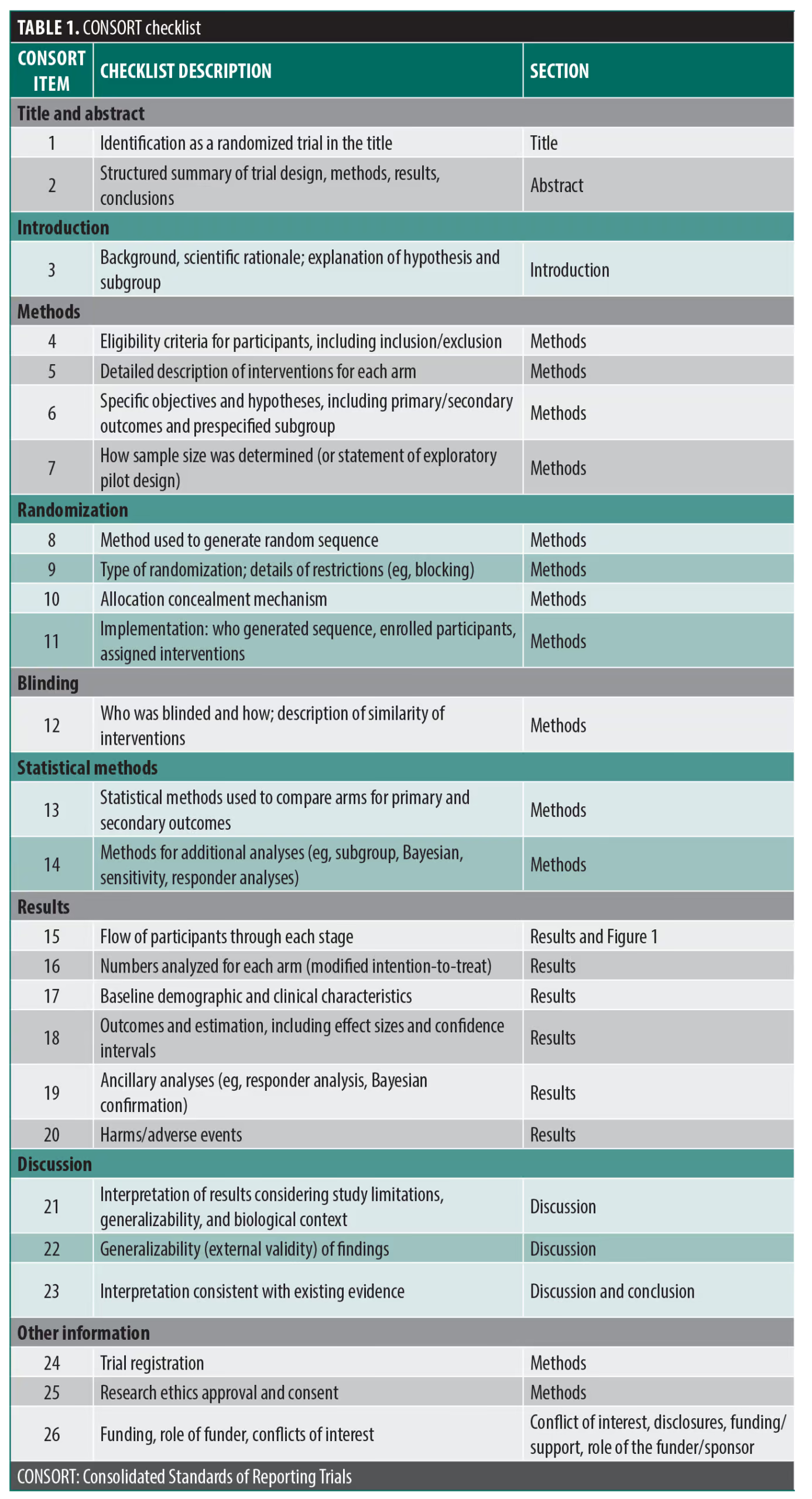
Fifty-two adults were enrolled: 48 were randomized, with 24 to the antioxidant-enriched sunscreen arm and 24 to the vehicle-only arm), and 4 entered a small nonrandomized comparator group (Figure 1; Table 1). Baseline demographic and clinical characteristics were similar between randomized arms. No serious adverse events occurred. The mITT population, therefore, comprised 41 participants with valid paired mtDNA data (antioxidant-enriched sunscreen, n=19; vehicle, n=18; nonrandomized comparator, n=4).
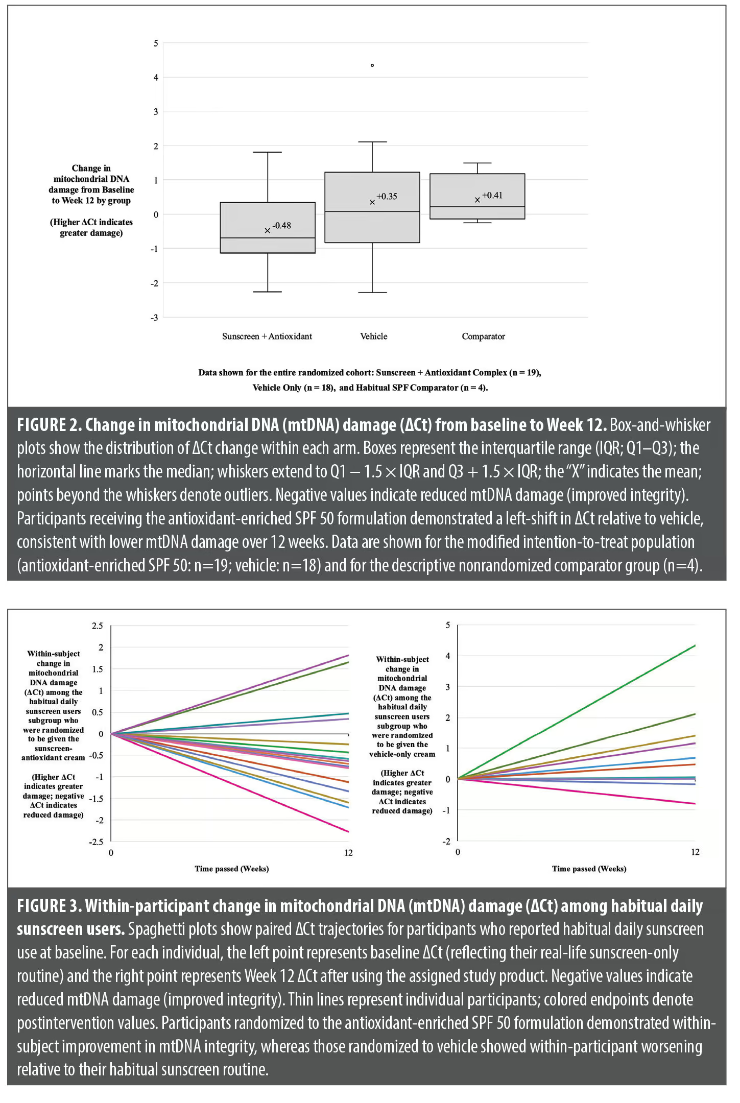
The primary outcome—the between-arm difference in change in ΔCt from baseline to Week 12—favored the antioxidant-enriched sunscreen. Mean ΔCt change was −0.48±1.05 in the antioxidant-enriched sunscreen arm and 0.35±1.52 in the vehicle arm, giving a between-arm difference of −0.83 (95% CI: −1.71 to 0.05; P=0.06; Figure 2). The standardized effect size was moderate (Hedges g=−0.62), with directionally concordant findings on Mann-Whitney U testing (P=0.07).
Responder analyses classified participants achieving improved mtDNA integrity (ΔCt<0) as biological responders. In the mITT population, 14 (73.7%) of 19 participants receiving the antioxidant-enriched sunscreen were responders compared to 7 (38.9%) of 18 in the vehicle arm (P=0.05, Fisher exact test). Among habitual daily sunscreen users, responder rates similarly favored the antioxidant-enriched sunscreen (n=12/16 [75%] vs n=2/8 [25%]; P=0.03; Figure 3), indicating a consistent individual-level pattern of protection.
Habitual daily sunscreen use was documented prospectively and defined a prespecified subgroup. Because baseline mtDNA integrity in these individuals reflected each participant’s habitual sunscreen-only routine, their baseline ΔCt served as a physiologically relevant benchmark. Among habitual sunscreen users randomized to the antioxidant-enriched sunscreen, within-individual mtDNA integrity improved significantly over 12 weeks (median ΔCt: −0.67; P=0.04). In contrast, habitual sunscreen users randomized to vehicle showed significant worsening relative to their usual sunscreen routine (median ΔCt: 0.58; P=0.03). Within this subgroup, both the within-subject changes in ΔCt and the proportion of responders favored the antioxidant-enriched sunscreen over vehicle (75% vs 25%; P=0.03).
A weakly informative Bayesian model estimating the baseline-to-Week 12 ΔCt difference yielded a posterior probability of 94% that the antioxidant-enriched sunscreen provided greater mtDNA protection than vehicle. The corresponding Bayes factor (BF10≈2.6) indicated that the observed data were roughly 2.5-fold more likely under a treatment-benefit model than under the null. These exploratory findings complemented the frequentist analyses and supported the consistent directional effect.
Sensitivity analyses, including nonparametric tests and outlier-robust estimators, produced directionally consistent results. Participant-level rankings showed a clear leftward shift in ΔCt change in the antioxidant-enriched sunscreen arm. The small nonrandomized comparator group (n=4) was evaluated descriptively to contextualize environmental variation and was not included in randomized comparisons.
Discussion
In this randomized, double-blind study, daily use of an encapsulated SPF 50 formulation containing a multi-antioxidant complex produced a consistent molecular signal of reduced mtDNA damage in facial skin compared to vehicle. Across the randomized cohort, the active formulation demonstrated a left-shift in ΔCt (mean change: −0.48 vs 0.35) and a substantially higher responder rate (73.7% vs 38.9%), indicating greater mitochondrial protection over 12 weeks of real-world exposure. Although the primary between-arm comparison narrowly missed conventional statistical significance, the magnitude of effect, responder distribution, and concordant results from nonparametric, sensitivity, and Bayesian analyses collectively support a biologically meaningful treatment effect.
Several factors may explain the presence of apparent responders within the vehicle arm. First, ambient UV and environmental exposures vary substantially between individuals, meaning some participants may have experienced lower cumulative oxidative stress during the study period irrespective of treatment allocation. Second, mtDNA integrity reflects a dynamic balance between damage and repair, and spontaneous biological variation may contribute to modest improvements over time in some individuals. Third, behavioral changes associated with study participation, including increased awareness of sun exposure and improved photoprotective habits outside of the assigned intervention, may have reduced mitochondrial damage. Finally, the highly sensitive qPCR assay is capable of detecting small biological fluctuations, and some individual responses may reflect normal biological variability. These factors likely account for the minority of vehicle-treated participants demonstrating improved mtDNA integrity despite the overall group trend toward worsening damage.
The between-arm effect corresponded to an absolute mean difference of −0.83 ΔCt. Because ΔCt is logarithmic (base 2), this difference is compatible with roughly a 2-fold greater preservation of intact mtDNA with the antioxidant-enriched sunscreen relative to vehicle, assuming typical qPCR amplification efficiencies. The 95% CI (−1.71 to 0.05) and moderate standardized effect size (Hedges g=−0.62) together support a meaningful biological signal consistent with enhanced photoprotection. Given the established role of mtDNA damage in driving ROS generation, mitochondrial dysfunction, cellular senescence, and extracellular matrix degradation, a 2-fold preservation of mtDNA integrity is likely to reflect a biologically meaningful reduction in cumulative photo-oxidative injury. However, as this study measured a molecular biomarker rather than downstream functional endpoints, no direct conclusions can be drawn regarding mitochondrial function, collagen preservation, skin aging, or clinical outcomes.
Habitual daily sunscreen use was recorded prospectively and defined a prespecified subgroup, allowing participants’ baseline ΔCt values to serve as biologically relevant benchmarks representing their real-world sunscreen-only routine. Within this subgroup, participants randomized to the antioxidant-enriched sunscreen demonstrated statistically significant improvement in mtDNA integrity relative to their baseline (median ΔCt: −0.67; P=0.04), whereas those randomized to vehicle showed statistically significant worsening relative to their usual sunscreen routine (median ΔCt: 0.58; P=0.03). These findings suggest that combining antioxidants with high-performance UV filters can improve mitochondrial protection beyond that achieved with sunscreen alone. However, because the study lacked a dedicated sunscreen-only randomized arm, the independent contributions of UV filters and antioxidants cannot be isolated, and the magnitude of any additive effects cannot be determined.
This design constraint arose from the proprietary encapsulated formulation, which precluded manufacturing a sunscreen-only version. As a single-site, 12-week trial with a modest sample size, statistical power and generalizability remain limited. Larger, multicenter studies with an additional sunscreen-only arm would strengthen mechanistic interpretation and allow clearer quantification of antioxidant contributions.
Nevertheless, taken together with existing literature on UV-A–induced mitochondrial oxidative damage, the role of ROS in photoaging,9,10 and prior evidence of photofilter-antioxidant synergy,25,26 the current findings provide in vivo support for the concept that antioxidants supplement rather than replace UV-filter protection. They appear to mitigate residual oxidative stress arising from UV-A, visible and infrared light, and pollutants, which are not fully addressed by UV filters alone.24,25 This extends previous laboratory and ex vivo observations7,8,33,54,55 to an in vivo mitochondrial biomarker measured under ambient conditions, demonstrating that a combined sunscreen-antioxidant formulation can produce measurable photoprotection within a short real-world timeframe.
The small nonrandomized comparator group served to identify potential seasonal or environmental confounding and showed no evidence of major background drift. The mtDNA long/short-amplicon qPCR assay used here is a validated biomarker of oxidative mtDNA injury, supported by extensive prior work from Birch-Machin and Moor.55 This assay captures cumulative oxidative damage with high sensitivity and has been widely used to quantify photodamage induced by UV, visible, and infrared wavelengths in mechanistic and ex vivo studies. Therefore, its application in the present randomized trial provides a robust translational bridge between laboratory photobiology and real-world human skin responses. By incorporating a physiologically relevant biomarker with established mechanistic relevance, this study offers an objective molecular readout of photoprotection in vivo and demonstrates the feasibility of using mtDNA integrity as an endpoint in future clinical trials of sunscreen and antioxidant interventions.
Conclusion
Daily use of an encapsulated SPF 50 formulation containing a multi-antioxidant complex produced a consistent molecular signal of reduced mtDNA damage compared to vehicle, with higher responder rates and directionally concordant sensitivity analyses. Among habitual sunscreen users, mtDNA integrity improved relative to each participant’s established sunscreen-only routine, suggesting additional benefits from combining antioxidants with high-performance UV filters. Although derived from a modest single-site pilot trial, these findings warrant further evaluation of combined sunscreen-antioxidant approaches.
Most importantly, this study establishes mtDNA integrity as a noninvasive biomarker capable of detecting biologically relevant differences in photoprotection in vivo under real-world conditions. Larger, multicenter trials with longer follow-up and additional comparator arms are needed to quantify the relationship between mtDNA integrity and photoprotection and to clarify the relative contributions of UV filters and antioxidants.
References
- Yuan X, Li H, Lee J, Lee DH. Role of mitochondrial dysfunction in UV-induced photoaging and skin cancers. Exp Dermatol. 2025;34(5):e70114.
- Wei M, He X, Liu N, Deng H. Role of reactive oxygen species in ultraviolet-induced photodamage of the skin. Cell Div. 2024;19(1):1.
- Pillai S, Oresajo C, Hayward J. Ultraviolet radiation and skin aging: roles of reactive oxygen species, inflammation and protease activation, and strategies for prevention of inflammation-induced matrix degradation. Int J Cosmet Sci. 2005;27(1):17–34.
- Tanveer M, Rashid H, Tasduq S. Molecular basis of skin photoaging and therapeutic interventions by plant-derived natural product ingredients: a comprehensive review. Heliyon. 2023;9(8):e13580.
- Ichihashi M, Ueda M, Budiyanto A, et al. UV-induced skin damage. Toxicology. 2003;189(1–2):21–39.
- Berneburg M, Grether-Beck S, Kürten V, et al. Singlet oxygen mediates the UVA-induced generation of the photoaging-associated mitochondrial common deletion. J Biol Chem. 1999;274(22):15345–15349.
- Hudson L, Rashdan E, Bonn C, et al. Individual and combined effects of the infrared, visible, and ultraviolet light components of solar radiation on damage biomarkers in human skin cells. FASEB J. 2020;34(3):3874–3883.
- Bowman A, Birch-Machin MA. Mitochondrial DNA as a sensitive biomarker of UV-induced cellular damage in human skin. Methods Mol Biol. 2021;2277:345–356.
- Liebel F, Kaur S, Ruvolo E, Kollias N, Southall MD. Irradiation of skin with visible light induces reactive oxygen species and matrix-degrading enzymes. J Invest Dermatol. 2012;132(7):1901–1907.
- Mancebo SE, Wang SQ. Recognizing the impact of ambient air pollution on skin health. J Eur Acad Dermatol Venereol. 2015;29(12):2326–2332.
- Quan T, Cho MK, Perry D, Quan T. Age-associated reduction of cell spreading induces mitochondrial DNA common deletion by oxidative stress in human skin dermal fibroblasts. J Biomed Sci. 2015;22:62.
- Majora M, Wittkampf T, Schuermann BA, et al. Functional consequences of mitochondrial DNA deletions in human skin fibroblasts: increased contractile strength in collagen lattices is due to oxidative stress-induced lysyl oxidase activity. Cell Commun Signal. 2009;7(Suppl 1):A65.
- Fahn HJ, Wang LS, Kao SH, et al. Smoking-associated mitochondrial DNA mutations and lipid peroxidation in human lung tissues. Am J Respir Cell Mol Biol. 1998;19(6):901–909.
- Briganti S, Picardo M. Antioxidant activity, lipid peroxidation and skin diseases. What’s new. J Eur Acad Dermatol Venereol. 2003;17(6):663–669.
- Addor FAS. Antioxidants in dermatology. An Bras Dermatol. 2017;92(3):356–362.
- Krutmann J, Schikowski T, Morita A, Berneburg M. Environmentally induced (extrinsic) skin aging: exposomal factors and underlying mechanisms. J Invest Dermatol. 2021;141(4 Suppl):1096–1103.
- Vierkötter A, Schikowski T, Ranft U, et al. Airborne particle exposure and extrinsic skin aging. J Invest Dermatol. 2010;130(12):2719–2726.
- Hüls A, Vierkötter A, Gao W, et al. Traffic-related air pollution contributes to development of facial lentigines: further epidemiological evidence from Caucasians and Asians. J Invest Dermatol. 2016;136(5):1053–1056.
- Lefebvre MA, Pham DM, Boussouira B, et al. Evaluation of the impact of urban pollution on the quality of skin: a multicentre study in Mexico. Int J Cosmet Sci. 2015;37(3):329–338.
- Krutmann J, Liu W, Li L, et al. Pollution and skin: from epidemiologic and mechanistic studies to clinical implications. J Dermatol Sci. 2014;76(3):163–168.
- Hughes MCB, Williams GM, Baker P, Green AC. Sunscreen and prevention of skin aging: a randomized trial. Ann Intern Med. 2013;158(11):781–790.
- Krutmann J, Schalka S, Watson REB, Wei L, Morita A. Daily photoprotection to prevent photoaging. Photodermatol Photoimmunol Photomed. 2021;37(6):482–489.
- Seité S, Fourtanier A, Moyal D, Young AR. Photodamage to human skin by suberythemal exposure to solar ultraviolet radiation can be attenuated by sunscreens: a review. Br J Dermatol. 2010;163(4):903–914.
- Haywood R, Wardman P, Sanders R, Linge C. Sunscreens inadequately protect against ultraviolet-A-induced free radicals in skin: implications for skin aging and melanoma? J Invest Dermatol. 2003;121(4):862–868.
- Zastrow L, Groth N, Klein F, et al. The missing link: light-induced (280–1600 nm) free radical formation in human skin. Skin Pharmacol Physiol. 2009;22(1):31–44.
- Dunaway S, Odin R, Zhou L, Ji L, Zhang Y, Kadekaro AL. Natural antioxidants: multiple mechanisms to protect skin from solar radiation. Front Pharmacol. 2018;9:392.
- Murray JC, Burch JA, Streilein RD, Iannacchione MA, Hall RP, Pinnell SR. A topical antioxidant solution containing vitamins C and E stabilized by ferulic acid provides protection against ultraviolet irradiation. J Am Acad Dermatol. 2008;59(3):418–425.
- Eicker J, Kürten V, Wild S, et al. Betacarotene supplementation protects from photoaging-associated mitochondrial DNA mutation. Photochem Photobiol Sci. 2003;2(6):655–659.
- Dijkhoff IM, Drasler B, Karakocak BB, et al. Impact of airborne particulate matter on skin: a systematic review from epidemiology to in vitro studies. Part Fibre Toxicol. 2020;17(1):35.
- Peng F, Xue C-H, Hwang S-K, Li W-H, Chen Z, Zhang JZ. Exposure to fine particulate matter associated with senile lentigo in Chinese women: a cross-sectional study. J Eur Acad Dermatol Venereol. 2016;31(2):355–360.
- Marionnet C, Pierrard C, Vioux-Chagnoleau C, et al. Interactions between fibroblasts and keratinocytes in a reconstructed skin model. J Invest Dermatol. 2006;126(5):971–979.
- Vioux-Chagnoleau C, Lejeune F, Sok J, et al. Reconstructed human skin: from photodamage to sunscreen photoprotection and anti-aging molecules. J Dermatol Sci Suppl. 2006;2(1):S1–S12.
- Moor J, Bowman A, Choudhary H, et al. Sun protection products protect against UV-induced mitochondrial DNA damage and blue light-induced cell decline in human dermal skin fibroblast cell viability. Cosmetics. 2025;12(3):128.
- Guan L, Lim HW, Mohammad TF. Sunscreens and photoaging: a review of current literature. Am J Clin Dermatol. 2021;22(6):819–828.
- He H, Li A, Li S, Tang J, Li L, Xiong L. Natural components in sunscreens: topical formulations with sun protection factor (SPF). Biomed Pharmacother. 2020;134:111161.
- Pinnell SR. Cutaneous photodamage, oxidative stress, and topical antioxidant protection. J Am Acad Dermatol. 2003;48(1):1–19.
- Matsui M, Hsia A, Miller J, et al. Non-sunscreen photoprotection: antioxidants add value to a sunscreen. J Investig Dermatol Symp Proc. 2009;14(1):56–59.
- Wu Y, Matsui M, Chen J, et al. Antioxidants add protection to a broad-spectrum sunscreen. Clin Exp Dermatol. 2011;36(2):178–187.
- Jesus A, Mota S, Torres A, et al. Antioxidants in sunscreens: which and what for? Antioxidants (Basel). 2023;12(1):138.
- Lin C, Lin M, Chung Y, Alalaiew A, Hung CF, Fang JY. Nano-based sunscreens and antioxidants for preventing and treating skin photoaging. Chemosphere. 2024;347:140702.
- Brand RM, Wipf P, Durham A, Epperly MW, Greenberger JS, Falo LD Jr. Targeting mitochondrial oxidative stress to mitigate UV-induced skin damage. Front Pharmacol. 2018;9:920.
- Bernasqué A, Faure C, Rezvani H, Cario M. A new eco-friendly and water-resistant sunscreen agent: Lecithin-based multilamellar liposomes. J Cosmet Dermatol. 2024;23(3):918–925.
- SunCat MTA II: encapsulated chemical sunscreens. BioNest. Accessed 30 Nov 2025. https://www.bio-nest.com/en-US/sp-SunCat_MTA_2.html
- Ghamarpoor R, Fallah A, Jamshidi M. Investigating the use of titanium dioxide (TiO2) nanoparticles on the amount of protection against UV irradiation. Sci Rep. 2023;13(1):9793.
- Faisal Z, Mazhar A, Batool SA, et al. Exploring the multimodal health-promoting properties of resveratrol: a comprehensive review. Food Sci Nutr. 2024;12(4):2240–2258.
- Tariq M, Qi H, Zakirullah, Zakareya, Liang H. Nano-encapsulation of glabridin using enzymatically hydrolyzed sodium caseinate for improved bioavailability and functional applications. Int J Biol Macromol. 2025;315(Pt 2):144541.
- Singh KN, Patil S, Barkate H. Protective effects of astaxanthin on skin: recent scientific evidence, possible mechanisms, and potential indications. J Cosmet Dermatol. 2020;19(1):22–27.
- Yousif HA, Al-Ani I, Hajleh MN, Matalqah S, Dayyih WA, Al-Dujaili EA. Preparation and evaluation of complexed ubiquinone (Coenzyme Q10) antiaging hyaluronic acid–vitamin C serum for skin care. J Cosmet Dermatol. 2025;24(1):e16706.
- Sailer J, Schmitt S, Zischka H, Gnaiger E. Direct effects of clinically relevant antibiotics on mitochondrial respiration. Int J Mol Sci. 2025;26(11):5379.
- Mollazadeh H, Tavana E, Fanni G, et al. Effects of statins on mitochondrial pathways. J Cachexia Sarcopenia Muscle. 2021;12(2):237–251.
- Bharath LP, Agrawal M, McCambridge G, et al. Metformin enhances autophagy and normalizes mitochondrial function to alleviate aging-associated inflammation. Cell Metab. 2020;32(1):44–55.e6.
- Lin LT, Li CJ, Lee YS, Tsui KH. Recombinant follicle-stimulating hormone and luteinizing hormone enhance mitochondrial function and metabolism in aging female reproductive cells. Int J Mol Sci. 2025;26(1):83.
- Stenberg C, Larkö O. Sunscreen application and its importance for the sun protection factor. Arch Dermatol. 1985;121(11):1400–1402.
- Birch-Machin MA, Tindall M, Turner R, Haldane F, Rees JL. Mitochondrial DNA deletions in human skin reflect photo- rather than chronological aging. J Invest Dermatol. 1998;110(2):149–152.
- Birch-Machin MA, Moor JA. Mitochondria as a skin health biomarker. Aesthet J. 2024;11(10):32–35.
From the 2nd Annual RAPIDS Immuno-Dermatology Conference: From Approval to Adoption—Translating Innovation Into Clinical Practice