
J Clin Aesthet Dermatol. 2026;19(7):16–22.
by Kanthi Bommareddy, MD; Sabrina Fabi, MD; and Mariana Muniz, MD
Dr. Bommareddy is with the Department of Dermatology, Larkin Health System, South Miami, Florida. Dr. Fabi is with Cosmetic Laser Dermatology and the University of California, San Diego, San Diego, California. Dr. Muniz is in dermatology private practice in São Paulo, Brazil.
FUNDING: No funding was provided for this article.
DISCLOSURES: The authors have no relevant conflicts of interest.
Abstract: Background: Glucagon-like peptide-1 receptor agonists (GLP-1 RAs) are increasingly prescribed for obesity and type 2 diabetes, resulting in rapid or massive weight loss (MWL) that can profoundly affect facial adipose tissue (AT) and skin homeostasis. Facial AT, comprising dermal, subcutaneous, and deep layers, plays a critical role in structural support, extracellular matrix (ECM) maintenance, and paracrine signaling to fibroblasts and keratinocytes. GLP-1 RA-MWL leads to AT depletion, with associated skin laxity, volume loss, and accelerated aging, particularly in periorbital and midfacial regions. Concurrently, improved glycemic control reduces advanced glycation end products, dampens inflammation, and preserves collagen integrity, highlighting the complex effects on the cutaneous microenvironment. Objective: To develop a regenerative, multifaceted approach for optimal aesthetic management. Results: Our proposed protocol includes multilayer fat grafting (millifat, microfat, nanofat) to restore AT and support fibroblast function, scaffolds such as calcium hydroxyapatite and poly-L-lactic acid to stimulate collagen and adipogenesis, hybrid hyaluronic acid complexes for volumization, and energy-based devices including microfocused ultrasound and fractional lasers to enhance ECM remodeling and skin tightening. Adjunctive biochemical cues such as polynucleotides, peptides, and platelet-rich plasma further promote tissue regeneration. Conclusion: A staged, individualized treatment plan, combined with lifestyle and nutritional optimization, ensures sustainable aesthetic outcomes for patients experiencing GLP-1 RA–associated facial changes. Keywords: GLP-1 receptor agonist-related weight loss, massive weight loss, regenerative aesthetics, energy-based devices
Introduction
Approximately 1.9 billion people are considered overweight, and approximately 650 million adults have obesity.1 Glucagon-like peptide-1 receptor agonists (GLP-1 RAs) are increasingly popular for managing type 2 diabetes and obesity. As of 2024, there are approximately 3.5 million prescriptions of GLP-1 RAs filled monthly in the United States (US).2 These medications, while effective for weight loss, have been associated with unintended effects on facial appearance, often referred to as “Ozempic face.” Patients taking GLP-1 RAs may experience rapid or massive weight loss (MWL), whereas patients using lifestyle modifications for weight loss may experience gradual, less pronounced weight loss. GLP-1 RA–associated MWL (GLP-1 RA-MWL) can lead to a reduction in facial fat, resulting in not only volume depletion but also changes in skin quality and laxity. As these medications become increasingly prescribed beyond the diabetes indication, it is imperative to understand the effects of GLP-1 RAs on the cutaneous microenvironment and provide effective aesthetic treatments to counteract these changes.
Adipose Tissue and Homeostasis
Facial adipose tissue (AT) has long been considered an inert, space-occupying lipid repository. Recent evidence, however, suggests that AT plays a role in cutaneous homeostasis, fitness, and appearance.3–6 Intrinsic aging and cellular senescence, UV-induced photodamage, and nutrition and exercise affect adipocyte quantity and quality and ultimately homeostatic ability.6 Cellular senescence and UV-induced inflammation result in greater regenerative signaling requirements and attenuated peroxisome proliferator-activated receptor γ expression, resulting in fewer differentiated, mature adipocytes.6–8 Less adipogenesis leads to reduced antioxidant ability in the skin and further susceptibility to UV damage. In vitro studies showed exacerbation in areas that are thinner and experience higher UV-A penetration.6,9
Anatomy and Physiology of Facial Adipose Tissue
Facial AT is composed of the dermal white adipose tissue (dWAT), subcutaneous white adipose tissue (sWAT), and deep white adipose tissue. The dWAT is located beneath the reticular dermis, sWAT is beneath the dWAT, and the deep white adipose tissue is beneath the sWAT, separated by the superficial musculoaponeurotic system (SMAS).6 With age, most individuals undergo fat hypertrophy in the deep white adipose tissue with generalized atrophy of dWAT.6,10 Facial compartments with thinner dermal layers, including the periorbital skin, undergo selective atrophy of the sWAT as well.6,10
AT functions in cutaneous homeostasis primarily in 3 ways: through paracrine signaling to reticular fibroblasts and keratinocytes, direct extracellular matrix (ECM) secretion, and structural support.6 Healthy dWAT secretes anti-inflammatory adipokines, such as leptin and adiponectin, that stimulate fibroblasts to synthesize collagen, elastin, and hyaluronic acid.11–13 Adiponectin stimulates keratinocytes to secrete natural moisturizing factor in vitro.14 Conversely, hypertrophied AT secretes inflammatory adipokines that attenuate fibroblast-mediated collagen production in vitro.6,11–13
Healthy AT contributes directly to the ECM.15–17 Adipocyte precursors secrete hepatocyte growth factor to facilitate proliferation, motility, pigmentation, and angiogenesis.18–23 Healthy adipocytes secrete matrix proteins and matrix-degrading enzymes, resulting in improved collagen structure. Hypertrophic AT secretes more matrix metalloproteinase 9, leading to elastin fiber and ECM degradation, thereby diminishing skin structural integrity and its ability to resist vertical gravitational vectors.6,24,25 Clinically, this manifests as depressed superior fat compartments with full inferior aspects encroaching on the inferior retaining ligament. In patients taking GLP-1 RAs, atrophied AT may deleteriously affect this tissue microenvironment.
Effect of Glycemic Control on Cutaneous Microenvironment
GLP-1 RAs limit gastric motility and promote better glycemic control.26 Lower circulating glucose reduces the rate of nonenzymatic glycation of collagen and ECM components, reducing the formation of advanced glycation end products (AGEs) in collagen.27 Better glycemic control through caloric restriction limits the accumulation of AGEs and reduces cross-linking that stiffens and damages collagen fibers.28 Caloric restriction stimulates autophagy of AGEs and damaged proteins, preserving collagen integrity and preventing premature aging.29 Additionally, caloric restriction can activate sirtuins (especially SIRT1), which are NAD+-dependent deacetylases that support fibroblast function, enhance collagen production, and promote ECM remodeling. In the skin, SIRT1 has been implicated in both intrinsic and stress-induced aging processes and may help preserve elasticity and firmness.30,31 Further, better glycemic control also dampens systemic inflammation and oxidative stress, both of which contribute to aging. Reduced inflammatory signaling protects dermal and epidermal cells from damage and slows matrix degradation.28,31 Finally, lower glycation levels preserve fibroblast viability and function, improving wound healing, matrix deposition, and angiogenesis. In animal models, caloric restriction is associated with more efficient skin wound repair and regeneration, possibly via enhanced vascular supply and growth factor signaling in the dermis.31–33
MWL and Its Impact on AT
Until recently, the literature focused on bariatric surgery–associated MWL (BS-MWL); however, GLP-1 RA-MWL is believed to differ from BS-MWL. Both experience excess skin laxity, reduced elasticity, and devolumization.34 Fat volume depletion in BS-MWL results from anatomical changes that restrict calorie intake and reduce nutrient absorption and through hormonal shifts and alterations in the gut microbiome that affect appetite, metabolism, and energy expenditure rather than adipocyte cell count depletion.17 BS-MWL exhibits increased fibrosis; decreased thick, organized collagen and elastin in the dermis; and vascular proliferation.35,36 Behavior of elastic fibers varies and can differ across body regions.37 GLP-1 RA-MWL has a direct lipolytic effect in addition to inhibiting lipid storage, reducing adipose-derived stem cell (ADSC) populations and attenuating adipocyte proliferation and differentiation.38–41
Facial Aesthetics After Weight Loss
MWL leads to significant pan-facial volume loss, starting with the superficial fat compartments before impacting deeper ones.42 Valente et al43 reported that BS-MWL contributes to accelerated perceived facial aging from facial fat deflation and laxity. Men older than 40 years, patients with a preoperative body mass index of 40 to 49.9 kg/m2, and those who lost more than 75% of excess weight experienced greater perceived aging. The periorbital region and temples showed atrophy, with 1 study reporting up to 41.8% reduction in superficial temporal fat pads.44 MWL leads to significant midfacial volume loss; the same study reported a reduction in cheek fat pads by 69.9% in GLP-1 RA-MWL.44 In a radiographic study of midface volume loss, Sharma et al45 reported a median fat loss of 11.0% in the superficial compartment and 7.0% in the deep compartment after GLP-1 RA therapy. Fat loss in the cheek and lips leads to lip atrophy, malar eminence flattening, and deep nasolabial folding.42,46 In the lower third, MWL is associated with platysmal band formation, increased submental and jowl laxity, and chin ptosis.42,46–48 Volume loss, which accentuated facial wrinkles, ligamentous laxity, and ECM degeneration, lead to a general downward sagging of the face and neck.42,47,49,50
Treatment Considerations for the Patient Taking a GLP-1 RA
Given the multifaceted role of AT in tissue homeostasis, a comprehensive regenerative approach should be considered for patients experiencing GLP-1 RA-MWL, rather than an approach focused on volumetric correction.
Why a Regenerative Approach?
Regenerative medicine uses the body’s natural reparative mechanisms to restore tissue function following injury, disease, trauma, congenital defects, and aging. This multidisciplinary field integrates cellular therapy, biochemical cues, scaffolds, and thermomechanical modulation through energy-based devices (EBDs) to target and rejuvenate adipocytes, fibroblasts, and keratinocytes. There are few large, randomized studies using regenerative aesthetics in patients taking GLP-1 RAs; however, we provide expert recommendations based on existing studies and clinical experience.
Fat Grafting
Replenishing the adipocyte reservoir through fat grafting could restore tissue homeostasis and protective mechanisms against facial aging. Injectable tissue replacement and regeneration is a standardized method of placing millifat (2.25 mm), microfat (1 mm), and nanofat (500 µm) into facial fat compartments.51 Deeper compartments are filled first followed by superficial ones, and larger parcel sizes are injected first before smaller ones. A regenerative approach to fat grafting involves placing fat in a multilayered approach based on anatomy and physiological need.51,52
Millifat is coarsely filtered and contains mature adipocytes, stromal vascular fraction, ADSCs, pericytes, fibroblasts, and ECM.53–56 Millifat is injected into the deep fat compartments for volumization and ligamentous support, providing structural integrity and minimizing MWL-related vertical tissue displacement.52,54 Microfat provides a modest volumization effect with more regenerative capacity. Microfat is placed in the deep white adipose tissue and sWAT.52 Nanofat contains primarily ADSCs along with growth factors, peptides, and cytokines that stimulate ECM remodeling, sustained angiogenesis, and cell turnover.52,57 ADSCs more efficiently stimulate adipogenesis through paracrine signaling of adiponectin, leptin, lipoprotein lipase (LPL), perilipin, and fatty acid-binding protein 1.57,58 Nanofat has a high regenerative capacity, enhancing overlying skin quality and fine wrinkles.52–56,59,60
Weight gain after discontinuation of a GLP-1 RA creates a theoretical risk of hypertrophy of mature adipocytes in grafted millifat and microfat, potentially causing excessive volumization.51 Placing nanofat, which contains no mature adipocytes, in superficial fat compartments can potentially prevent contour irregularities and overfilling.52,60 However, it is unknown to the authors if the regenerative properties of nanofat are impacted by GLP-1 RAs, which could raise a concern regarding early use of this modality.
Biochemical Cues
Biochemical cues are molecular signals that regulate cellular processes by interacting with specific receptors or signaling cascades. Polynucleotide (PN) is a general term referring to DNA or RNA fragments derived from salmon sperm (eg, Oncorhynchus mykiss or Oncorhynchus keta) that enter cells via pinocytosis, hydrate tissues, and serve as a source of nucleotide bases.61 PNs generally refer to longer chains (>1500 kDa) that serve as biochemical cues and scaffolds. Polydeoxyribonucleotides are smaller (<1500 kDa) polymers of DNA fragments that function as biochemical cues. Though their exact mechanism is unclear, proposed actions include activating DNA repair pathways, stimulating adenosine A2A receptors, and enhancing extracellular signal–regulated kinase/mitogen-activated protein kinase signaling, processes that boost collagen production and reduce tissue breakdown.62,63 PNs also support fibroblast activity, increasing production of structural and ECM components.64 In case studies, PNs have been shown to improve skin tone, hydration, and elasticity and reduce wrinkles, particularly around the eyes.64–67 Additional studies suggest benefits in reducing facial redness and improving scar quality.68–70
Platelet-rich plasma (PRP) is an autologous concentrate rich in growth factors that promote angiogenesis, collagen production, and tissue regeneration.71 Platelet-rich fibrin contains a fibrin matrix with platelets, leukocytes, and cytokines for sustained release of growth factors.72 While there is a lack of large randomized trials, in smaller studies, PRP enhanced skin vitality by boosting collagen, elastin, and hyaluronic acid and improving tone, texture, and vascularity. Smaller studies have shown efficacy in reducing fine lines, enlarged pores, and uneven skin tone.71,73–75 PRP as an adjunct therapy for fat grafting can potentially enhance graft survival, decrease bruising and inflammation, and stimulate tissue repair and regeneration.76–78
Exosomes are extracellular vesicles (EV) (30–150 nm in size), released by most cell types, that carry proteins, lipids, and nucleic acids. They show promise in immunomodulation, regeneration, drug delivery, and aesthetics.79 ADSC-EVs have been shown to upregulate type 1 and type 3 collagen via the PI3K/Akt pathway and stimulate angiogenesis, supporting their potential in dermal remodeling.80–82 However, clinical use faces hurdles due to variability in sourcing, isolation methods, and stability. Production at scale is costly and slow, and research lacks standardization, with most data still preclinical. These issues hinder reproducibility, validation, and regulatory approval.83
Peptides are short-chain (3–30 amino acids) molecules with potential anti-oxidant, antimicrobial, and anti-inflammatory functions important for tissue homeostasis and inhibiting age-related enzymes.84 Peptides are classified based on their function: signal, carrier, neurotransmitter inhibitor, and enzyme inhibitor peptides. As a cosmeceutical, peptides act on fibroblasts to stimulate collagen and neuromuscular junctions to relax facial muscles while also providing ample essential trace elements to promote tissue repair and prevent collagen breakdown, all potentially attenuating rhytids.85,86 Recent literature supports the role of peptides in augmenting cell communication, reducing inflammation, and stimulating angiogenesis.85,87 A multicenter study of a proprietary peptide blend reformulated with octapeptide-45 and magnolol demonstrated significant improvements in collagen, elastin, hyaluronic acid (HA), and adipocyte maturation as evidenced by increased proportion of perilipin-1 staining.88 Stability and permeability are major concerns for peptides. Environmental factors and proteases degrade peptides before reaching their target, and peptides poorly penetrate cell membranes. Effective delivery systems are required for topical use.86 Despite its potential, further large randomized trials with robust statistical power are needed.
Scaffolds
Calcium hydroxyapatite (CaHA) is a biostimulator made of synthetic microspheres that stimulates collagen, elastin, and proteoglycan production, as well as neovascularization and dermal remodeling through fibroblast recruitment and proliferation.89 It enhances skin thickness and elasticity and is approved by the US Food and Drug Administration (FDA) for treating facial wrinkles, HIV-associated lipoatrophy, hand volume loss, and jawline resorption.89 Diluted CaHA is widely used for skin rejuvenation, improving hydration, texture, and fine lines.90 When combined with treatments such as micronutrient mesotherapy or HA fillers, CaHA shows potential synergistic effects, enhancing skin firmness, luminosity, pigmentation, and volume restoration.91
Poly-L-lactic acid (PLLA) is a synthetic, biodegradable polymer that has been shown to stimulate ADSCs, which go on to further differentiate into fibroblasts and preadipocytes, resulting in a 27% increase in superficial adipose tissue and 26.1% increase in thickness of dermal tissue.92 In vitro studies and gene expression analysis support the ability of PLLA to stimulate adipokine expression, type 4 and 6 collagen production, and greater adipogenesis compared to CaHA.93,94 After injection, PLLA is slowly broken down by macrophages, with peak collagen synthesis around 6 months and continued activity up to 2 years.95 Approved by the FDA for HIV-related facial lipoatrophy, PLLA has been shown to improve skin thickness, elasticity, and hydration and reduce wrinkles, redness, and pigmentation.96–100
Hybrid cooperative complexes (HCCs) of high- and low-molecular weight hyaluronan is a HA-based medical device that delivers HA for a precise, natural, and less invasive alternative for adipose tissue restoration.101,102 In vitro studies support the ability of HCCs to stimulate elastin and collagen synthesis in fibroblast and keratinocyte cultures, potentially improving fibroblast function and skin elasticity.102 In an in vitro study with cultured ADSCs treated with HCCs and linear or cross-linked HA, HCCs improved adipogenic differentiation and viability as evidenced by a greater increase in LPL expression at Day 14 (40-fold compared to baseline), greater adiponectin immunofluorescence staining (Day 14), and improved Oil Red O staining (Day 14).103,104
Thermal-Mechanical EBD
Several EBDs deliver thermal-mechanical energy to superficial and deep layers to improve skin quality, laxity, and appearance. Microfocused ultrasound with visualization (MFU-V) targets multiple skin layers and the SMAS with ultrasound energy.105,106 Ultrasound energy creates localized thermal coagulation points, which initiate fibroblast recruitment and collagen and elastin expression.107,108 MFU-V results in improved skin firmness, biomechanics, and appearance.105 Combination therapy with HA, CaHA, and PLLA has been explored for the synergistic effects on multiplane fibroblast function while providing immediate volumetric improvement.105,109–114
Other EBDs using thermal energy or controlled wounding also provide similar regenerative effects on fibroblast function and ECM remodeling. Fractional lasers create thermal injury, which stimulates collagen production and remodeling.115–117 Recent literature suggests that EBDs also modulate epigenetic changes that lead to sustained rejuvenation and remodeling.118–120 For example, EBD therapy attenuates matrix metalloproteinase and heat shock protein expression through epigenetic modification.121 EBD can upregulate DNA methylation, which silences genes associated with skin aging.122 EBDs also modulate histone accessibility through acetylation and methylation, promoting collagen synthesis and cell proliferation.117,122,123
A Staged Therapeutic Recommendation
GLP-1 RA-MWL requires a multistep treatment plan that addresses each component of aging. The initial consultation involves a thorough history, including length of GLP-1 RA treatment, estimated weight loss, future treatment plans, history of cosmetic procedures, comorbidities, and medication reconciliation. The authors propose a staged treatment plan that takes into consideration the timing of the treatment (Table 1).
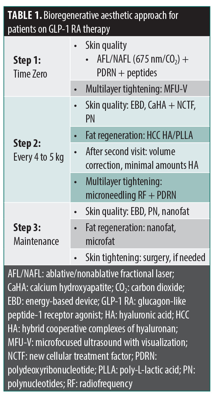
Time zero. Patients are not at an ideal body weight when starting a GLP-1 RA; therefore, injecting an HA-based filler or biostimulators might cause excessive volumization. The goal is to target fibroblasts and keratinocytes to improve skin quality and laxity. Multilayer tightening using MFU-V recruits fibroblasts to initiate ECM remodeling, especially elastin, that can contribute to a better recoil of the skin during the deflation of fat pads. A combination of ablative and nonablative fractional lasers assists with superficial epidermal resurfacing and dermal collagen and elastin production that can be impaired due to AT changes. Topical PN and peptides help improve skin tone, rhytids, hydration, and elasticity.
Weight loss phase. For every 4 to 5 kg of weight lost, patients require follow-up visits to assess skin quality and volumetric changes. Skin quality can be addressed with EBDs along with topical therapy. Scaffolds such as CaHA alone or in combination with peptides and micronutrients can be used, especially if skin density needs to be enhanced. Volume loss is addressed with HCCs and/or PLLA. HA filler can be used in follow-up appointments to address stubborn areas of volume loss.
Maintenance. After desired weight is achieved and stable, visits should occur every 6 to 12 months. Along with all the previous recommendations, fat grafting can be considered; however, careful placement of appropriate fat products is crucial for a natural appearance. Skin tightening should be addressed at every appointment. MFU-V or microneedling with radiofrequency (RF) can be employed to lift and tighten dermal and subdermal planes. Microneedling-assisted drug delivery with PNs can potentially amplify dermal tightening and remodeling.
Final considerations. Cost, accessibility, and safety are important considerations with regenerative modalities. These therapies can be cost-prohibitive and may require multiple sessions, limiting accessibility for a broader patient population. Additionally, variability in provider expertise and device availability can impact treatment outcomes and equity of care. Given the cost and accessibility parameters of these elective procedures, clinicians should counsel patients regarding expected results and prioritize safety. Care must be taken to avoid infections, granulomas, and vascular occlusion with biostimulators and fillers. EBDs can potentially lead to pigmentary changes, scarring, infections, and burns.
Lifestyle modifications are essential for managing patients taking GLP-1 RAs. Adequate protein and nutrition are essential for tissue regeneration and repair. Vitamins A, C, and D are enzymatic cofactors that facilitate wound healing, collagen synthesis, and remodeling. Zinc, selenium, and iron are necessary for fibroblast proliferation, collagen synthesis, and epithelialization.124–129
Conclusion
GLP-1 RA-MWL significantly affects multiple facial planes, leading to visible volume loss, skin laxity, and signs of accelerated aging. These changes reflect both structural and homeostatic disruptions, requiring a regenerative, multifaceted approach to aesthetic treatment. Skin laxity is addressed through MFU-V or microneedling with RF; fractional lasers, CaHA, and topical PNs and peptides improve skin quality; and,finally, PLLA, HCCs, and HA filler are useful for volume repletion. Ongoing monitoring and tailored interventions are critical, especially as patients may experience fluctuating weight or continued treatment with GLP-1 RAs. Finally, incorporating nutritional support and lifestyle optimization ensures more durable aesthetic and functional outcomes in this growing field.
References
- Iqbal J, Wu HX, Hu N, et al. Effect of glucagon-like peptide-1 receptor agonists on body weight in adults with obesity without diabetes mellitus—a systematic review and meta-analysis of randomized control trials. Obes Rev. 2022;23(6):e13435.
- Berning P, Adhikari R, Schroer AE, et al. Longitudinal analysis of obesity drug use and public awareness. JAMA Netw Open. 2025;8(1):e2457232.
- Zwick RK, Guerrero-Juarez CF, Horsley V, Plikus MV. Anatomical, physiological, and functional diversity of adipose tissue. Cell Metab. 2018;27(1):68–83.
- Longo M, Zatterale F, Naderi J, et al. Adipose tissue dysfunction as determinant of obesity-associated metabolic complications. Int J Mol Sci. 2019;20(9):2358.
- Alexander CM, Kasza I, Yen CLE, et al. Dermal white adipose tissue: a new component of the thermogenic response. J Lipid Res. 2015;56(11):2061–2069.
- Galanin I, Nicu C, Tower JI. Facial fat fitness: a new paradigm to understand facial aging and aesthetics. Aesthetic Plast Surg. 2021;45(1):151–163.
- Kim EJ, Kim YK, Kim JE, et al. UV modulation of subcutaneous fat metabolism. J Invest Dermatol. 2011;131(8):1720–1726.
- Cartwright MJ, Tchkonia T, Kirkland JL. Aging in adipocytes: potential impact of inherent, depot-specific mechanisms. Exp Gerontol. 2007;42(6):462–471.
- Fang CL, Huang LH, Tsai HY, Chang HI. Dermal lipogenesis inhibits adiponectin production in human dermal fibroblasts while exogenous adiponectin administration prevents against UVA-induced dermal matrix degradation in human skin. Int J Mol Sci. 2016;17(7):1129.
- Tower JI, Gordon NA, Paskhover B. Deep cheek fat volumes and midfacial aging. Aesthet Surg J. 2020;40(5):467–475.
- Allingham PG, Brownlee GR, Harper GS, ePho M, Nilsson SK, Brown TJ. Gene expression, synthesis and degradation of hyaluronan during differentiation of 3T3-L1 adipocytes. Arch Biochem Biophys. 2006;452(1):83–91.
- Ezure T, Amano S. Negative regulation of dermal fibroblasts by enlarged adipocytes through release of free fatty acids. J Invest Dermatol. 2011;131(10):2004–2009.
- Ezure T, Amano S. Adiponectin and leptin up-regulate extracellular matrix production by dermal fibroblasts. Biofactors. 2007;31(3–4):229–236.
- Jin T, Park KY, Seo SJ. Adiponectin upregulates filaggrin expression via SIRT1-mediated signaling in human normal keratinocytes. Ann Dermatol. 2017;29(4):407–413.
- Zhang Z, Shao M, Hepler C, et al. Dermal adipose tissue has high plasticity and undergoes reversible dedifferentiation in mice. J Clin Invest. 2019;129(12):5327–5342.
- Peng Y, Cheong S, Lu F, He Y. Dermal white adipose tissue: development and impact on hair follicles, skin defense, and fibrosis. FASEB J. 2024;38(18):e70047.
- Spalding KL, Arner E, Westermark PO, et al. Dynamics of fat cell turnover in humans. Nature. 2008;453(7196):783–787.
- Seeger MA, Paller AS. The roles of growth factors in keratinocyte migration. Adv Wound Care (New Rochelle). 2015;4(4):213–224.
- Hirobe T, Osawa M, Nishikawa SI. Hepatocyte growth factor controls the proliferation of cultured epidermal melanoblasts and melanocytes from newborn mice. Pigment Cell Res. 2004;17(1):51–61.
- Wolnicka-Glubisz A, Pecio A, Podkowa D, Plonka PM, Grabacka M. HGF/SF increases number of skin melanocytes but does not alter quality or quantity of follicular melanogenesis. PLoS One. 2013;8(11):e74883.
- Gille J, Khalik M, König V, Kaufmann R. Hepatocyte growth factor/scatter factor (HGF/SF) induces vascular permeability factor (VPF/VEGF) expression by cultured keratinocytes. J Invest Dermatol. 1998;111(6):1160–1165.
- Dunsmore SE, Rubin JS, Kovacs SO, Chedid M, Parks WC, Welgus HG. Mechanisms of hepatocyte growth factor stimulation of keratinocyte metalloproteinase production. J Biol Chem. 1996;271(40):24576–24582.
- Bell LN, Cai L, Johnstone BH, Traktuev DO, March KL, Considine RV. A central role for hepatocyte growth factor in adipose tissue angiogenesis. Am J Physiol Endocrinol Metab. 2008;294(2):E336–E344.
- Chun TH, Inoue M. 3-D adipocyte differentiation and peri-adipocyte collagen turnover. Methods Enzymol. 2014;538:15–34.
- Ezure T, Amano S. Increment of subcutaneous adipose tissue is associated with decrease of elastic fibres in the dermal layer. Exp Dermatol. 2015;24(12):924–929.
- Jalleh RJ, Marathe CS, Rayner CK, et al. Physiology and pharmacology of effects of GLP-1-based therapies on gastric, biliary and intestinal motility. Endocrinology. 2024;166(1):bqae155.
- Sell DR, Lane MA, Obrenovich ME, et al. The effect of caloric restriction on glycation and glycoxidation in skin collagen of nonhuman primates. J Gerontol A Biol Sci Med Sci. 2003;58(6):508–516.
- Chen CY, Zhang JQ, Li L, et al. Advanced glycation end products in the skin: molecular mechanisms, methods of measurement, and inhibitory pathways. Front Med (Lausanne). 2022;9:837222.
- Chung KW, Chung HY. The effects of calorie restriction on autophagy: role on aging intervention. Nutrients. 2019;11(12):2923.
- Bielach-Bazyluk A, Zbroch E, Mysliwiec H, et al. Sirtuin 1 and skin: implications in intrinsic and extrinsic aging—a systematic review. Cells. 2021;10(4):813.
- Choi YJ. Shedding light on the effects of calorie restriction and its mimetics on skin biology. Nutrients. 2020;12(5):1529.
- Hunt ND, Li GD, Zhu M, et al. Effect of calorie restriction and refeeding on skin wound healing in the rat. Age (Dordr). 2012;34(6):1453–1458.
- Ren H, Zhao F, Zhang Q, Huang X, Wang Z. Autophagy and skin wound healing. Burns Trauma. 2022;10:tkac003.
- Cabbabe SW. Plastic surgery after massive weight loss. Mo Med. 2016;113(3):202–206.
- Cálix M, Menéndez R, Baley M, Cadena A, Carrillo C, García-Jiménez J. Histological changes in skin and subcutaneous cellular tissue in patients with massive weight loss after bariatric surgery. Aesthetic Plast Surg. 2024;48(23):5060–5066.
- Rocha RI, Junior WC, Modolin MLA, Takahashi GG, Caldini ETEG, Gemperli R. Skin changes due to massive weight loss: histological changes and the causes of the limited results of contouring surgeries. Obes Surg. 2021;31(4):1505–1513.
- Hany M, Zidan A, Ghozlan NA, et al. Comparison of histological skin changes after massive weight loss in post-bariatric and non-bariatric patients. Obes Surg. 2024;34(3):855–865.
- Villanueva-Peñcarrillo ML, Márquez L, González N, Díaz-Miguel M, Valverde I. Effect of GLP-1 on lipid metabolism in human adipocytes. Horm Metab Res. 2001;33(2):73–77.
- Ridha Z, Fabi SG, Zubar R, Dayan SH. Decoding the implications of glucagon-like peptide-1 receptor agonists on accelerated facial and skin aging. Aesthet Surg J. 2024;44(11):NP809–NP818.
- Nikolis A, Enright KM, Fabi SG, et al. Consensus statements on managing aesthetic needs in prescription medication-driven weight loss patients: an international, multidisciplinary Delphi study. J Cosmet Dermatol. 2025;24(4):e70094.
- Firsowicz M, Kamrani P, Zubair R, et al. Cutaneous variations in stem-cell population in those on GLP1-receptor agonists: a comparative controlled study. Dermatol Surg. 2026;52(6S):S39–S44.
- Jafar AB, Jacob J, Kao WK, Ho T. Soft tissue facial changes following massive weight loss secondary to medical and surgical bariatric interventions: a systematic review. Aesthet Surg J Open Forum. 2024;6:ojae069.
- Valente DS, da Silva JB, Mottin CC, et al. Influence of massive weight loss on the perception of facial age: the facial age perceptions cohort. Plast Reconstr Surg. 2018;142(4):481e–488e.
- Deot N, Hughes E, Hatala A, Debick N, Suryadevara A. Patterns of midfacial Fat volume changes with semaglutide. Poster presented at: AAFPRS Annual Meeting 2023; 25–28 Oct 2023; Las Vegas, NV. Poster 56.
- Sharma RK, Vittetoe KL, Barna AJ, et al. Radiographic midfacial volume changes in patients on GLP-1 agonists. Otolaryngol Head Neck Surg. 2025;173(2):360–366.
- Sclafani AP. Restoration of the jawline and the neck after bariatric surgery. Facial Plast Surg. 2005;21(1):28–32.
- Narasimhan K, Ramanadham S, Rohrich RJ. Face lifting in the massive weight loss patient: modifications of our technique for this population. Plast Reconstr Surg. 2015;135(2):397–405.
- Biörserud C, Fagevik Olsén M, Elander A, Wiklund M. Objective measurements of excess skin in post bariatric patients—inter-rater reliability. J Plast Surg Hand Surg. 2016;50(2):68–73.
- Tay JQ. Ozempic face: a new challenge for facial plastic surgeons. J Plast Reconstr Aesthet Surg. 2023;81:97–98.
- Humphrey CD, Lawrence AC. Implications of Ozempic and other semaglutide medications for facial plastic surgeons. Facial Plast Surg. 2023;39(6):719–721.
- Cohen SR, Womack H, Ghanem A. Fat grafting for facial rejuvenation through injectable tissue replacement and regeneration: a differential, standardized, anatomic approach. Clin Plast Surg. 2020;47(1):31–41.
- Hu H, Shi Y, Qian Y, et al. Pyramidal multiple-theory (multi-type, multi-method and multi-layer) for facial fat grafting. J Cosmet Dermatol. 2023;22(3):937–944.
- Tang HH, Han XL, Debels H, et al. Fate of free fat grafts with or without adipogenic adjuncts to enhance graft outcomes. Plast Reconstr Surg. 2018;142(4):939–950.
- Coleman SR. Structural fat grafting: more than a permanent filler. Plast Reconstr Surg. 2006;118(3 Suppl)L108S–120S.
- Mojallal A, Lequeux C, Shipkov C, et al. Improvement of skin quality after fat grafting: clinical observation and an animal study. Plast Reconstr Surg. 2009;124(3):765–774.
- Uyulmaz S, Sanchez Macedo N, Rezaeian F, Giovanoli P, Lindenblatt N. Nanofat grafting for scar treatment and skin quality improvement. Aesthet Surg J. 2018;38(4):412–428.
- Jeyaraman M, Muthu S, Sharma S, Ganta C, Ranjan R, Jha SK. Nanofat: a therapeutic paradigm in regenerative medicine. World J Stem Cells. 2021;13(11):1733–1746.
- Guneta V, Tan NS, Chan SKJ, et al. Comparative study of adipose-derived stem cells and bone marrow-derived stem cells in similar microenvironmental conditions. Exp Cell Res. 2016;348(2):155–164.
- Zuk PA, Zhu M, Mizuno H, et al. Multilineage cells from human adipose tissue: implications for cell-based therapies. Tissue Eng. 2001;7(2):211–228.
- Friji MT. Nanofat grafting: basic research and clinical applications. Plast Reconstr Surg. 2014;134(2):333e-334e.
- Urso Pitassi LH, Pearson K, Antônio de Assis L, et al. Polynucleotides in skin regeneration: targeting the adenosine A2A receptor and salvage pathway. Dermatol Surg. 2024;50(11S):S131–S134.
- Shin SM, Baek EJ, Kim KH, Kim KJ, Park EJ. Polydeoxyribonucleotide exerts opposing effects on ERK activity in human skin keratinocytes and fibroblasts. Mol Med Rep. 2023;28(2):148.
- Lee KWA, Chan KWL, Lee A, et al. Polynucleotides in aesthetic medicine: a review of urrent practices and perceived effectiveness. Int J Mol Sci. 2024;25(15):8224.
- Park KY, Seok J, Rho NK, Kim BJ, Kim MN. Long-chain polynucleotide filler for skin rejuvenation: efficacy and complications in five patients. Dermatol Ther. 2016;29(1):37–40.
- Lis A, Kawałkiewicz W, Kubisz L. A comparison of the efficacy of reducing the signs of ageing in the neck skin after polynucleotides injections in regimens with different dosing intervals – a case study. J Face Aesthet. 2023;6(1):28–45.
- Brandi C, Cuomo R, Nisi G, Grimaldi L, D’Aniello C. Face rejuvenation: a new combinated protocol for biorevitalization. Acta Biomed. 2018;89(3):400–405.
- Lim TS, Liew S, Tee XJ, et al. Polynucleotides HPT for Asian skin regeneration and rejuvenation. Clin Cosmet Investig Dermatol. 2024;17:417–431.
- Lee D, Kim MJ, Park HJ, et al. Current practices and perceived effectiveness of polynucleotides for treatment of facial erythema by cosmetic physicians. Skin Res Technol. 2023;29(9):e13466.
- Kim JH, Jeong JJ, Lee YI, et al. Preventive effect of polynucleotide on post-thyroidectomy scars: a randomized, double-blinded, controlled trial. Lasers Surg Med. Published online 25 Mar 2018.
- Araco A, Araco F. Preliminary prospective and randomized study of highly purified polynucleotide vs placebo in treatment of moderate to severe acne scars. Aesthet Surg J. 2021;41(7):NP866–NP874.
- Merchán WH, Gómez LA, Chasoy ME, Alfonso-Rodríguez CA, Muñoz AL. Platelet-rich plasma, a powerful tool in dermatology. J Tissue Eng Regen Med. 2019;13(5):892–901.
- Acebes-Huerta A, Arias-Fernández T, Bernardo Á, et al. Platelet-derived bio-products: classification update, applications, concerns and new perspectives. Transfus Apher Sci. 2020;59(1):102716.
- Alam M, Hughart R, Champlain A, et al. Effect of platelet-rich plasma injection for rejuvenation of photoaged facial skin: a randomized clinical trial. JAMA Dermatol. 2018;154(12):1447–1452.
- Shen J, Yu X, Zhuang Y, Kong Y, Zhao J, Yao Z. Clinical efficacy and safety of targeted injection of PRP with skin booster in the treatment of aging face. Biotechnol Genet Eng Rev. 2024;40(3):2196–2212.
- Ulusal BG. Platelet-rich plasma and hyaluronic acid – an efficient biostimulation method for face rejuvenation. J Cosmet Dermatol. 2017;16(1):112–119.
- Modarressi A. Platlet rich plasma (PRP) improves fat grafting outcomes. World J Plast Surg. 2013;2(1):6–13.
- Debuc B, Gendron N, Cras A, et al. Improving autologous fat grafting in regenerative surgery through stem cell-assisted lipotransfer. Stem Cell Rev Rep. 2023;19(6):1726–1754.
- Liao HT, Marra KG, Rubin JP. Application of platelet-rich plasma and platelet-rich fibrin in fat grafting: basic science and literature review. Tissue Eng Part B Rev. 2014;20(4):267–276.
- Wu D, Zhao X, Xie J, et al. Physical modulation of mesenchymal stem cell exosomes: a new perspective for regenerative medicine. Cell Prolif. 2024;57(8):e13630.
- Zhang W, Bai X, Zhao B, et al. Cell-free therapy based on adipose tissue stem cell-derived exosomes promotes wound healing via the PI3K/Akt signaling pathway. Exp Cell Res. 2018;370(2):333–342.
- Xiong M, Zhang Q, Hu W, et al. The novel mechanisms and applications of exosomes in dermatology and cutaneous medical aesthetics. Pharmacol Res. 2021;166:105490.
- An Y, Lin S, Tan X, et al. Exosomes from adipose-derived stem cells and application to skin wound healing. Cell Prolif. 2021;54(3):e12993.
- Beer L, Mildner M, Ankersmit HJ. Cell secretome based drug substances in regenerative medicine: when regulatory affairs meet basic science. Ann Transl Med. 2017;5(7):170.
- Aguilar-Toalá JE, Hernández-Mendoza A, González-Córdova AF, Vallejo-Cordoba B, Liceaga AM. Potential role of natural bioactive peptides for development of cosmeceutical skin products. Peptides. 2019;122:170170.
- Lupo MP, Cole AL. Cosmeceutical peptides. Dermatol Ther. 2007;20(5):343–349.
- Pintea A, Manea A, Pintea C, et al. Peptides: emerging candidates for the prevention and treatment of skin senescence: a review. Biomolecules. 2025;15(1):88.
- Zhang L, Falla TJ. Cosmeceuticals and peptides. Clin Dermatol. 2009;27(5):485–494.
- Gold M, Boyd C, Mraz D, Robison T, Shafiq F, Widgerow AD. A multi-center clinical trial to evaluate the efficacy of the next generation TriHex technology antiaging regimen. J Cosmet Dermatol. 2025;24(4):e70192.
- Aguilera SB, McCarthy A, Khalifian S, Lorenc ZP, Goldie K, Chernoff WG. The role of calcium hydroxylapatite (Radiesse) as a regenerative aesthetic treatment: a narrative review. Aesthet Surg J. 2023;43(10):1063–1090.
- de Almeida AT, Figueredo V, da Cunha ALG, et al. Consensus recommendations for the use of hyperdiluted calcium hydroxyapatite (Radiesse) as a face and body biostimulatory agent. Plast Reconstr Surg Glob Open. 2019;7(3):e2160.
- Theodorakopoulou E, McCarthy A, Perico V, Aguilera SB. Optimizing skin regenerative response to calcium hydroxylapatite microspheres via poly-micronutrient priming. J Drugs Dermatol. 2023;22(9):925–934.
- Zubair R, Ishii L, Loyal J, Hartman N, Fabi SG. SPLASH: split-body randomized clinical trial of poly- L -lactic acid for adipogenesis and volumization of the hip dell. Dermatol Surg. 2024;50(12):1155–1162.
- Kim HW, Jung YA, Yun JM, et al. Effects of poly-L-lactic acid on adipogenesis and collagen gene expression in cultured adipocytes irradiated with ultraviolet B rays. Ann Dermatol. 2023;35(6):424–431.
- Waibel J, Ziegler M, Nguyen TQ, et al. Comparative bulk RNA-seq analysis of poly-L-lactic acid versus calcium hydroxylapatite reveals a novel, adipocyte-mediated regenerative mechanism of action unique to PLLA. Dermatol Surg. 2024;50(11S):S166–S171.
- Ao YJ, Yi Y, Wu GH. Application of PLLA (poly-L-lactic acid) for rejuvenation and reproduction of facial cutaneous tissue in aesthetics: a review. Medicine (Baltimore). 2024;103(11):e37506.
- Fitzgerald R, Bass LM, Goldberg DJ, Graivier MH, Lorenc ZP. Physiochemical characteristics of poly-L-lactic acid (PLLA). Aesthet Surg J. 2018;38(suppl 1):S13–S17.
- Bohnert K, Dorizas A, Lorenc P, Sadick NS. Randomized, controlled, multicentered, double-blind investigation of injectable poly-L-lactic acid for improving skin quality. Dermatol Surg. 2019;45(5):718–724.
- Wilkerson EC, Goldberg DJ. Poly-L-lactic acid for the improvement of photodamage and rhytids of the décolletage. J Cosmet Dermatol. 2018;17(4):606–610.
- Baroni E do RV, Biondo-Simões M de LP, Auersvald A, et al. Influence of aging on the quality of the skin of white women: the role of collagen. Acta Cir Bras. 2012;27(10):736–740.
- Jabbar A, Arruda S, Sadick N. Off face usage of poly-L-lactic acid for body rejuvenation. J Drugs Dermatol. 2017;16(5):489–494.
- Cassuto D, Cigni C, Bellia G, Schiraldi C. Restoring adipose tissue homeostasis in response to aging: initial clinical experience with Profhilo Structura®. Gels. 2023;9(8):614.
- Stellavato A, Corsuto L, D’Agostino A, et al. Hyaluronan hybrid cooperative complexes as a novel frontier for cellular bioprocesses re-activation. PLoS One. 2016;11(10):e0163510.
- Stellavato A, La Noce M, Corsuto L, et al. Hybrid complexes of high and low molecular weight hyaluronans highly enhance hascs differentiation: Implication for facial bioremodelling. Cell Physiol Biochem. 2017;44(3):1078–1092.
- Alessio N, Stellavato A, Squillaro T, et al. Hybrid complexes of high and low molecular weight hyaluronan delay in vitro replicative senescence of mesenchymal stromal cells: a pilot study for future therapeutic application. Aging. 2018;10(7):1575–1585.
- Vachiramon V, Pavicic T, Casabona G, et al. Microfocused ultrasound in regenerative aesthetics: a narrative review on mechanisms of action and clinical outcomes. J Cosmet Dermatol. 2025;24(2):e16658.
- White WM, Makin IRS, Barthe PG, Slayton MH, Gliklich RE. Selective creation of thermal injury zones in the superficial musculoaponeurotic system using intense ultrasound therapy: a new target for noninvasive facial rejuvenation. Arch Facial Plast Surg. 2007;9(1):22–29.
- Suh DH, Choi JH, Lee SJ, Jeong KH, Song KY, Shin MK. Comparative histometric analysis of the effects of high-intensity focused ultrasound and radiofrequency on skin. J Cosmet Laser Ther. 2015;17(5):230–236.
- Marquardt K, Hartmann C, Wegener F, et al. Microfocused ultrasound with visualization induces remodeling of collagen and elastin within the skin. J Cosmet Dermatol. 2025;24(1):e16638.
- McCarthy AD, Soares DJ, Chandawarkar A, El-Banna R, Hagedorn N. Dilutional rheology of Radiesse: implications for regeneration and vascular safety. J Cosmet Dermatol. 2024;23(6):1973–1984.
- Casabona G, Kaye K, Cotofana S, Davidovic K, Alfertshofer M, Freytag L. Histological effects of a combined collagen stimulation procedure consisting of microfocused ultrasound, soft tissue filler, and Ca-HA injections. J Cosmet Dermatol. 2023;22(6):1724–1730.
- Goldberg D, Guana A, Volk A, Daro-Kaftan E. Single-arm study for the characterization of human tissue response to injectable poly-L-lactic acid. Dermatolog Surg. 2013;39(6):915–922.
- Hart DR, Fabi SG, White WM, Fitzgerald R, Goldman MP. Current concepts in the use of PLLA: clinical synergy noted with combined use of microfocused ultrasound and poly-l-lactic acid on the face, neck, and décolletage. Plast Reconstr Surg. 2015;136(5 Suppl):180S–187S.
- Friedmann DP, Fabi SG, Goldman MP. Combination of intense pulsed light, Sculptra, and Ultherapy for treatment of the aging face. J Cosmet Dermatol. 2014;13(2):109–118.
- Jerdan K, Fabi SG. A combination approach with lasers, light, and energy-based devices for ultimate rejuvenation of the aging face. Curr Dermatol Rep. 2016;5(3):208–216.
- Raddatz G, Hagemann S, Aran D, et al. Aging is associated with highly defined epigenetic changes in the human epidermis. Epigenetics Chromatin. 2013;6(1):36.
- Falckenhayn C, Bienkowska A, Söhle J, et al. Identification of dihydromyricetin as a natural DNA methylation inhibitor with rejuvenating activity in human skin. Front Aging. 2023;4:1258184.
- Lu AT, Quach A, Wilson JG, et al. DNA methylation GrimAge strongly predicts lifespan and healthspan. Aging (Albany NY). 2019;11(2):303–327.
- Wang K, Liu H, Hu Q, et al. Epigenetic regulation of aging: implications for interventions of aging and diseases. Signal Transduct Target Ther. 2022;7(1):374.
- Zamani ARN, Saberianpour S, Geranmayeh MH, Bani F, Haghighi L, Rahbarghazi R. Modulatory effect of photobiomodulation on stem cell epigenetic memory: a highlight on differentiation capacity. Lasers Med Sci. 2020;35(2):299–306.
- Basu A, Tiwari VK. Epigenetic reprogramming of cell identity: lessons from development for regenerative medicine. Clin Epigenetics. 2021;13(1):144.
- Beigvand HH, Razzaghi M, Rostami-Nejad M, et al. Assessment of laser effects on skin rejuvenation. J Lasers Med Sci. 2020;11(2):212–219.
- Haykal D, Will F, Cartier H, Dahan S. Epigenetic modifications and the role of medical lasers in enhancing skin regeneration. J Cosmet Dermatol. 2025;24(1):e16780.
- Marioni RE, Belsky DW, Deary IJ, Wagner W. Association of facial ageing with DNA methylation and epigenetic age predictions. Clin Epigenetics. 2018;10(1):140.
- Ghaly P, Iliopoulos J, Ahmad M. The role of nutrition in wound healing: an overview. Br J Nurs. 2021;30(5):S38–S42.
- Quain AM, Khardori NM. Nutrition in wound care management: a comprehensive overview. Wounds. 2015;27(12):327–335.
- Molnar JA, Underdown MJ, Clark WA. Nutrition and chronic wounds. Adv Wound Care (New Rochelle). 2014;3(11):663–681.
- Acton C. The importance of nutrition in wound healing. Wounds UK. 2013;9(3):61–64.
- Lansdown ABG, Mirastschijski U, Stubbs N, Scanlon E, Ågren MS. Zinc in wound healing: theoretical, experimental, and clinical aspects. Wound Repair Regen. 2007;15(1):2–16.
- Johnston E. The role of nutrition in tissue viability. Wound Essentials. 2007;2:10–21.
From the 2nd Annual RAPIDS Immuno-Dermatology Conference: From Approval to Adoption—Translating Innovation Into Clinical Practice