
J Clin Aesthet Dermatol. 2026;19(7–8 Suppl 1):S20–S23.
Katie L. Frederickson, MSN, FNP-BC; Tammy E. Ehimwenma-Point Du Jour, MBS; Sarah P. Pourali, MD; and Chike Nzerue, MD
Msses. Frederickson and Ehimwenma-Point Du Jour and Dr. Nzerue are with Meharry Medical College, School of Medicine, Nashville, Tennessee. Dr. Pourali is with Vanderbilt University Medical Center, Department of Dermatology, Nashville, Tennessee.
Funding: No funding was provided for this article.
Disclosures: The authors have no relevant conflicts of interest.
Abstract: This case report represents a 62‐year‐old African American female patient with diabetic kidney disease who presented with a suspected pityriasis rubra pilaris–like eruption after starting oral empagliflozin for 1 week. The patient presented with a severely pruritic rash, erythematous plaques, hyperpigmented patches, and follicular papules on the scalp, face, trunk, and extremities. The patient did not respond to initial treatment including topical mometasone or oral prednisone. Gabapentin, topical triamcinolone, clobetasol, and tacrolimus were additionally prescribed for symptomatic relief. Acitretin was initiated and slowly titrated from 10 to 25 to 30 mg daily. Aprelimast was added at the 6-month follow-up to further increase clearance. Slow yet progressive improvement of the rash and pruritus was observed. This clinical vignette highlights the importance of early clinical recognition and education about the adverse effects of temporally associated empagliflozin pityriasis rubra pilaris–like eruptions. Keywords: General dermatology, medical dermatology, SGLT2 inhibitors, pityriasis rubra pilaris, drug eruption
Introduction
Pityriasis rubra pilaris (PRP) is a papulosquamous inflammatory dermatosis characterized by hyperkeratotic follicular papules coalescing into orange and red scaly plaques demonstrating islands of sparing and palmoplantar keratoderma.1 Due to the overlapping features of PRP with psoriasis and other cutaneous eruptions, establishing a clinical diagnosis may create diagnostic challenges. Given the rising curiosity of acute drug-related dermatoses in dermatology, our objectives are to 1) provide a detailed clinical case of suspected PRP precipitated by sodium glucose transporter 2 (SGLT2) inhibitors; 2) assist healthcare providers in preemptive identification of PRP; and 3) discuss effective treatment options to manage PRP while prioritizing patient comfort.
Case Report
A 62-year-old African American female patient with a past medical history of hypertension, type 2 diabetes mellitus, stage 3 chronic kidney disease (CKD), hyperlipidemia, and schizophrenia presented to the emergency department on March 21, 2025, with a 7-day history of a severely pruritic exanthem. Two weeks prior to the onset, a 1-week sample of empagliflozin 10 mg daily was initiated. The eruption presented as erythematous, scaling patches in a cephalocaudal fashion of the eyebrows, temples, scalp, and ears, quickly progressing to the palms, trunk, extremities, and soles of the feet. The patient denied fever, chills, or joint pain. Empagliflozin was discontinued at this time, and topical mometasone 0.05% cream was initiated twice daily with a 10-day course of oral prednisone.
On March 24, 2025, in the nephrology clinic, the rash was consistent with the previous presentation, with no subjective or objective improvement. Subsequent management included a second 10-day course of oral prednisone and continuation of topical mometasone 0.05% cream twice daily. An emergent dermatology referral was initiated.
At the initial dermatology evaluation on April 28, 2025, the patient reported no improvement of symptoms despite diligent adherence to topical steroids and oral prednisone. Physical exam revealed well-defined erythematous plaques with thick scale on the scalp, hyperpigmented patches with fine scale on the face, and follicular-based papules with scaly plaques on the trunk and extremities with islands of sparing. The elbows and knees revealed distinct hyperpigmented, erythematous plaques. Palms and soles were notable for extensive orange hyperkeratosis (Figures 1 and 2).
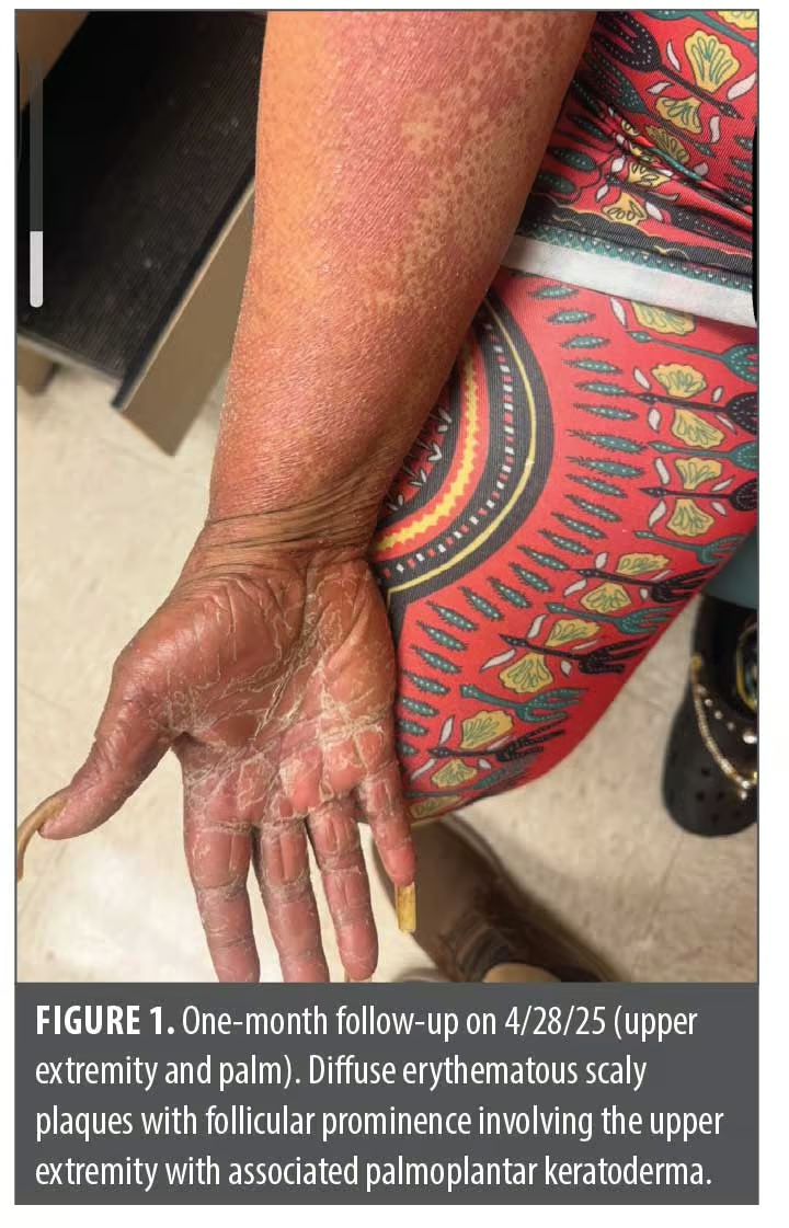
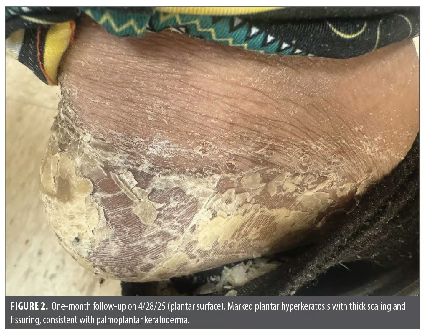
The patient opted against a skin biopsy due to extreme fear of needles. Therefore, the patient was diagnosed clinically with suspected type I PRP, and the eruption was not confirmed histologically. Differential diagnoses included other papulosquamous disorders, such as psoriasis vulgaris, palmoplantar psoriasis, exanthematous drug eruption, and acrodermatitis continua of hallopeau. The clinical assessment findings led to a probable diagnosis of suspected type I PRP due to the classic morphology of hyperkeratosis with orange-red plaques with well-demarcated borders and islands of sparing.
Initial systemic therapy options were discussed, including retinoids, immune suppressants such as methotrexate, and interleukin (IL)-12/IL-23 inhibitors. Oral methotrexate and IL-12/23 inhibitors were considered; however, due to the patient’s extreme fear of needles and history of stage 3 CKD, these drugs were not a first-line option. Moreover, initial therapy with apremilast was excluded due to a history of schizophrenia with the risk of potential worsening of psychiatric symptoms. Medication additions included clobetasol 0.05% ointment twice daily to the thickest plaques on the elbows, knees, palms, and soles; triamcinolone 0.05% ointment twice daily to the trunk and extremities; and tacrolimus 0.1% topical ointment to the facial skin twice daily. Mometasone was discontinued at this time. Urea 10% topical cream was also recommended twice daily for dry skin.
At the subsequent dermatology follow-up visit on June 17, 2025, systemic therapy was initiated due to intractable pruritus and difficulty sleeping. Acitretin 10 mg daily was selected to accommodate the patient’s preference for oral therapy in addition to her history of stage 3 CKD and schizophrenia. A comprehensive metabolic panel and estimated glomerular filtration rate (eGFR) were obtained and monitored throughout the treatment course. Baseline eGFR was 38 mL/min, blood urea nitrogen (BUN) was 17, and creatinine was 1.55 mg/dL (Table 1).
Symptomatic management included gabapentin 100 mg at bedtime for pruritus, with continuation of topical steroids and tacrolimus. The patient was counseled on the risks, benefits, and side effects of all medications and the importance of adherence to therapy, regular lab evaluation, and follow-up appointments.
The patient returned to the clinic on July 15, 2025, reporting a mild improvement of the rash and pruritis. Acitretin was increased to 25 mg daily at this time, and all other prescribed medications were continued. No adverse effects of apremilast were reported. The patient’s eGFR was 43 mL/min, BUN was 11, and creatinine was 1.38 mg/dL (Table 1).
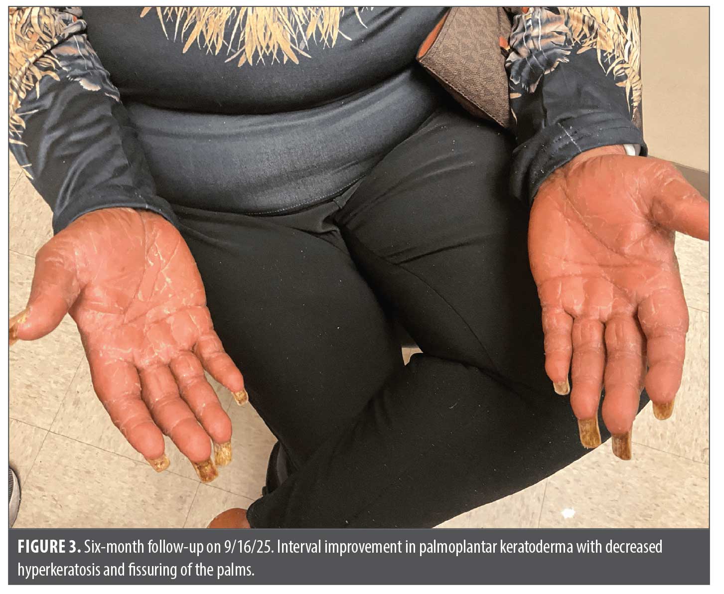
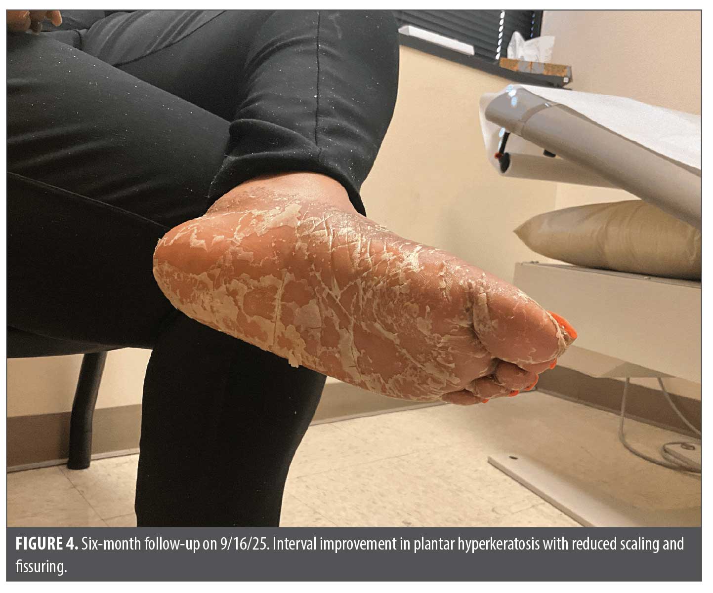
On September 15, 2025, clinical reassessment demonstrated a significant subjective reduction in pruritus, scaling, and softening of the plaques. Erythema and scaling were noticeably decreased, and the patient reported a significant improvement in sleep quality (Figures 3 and 4). Acitretin was increased to 30 mg daily and apremilast 10 mg daily was initiated. The tacrolimus 0.1% was replaced at this time with hydrocortisone 2.5% cream due to a persistent burning sensation. The risks, benefits, and side effects of apremilast were thoroughly reviewed, and the benefit of rash clearance outweighed the risk of potential worsening of psychiatric symptoms. The patient continued her regular dose of aripiprazole, and mood changes were closely monitored.
The patient was unable to attend her subsequent office visit. However, she reported an update over the phone of consistent, gradual improvement of the skin eruption and pruritus.
Limitations of this case include lack of biopsy and histopathology confirmation, limited exclusion of differential diagnoses, the remote phone call follow-up appointment, and the inability to establish causation from a single case.
Discussion
PRP is a papulosquamous inflammatory dermatosis characterized by hyperkeratotic follicular papules coalescing into orange and red scaly plaques demonstrating islands of sparing and palmoplantar keratoderma.1 Although the mechanisms underlying the development remain uncertain, PRP has been associated with infections, malignancy, autoimmune disease, and medications, most notably tyrosine kinase inhibitors and toll-like receptor 7 agonists.1–3 Importantly, PRP has not been associated with SGLT2 inhibitors, such as empagliflozin.
Distinguishing PRP from psoriasis might be clinically challenging because of overlapping papulosquamous features.4 In the absence of histopathologic confirmation, recognition of characteristic clinical findings, including follicular hyperkeratosis, palmoplantar keratoderma, orange-red plaques, and islands of sparing, might help support the diagnosis.
SGLT2 inhibitors promote glycemic control through reduced reabsorption of glucose at the proximal convoluted tubule, thereby encouraging glucose excretion. The most common adverse effects associated with these drugs include urinary tract infections, vaginal candidiasis, polyuria, nausea, constipation,5 and drug-induced cutaneous events; these range from pruritus and fixed drug eruptions to psoriasiform eruptions, and, rarely, severe entities such as Fournier’s gangrene, and Stevens-Johnson syndrome.5,6 Individual case reports have also described empagliflozin-associated cutaneous drug eruptions, further supporting the diverse spectrum of dermatologic adverse events reported with this drug class.6 Patients with diabetic vascular disease have been described as particularly vulnerable to cutaneous drug reactions, reflecting impaired skin perfusion, neuropathy, and immune dysfunction, with a heightened susceptibility to hypersensitivity reactions. Although CKD has not been broadly linked to all cutaneous adverse drug reactions, it has been recognized as a susceptibility factor in severe T cell–mediated reactions, such as allopurinol-induced drug reaction with eosinophilia and systemic symptoms, where impaired renal clearance of drug metabolites increases the risk of hypersensitivity.7
Building on these clinical observations, the relationship between SGLT2 inhibition and inflammatory skin disease remains incompletely understood. In the present case, it is possible that SGLT2 inhibition contributed to the development of a PRP-like eruption through modulation of inflammatory pathways. Experimental studies have demonstrated that empagliflozin could influence the IL-17/IL-23 axis via the AMPK/mTOR/autophagy pathway,8 typically resulting in downregulation of proinflammatory signaling. However, the clinical relevance of these immunologic effects in cutaneous disease remains unclear. In the absence of histopathological confirmation or molecular evaluation in this patient, this proposed mechanism remains speculative. Further research is needed to better define the immunological pathways underlying medication-associated PRP-like eruptions.
This case is unique given the development of suspected adult-onset PRP-like eruption following the initiation of empagliflozin in a patient with stage 3 CKD and no personal or family history of psoriasis or other chronic dermatoses. The eruption was extensive, clinically consistent with a type I PRP-like eruption, and required escalation from topical therapy to systemic agents, including acitretin and apremilast. This presentation distinguishes our case from previously reported SGLT2 inhibitor–associated reactions, as most of them reported in the literature involved dapagliflozin and presented as psoriasiform or fixed drug eruptions.5,6 Furthermore, prior reported SGLT2 inhibitor–associated psoriasiform eruptions often occurred in patients with a history of psoriasis. Viewing this suspected empagliflozin-associated PRP-like eruption in the context of dapagliflozin-associated psoriasiform eruptions underscores the heterogeneity of SGLT2 inhibitor–related cutaneous reactions and suggests that host factors such as CKD and immune dysregulation might modulate the clinical phenotype.
Conclusion
This case highlights a potential association between SGLT2 inhibitor therapy and the development of a PRP-like eruption, emphasizing the importance of maintaining a high index of suspicion for medication-induced papulosquamous disorders. Dermatology providers should carefully review recent medication changes in patients presenting with new onset or atypical PRP-like eruptions, even in the absence of prior dermatologic disease. Early recognition, discontinuation of the suspected agent, and timely initiation of appropriate therapy can help reduce disease severity and improve clinical outcomes.
Ethics statement
The patient in this manuscript has given written informed consent for participation in the study and the use of their de-identified, anonymized, aggregated data and their case details (including photographs) for publication.
References
- Joshi TP, Duvic M. Pityriasis rubra pilaris: an updated review of clinical presentation, etiopathogenesis, and treatment options. Am J Clin Dermatol. 2024;25:243–259.
- Mufti A, Lytvyn Y, Sachdeva M, et al. Drugs associated with development of pityriasis rubra pilaris: a systematic review. J Am Acad Dermatol. 2021;84(4):1071–1081.
- Weber I, Gloss C, Castillo B, Smith EH, et al. Ponatinib-associated cutaneous eruptions: a case series and review of clinicopathologic findings. Am J Dermatopathol. 2023;45(1):64–68.
- Ross NA, Chung HJ, Li Q, et al. Epidemiologic, clinicopathologic, diagnostic, and management challenges of pityriasis rubra pilaris: a case series of 100 patients. JAMA Dermatol. 2016;152(6):670–675.
- Qiu M, Ding L, Zeng Y,et al. Comprehensive analysis of adverse events associated with SGLT2 inhibitors. Front Endocrinol. 2021;12:743807.
- Mederle AL, Dumitrescu P, Borza C, Kundnani NR. Cutaneous adverse drug reactions associated with SGLT2 inhibitors. J Clin Med. 2024;14(1):188.
- Ng C-Y, Yeh Y-T, Wang C-W, et sl. Impact of the HLA-B*58:01 allele and renal impairment on allopurinol-induced cutaneous adverse reactions. J Invest Dermatol. 2016;136(7):1373–1377.
- Meng Z, Liu X, Li T, et al. The SGLT2 inhibitor empagliflozin negatively regulates IL-17/IL-23 axis-mediated inflammatory responses in T2DM with NAFLD via the AMPK/mTOR/autophagy pathway. Int Immunopharmacol. 2021;4(107492):107492.
From the 2nd Annual RAPIDS Immuno-Dermatology Conference: From Approval to Adoption—Translating Innovation Into Clinical Practice