
J Clin Aesthet Dermatol. 2026;19(9):42–48.
Rebecca Olsen, DO, MS; Aarushi Gulati, BS; Rachna Sahasrabudhe, DO; and Abdul Rahim Jaber, MD
Dr. Olsen is with Northwest Medical Center, Tucson, Arizona. Ms. Gulati is with A.T. Still University School of Osteopathic Medicine in Arizona, Mesa, Arizona. Dr. Sahasrabudhe is with Trinity Health Ann Arbor, Ann Arbor, Michigan. Dr. Jaber is with American University of Anguilla School of Medicine, Anguilla.
FUNDING: No funding was provided for this article.
DISCLOSURES: The authors have no relevant conflicts of interest.
Abstract: BACKGROUND: Animal- and human-derived materials are commonly used in dermatologic and cosmetic products, yet their presence is often unclear to clinicians and patients. OBJECTIVE: To examine ethical and informed-consent implications of these materials, particularly when they conflict with patients’ religious, cultural, or personal values. METHODS: A narrative literature search and product formulary audit were conducted on PubMed through February 2025, augmented by United States Food and Drug Administration summaries and manufacturer dossiers. Data extraction focused on material origin, clinical use, outcomes, adverse events, and labeling transparency. RESULTS: Numerous dermatologic products contain animal- or human-derived components, such as rooster-comb–derived hyaluronic acid, bovine collagen grafts, shark-derived glycosaminoglycans, and human-derived materials. The source of these materials is frequently absent or inconsistently reported in labeling, limiting clinicians’ ability to counsel patients effectively. Studies demonstrate that when informed and given alternatives, most patients prefer nonanimal-derived options. Advances in synthetic and bioengineered materials make such alternatives increasingly feasible. LIMITATIONS: This review is not exhaustive. Given the extent of dermatologic and cosmetic products on the market and variability in ingredient disclosure, some animal- or human-derived materials may not have been captured. Rapid product innovation and inconsistent reporting of material origins further limit comprehensive identification. CONCLUSION: Improved transparency regarding product origins is essential for meaningful informed consent in dermatology. Incorporating brief intake screening questions and updating consent forms to disclose material sources may enhance shared decision-making, patient trust, and ethically aligned, patient-centered care. Keywords: Animal-derived materials, ethics, informed consent, surgery, religion, surgical products
Introduciton
According to a 2019 study, most patients prefer to be informed if animal-derived products are used in their dermatologic procedures (n=75/101; 74%).1 The study surveyed patients on their thoughts regarding use of animal-derived sutures based on their dietary preferences. Data indicated that 33% of patients would decline the use of sutures that contained animal byproducts (n=32/96) and 47% would decline animal-derived sutures even if that meant they would have to return for another visit to remove the stitches (n=31/55; 47%). Interestingly, the response to this survey was not statistically significant between vegetarians and nonvegetarians.1 In 2013, spiritual leaders representing the 6 largest religions worldwide were surveyed on the use of products containing materials derived from animal sources. This survey found that animal-derived products (predominantly porcine ± bovine) are generally not allowed in the Islam (Sunni and Shiite), Sikhism, and Vaishnavism (a branch of Hinduism) religions.2 Although clinical treatment decisions may be influenced by religious and/or cultural beliefs, individual treatment preferences may vary. For example, attitudes and beliefs about animal product use can also impact dietary choices. In strict vegetarianism, meat products are avoided with periodic consumption of nonmeat animal products, which contrasts with strict veganism, where no animal products are consumed.3 Informed consent is used to protect patient autonomy for shared decision-making relationships.
It is a physician’s responsibility to recognize materials that contain animal byproducts, as these products may potentially conflict with the patient’s religious and/or cultural beliefs. Informed consent is an ethical and legal requirement integral to modern surgical practice. Clinicians have to consider, disclose, and discuss risks and concerns relevant to each patient. With advances in medicine, animal-derived products and adjuncts are available for use in dermatology. For individuals to give informed consent, it is essential to provide clear and comprehensive information regarding products that will be used in their clinical treatment.4 To ensure informed consent and shared decision-making, it is important to address each patient individually to determine any relevant beliefs or views regarding the use of animal-derived products so that concerns are identified and appropriate alternatives are offered.4
This literature review will discuss hemostatic agents, sutures, glues, skin substitutes, and fillers used in dermatology. It will highlight which products are derived from animal or human sources in the hopes of increasing physician awareness of product formulations.
Discussion
Drugs. Mechanical hemostatic agents act as a barrier by swelling or acting as molecular sieves to concentrate solid blood components. Swelling causes pressure at the site while physically blocking the bleed and provides a foundation for the first phase of coagulation and platelet aggregation. Passive substrates such as collagen, gelatin, regenerated oxidized cellulose, and polysaccharide spheres induce platelet activation and aggregation for rapid clot formation. These are primarily derived from porcine, bovine, equine, or vegetable origins and are available in many forms including powder, sheet, and gauze.5
Absorbable gelatin and gelatin-based sponges (Table 1) are derived from porcine collagen, absorbing blood and other fluids to create a framework to promote clot formation. They are commonly used in cutaneous dermatologic surgical procedures to achieve local hemostasis. These products can be used alone or in combination with topical thrombin (human, bovine, or recombinant origin) to enhance hemostatic efficacy. They should be avoided in tight, confined spaces due to the risk of expansion and compressive necrosis.6 These gelatin sponges are usually absorbed over 4 to 6 weeks and should be avoided in patients with severe coagulopathies, as the agents require an intact coagulation cascade to properly function.6 Other risks include development of infection, abscess, foreign body reaction, and granuloma formation.6–9
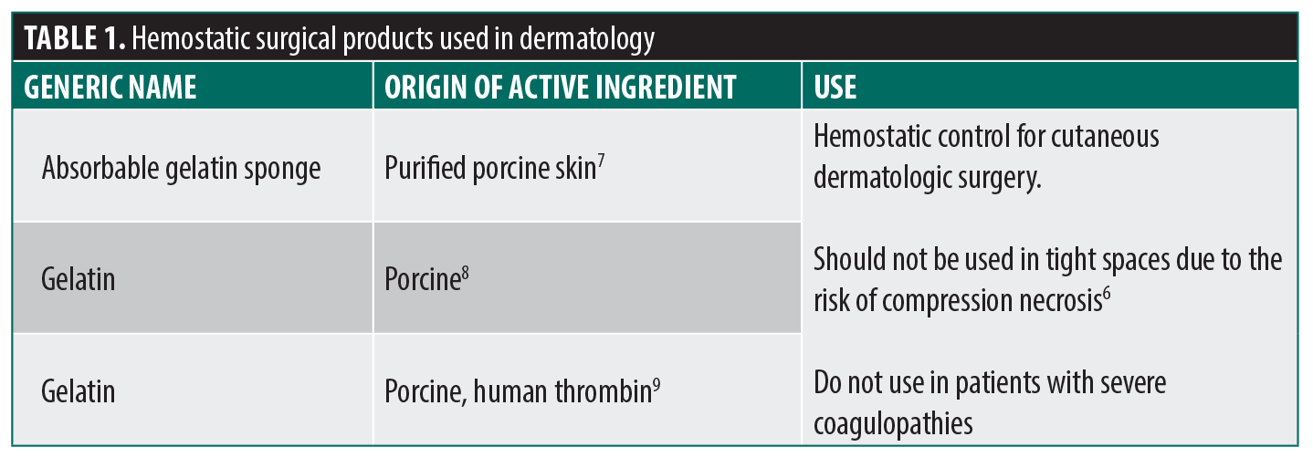
- There are several nonanimal/human-derived alternatives available (Table 2):
- Cellulose-based products are plant-derived oxidized regenerated cellulose that induce the extrinsic coagulation cascade and provide a physical matrix and acidic environment to promote clotting and inhibit bacterial growth. These are typically absorbed within 1 to 2 weeks.6 It is not recommended that cellulose-based products be combined with topical thrombin because the acidic environment can inactivate thrombin,6 and the combination may cause unnecessary additional tissue inflammation. Risks of use include infections, abscesses, foreign-body reactions, and granuloma formation. The agents are fairly slow to absorb (1–2 weeks) and, although rare, have been reported to persist up to 15 months postoperatively.6
- Microporous polysaccharide hemospheres are composed of plant-derived polysaccharide spheres that rapidly absorb water, thereby concentrating clotting factors at the bleeding site.6 The product absorbs quickly (48 hours) but should be used with caution in patients with diabetes, as it can alter serum glucose levels. One adverse effect of this product is immediate swelling of the application site, potentially damaging surrounding structures.5,6
- Polyethylene glycol hydrogel is used as a sealant rather than a primary hemostatic agent in cutaneous dermatologic surgery. It induces polyethylene glycol polymerization, forms tissue crosslinks, and increases platelet adherence. A notable adverse effect is significant tissue swelling, which can also potentially damage surrounding tissues.5,10,11
- There is also a hydrophilic polymer and potassium salt–based powder that is infrequently used in dermatology. The polymers cause a dehydration effect on the blood, and the potassium salt binds to positively charged red blood cells, leading to the formation of an eschar within 1 minute of application. It is an inexpensive, fast-acting agent with very few adverse effects.5
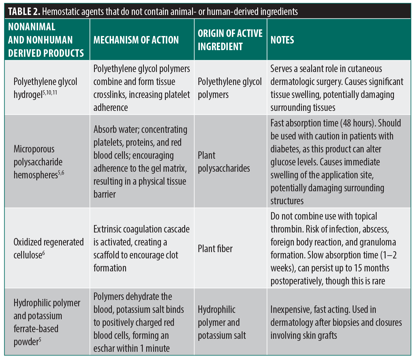
Topical caustic hemostatic agents (Table 3) are frequently used in dermatologic procedures to achieve rapid hemostasis, especially in settings where patients are on anticoagulants or electrocautery is contraindicated. There are 3 main topical hemostatic agents: aluminum chloride, ferric subsulfate (Monsel’s solution), and silver nitrate. Two additional agents have historically been used but are now less common in current practice (Supplemental Table 1). This class of hemostatic agents causes coagulation of proteins, leading to localized tissue necrosis, eschar formation, and thrombus formation.12 The topical agents described are available at low cost and can be stored at room temperature. These agents achieve rapid hemostasis and are easy to use, but many carry the risk of tissue necrosis, tissue staining, and delayed wound healing.12
Monsel’s solution (20% ferric subsulfate solution) is used in the setting of capillary and small-vessel bleeding of small wounds on skin or mucosal surfaces. It is applied directly to the wound on a cotton swab with light pressure and may be used prior to electrodesiccation. Due to the acidic environment of this solution, there is minimal bacterial growth in the solution. The most common adverse risk associated with Monsel’s solution is persistent gray-to-brown dyspigmentation due to dermal deposits of iron particles, which may also complicate histopathologic interpretation. Other risks include postinflammatory hyperpigmentation, erythema, dermal fibrosis, treatment-site infections, and impaired re-epithelization of the wound.12
Aluminum chloride (concentration 20%–40%) is commonly used for superficial postsurgical bleeding such as shave biopsies. It is applied with a cotton-tipped applicator using light pressure in a twisting motion perpendicular to the cutaneous surface, rapidly achieving hemostasis. It is a popular choice among many dermatologists due to its minimal risk of tissue discoloration. The solution does have notable adverse effects including tissue irritation, painful local paresthesia, and, if used excessively, delayed wound re-epithelialization.12
Silver nitrate sticks are another low-cost option used for pinpoint bleeding and the treatment of hypergranulation.14 By coagulating surface proteins to form a thin eschar, silver nitrate can cause hyperpigmentation, which typically resolves within a few days. However, there is a slight risk of tattooing from silver deposition within skin, particularly if large surface areas are treated. Additional risks include tissue irritation and delayed re-epithelialization.12,13
Sutures. Sutures, both absorbable and nonabsorbable, are used in dermatologic procedures such as punch biopsies, excisions, and Mohs closures. Multiple options exist for each type of suture. Animal-derived and alternative suture options are depicted in Table 4. Absorbable sutures, commonly used for dermal closure, are predominantly synthetic. However, gut suture subtypes are derived from sheep or bovine sources. Nonabsorbable sutures, which are typically used for superficial closures and require manual removal, are also largely synthetic. An important exception is silk suture, which is derived from silkworm larval protein.15
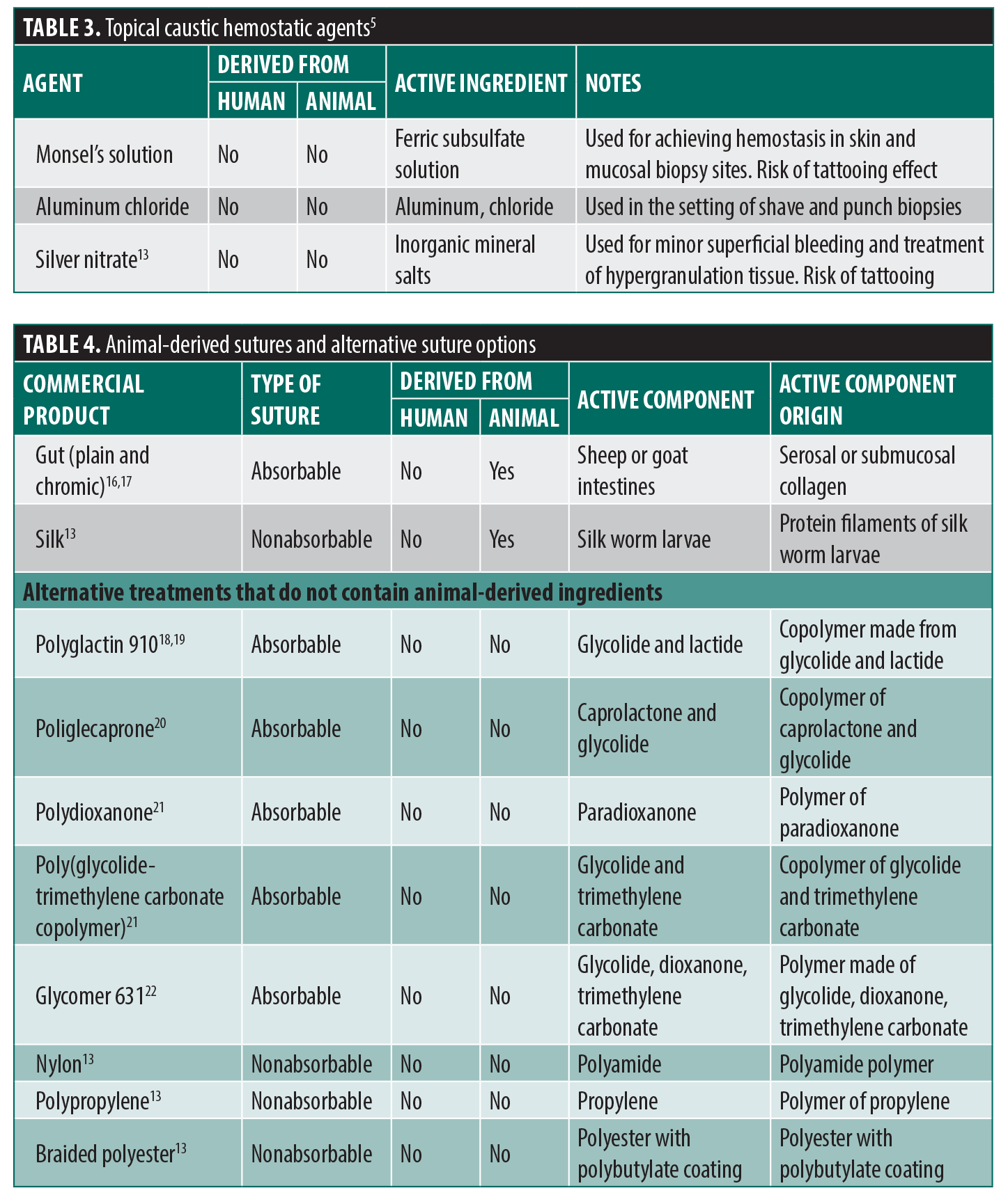
Glues. Three main surgical adhesives are used in dermatologic procedures. All included in this review are synthetically derived, as shown in Table 5. While other animal-derived glues are used in surgery, these seem to be more prevalent in artery closure, which is not commonly encountered in dermatologic procedures.23
Skin substitutes (grafts). Skin substitutes are valuable tools for the management of burns, chronic wounds, and complex reconstructive procedures. In dermatology, these products are encountered in surgery and Mohs reconstruction. A list of skin substitutes approved by the United States Food and Drug Administration (FDA) is shown in Table 6.
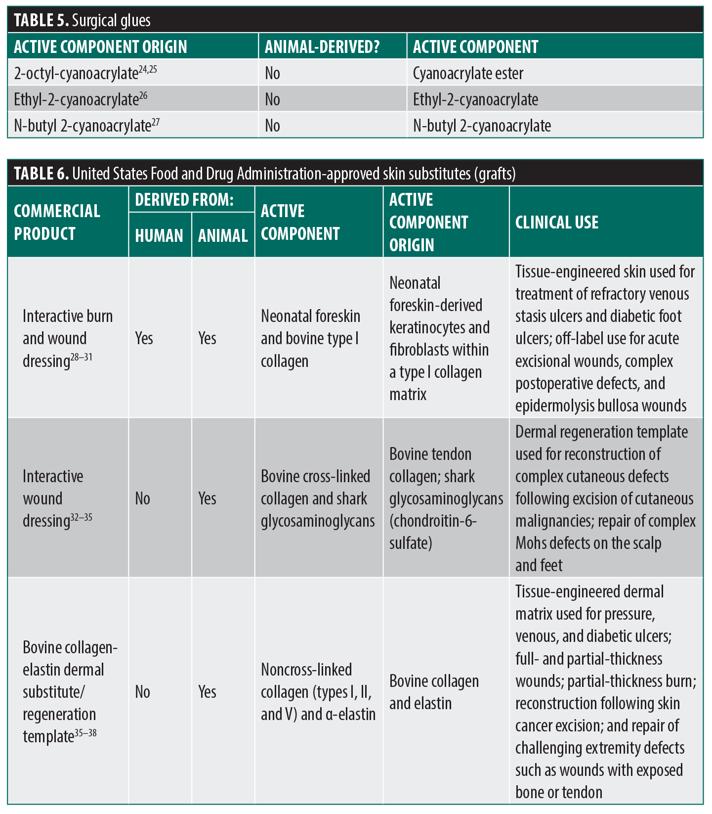
Interactive burn and wound dressing is a bilayered skin substitute composed of neonatal foreskin-derived keratinocytes and fibroblasts embedded in bovine type I collagen. It is FDA approved for the treatment of venous stasis ulcers and diabetic foot ulcers, offering an effective option for chronic wound treatment.28,29 It has also been used off label for enhanced healing of acute excisional wounds, complex surgical defects, and epidermolysis bullosa wounds.30,31
Interactive wound dressing is a dermal regeneration template composed of bovine collagen and shark-derived glycosaminoglycans that provides a scaffold for tissue regeneration. It is used in dermatologic surgery for reconstruction of complex cutaneous defects following the excision of cutaneous malignancies13,32 and is also used in the repair of complex Mohs defects on the scalp and feet.33
The bovine collagen-elastin dermal substitute/regeneration template has demonstrated efficacy in the treatment of full- and partial-thickness wounds, burns, and ulcers.34,36 Potential complications include localized inflammatory responses and foreign body reactions.34,36 It has also been used for reconstruction following skin cancer excision and in the treatment of challenging extremity defects, including wounds with exposed bone, tendon, or other critical structures.37,38 Despite their widespread use and clinical benefits, animal-derived skin substitutes frequently lack clear labeling regarding their sources, making it difficult for physicians and patients to make informed decisions on clinical care.39
Fillers. In 2020, an estimated 3.4 million soft tissue filler injections were performed in the United States.40 Currently, there are more than 500 FDA-approved facial fillers, which are frequently used to restore facial volume, simulate or support collagen production, and soften facial lines. There are 2 main categories of fillers: physical and biostimulatory fillers.41 Physical fillers include hyaluronic acid (HA) and collagen-based fillers. Biostimulatory fillers and semipermanent fillers include calcium hydroxyapatite (CaHA), poly-L-lactic acid, polycaprolactone, and polymethyl methacrylate–based products.41
HA-based fillers are among the most widely used injectable fillers. In 2024, there were 28 FDA-approved HA dermal fillers on the market.42 HA is a glycosaminoglycan found in the extracellular matrix (ECM) of the dermis that binds and retains water, yielding hydration and volume.43 Early HA fillers were derived from rooster combs. Most modern HA fillers are produced through bacterial fermentation and are chemically cross-linked to slow enzymatic degradation after injection into the dermis, prolonging their clinical effect.42 This transition was largely complete by 2006, with bacterial fermentation becoming the predominant method for producing injectable filler materials and animal- and human-derived collagen fillers comprising a minority of products in clinical use.44 Modern HA fillers are categorized within multiple contemporary product families which are available for different anatomic indications and rheologic properties; these products are typically acquired through microbial fermentation only.
Collagen makes up the majority of the dermal ECM, supporting tissue strength and interacting with proteins and enzymes involved in tissue repair and remodeling. With aging, collagen becomes less cohesive and organized, such that there is less resistance to mechanical forces resulting in the creation of wrinkles and loss of facial volume.
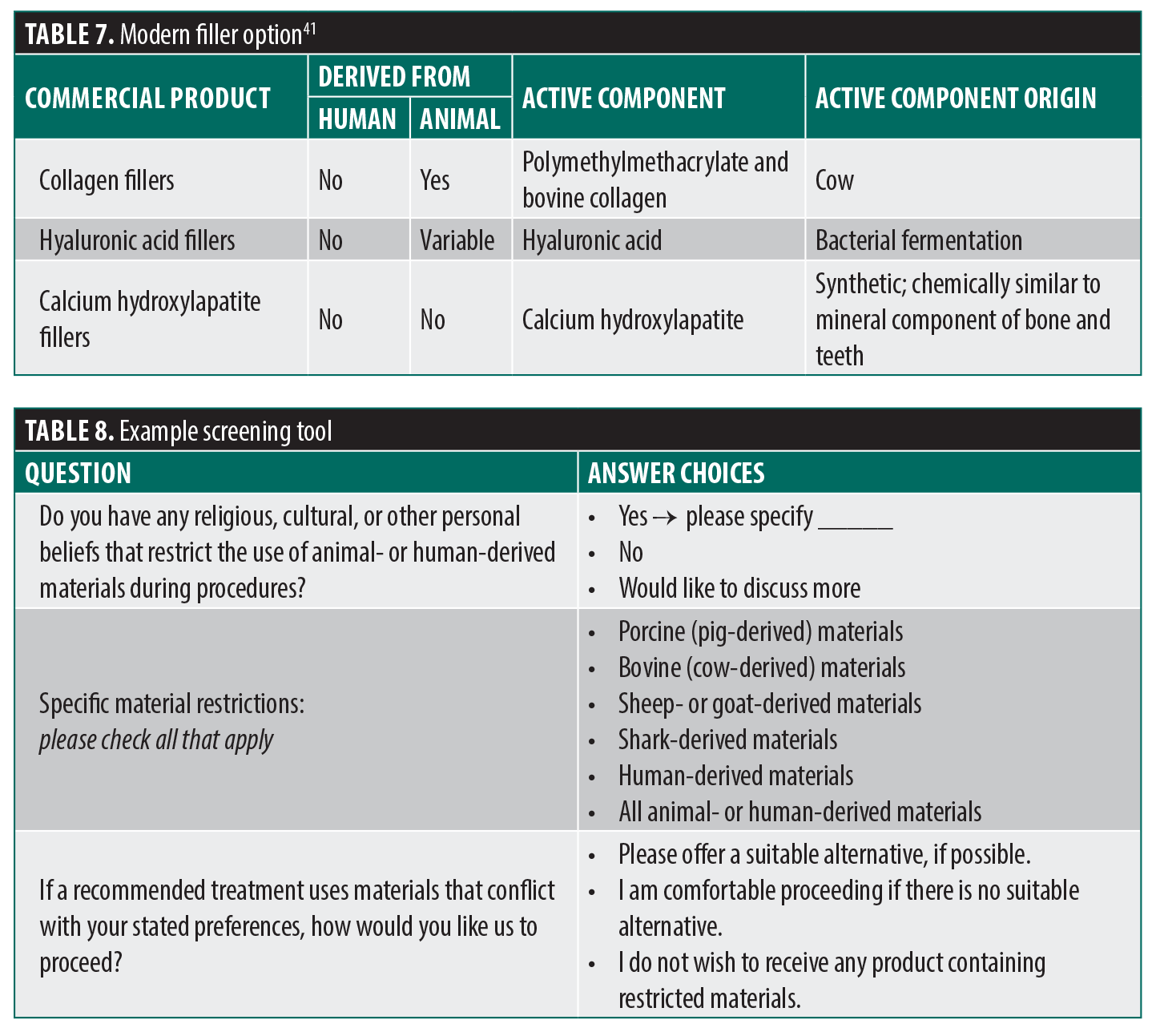
Collagen-based injectable fillers have a long clinical history. The FDA approved the first collagen filler, a purified bovine dermal collagen, in 1981, and the glutaraldehyde-cross-linked collagen filler in 1985. For nearly 2 decades, bovine collagen fillers were the most popular injectable implants in the United States. However, bovine collagen can potentially expose patients to zoonotic diseases, particularly bovine spongiform encephalopathies and foot-and-mouth disease.45 Intradermal allergy testing is recommended prior to injection of bovine collagen products due to the risk of hypersensitivity (approximately 2% to 4% of the population).45,46 To reduce immunogenicity concerns associated with animal-sourced collagen, human-derived collagen fillers were developed (dermal collagen implant, aesthetic dermal filler, dermal collagen implant/injectable facial filler, human-based dermal collagen implant). Collagen fibers and ECM are carefully acquired from human cadaveric tissue, tested for safety, and sterilized to create autologous human collagen, micronized acellular human dermis, and preserved particulate fascia lata or micronized human fascia, although these products are no longer commercially available in the United States.41 Most biostimulatory fillers do not contain any animal byproducts. Fillers containing CaHA are chemically similar to the mineral component of human bone and teeth. As it is human derived, allergy testing is not needed. There is also a synthetic CaHA filler available which is made with synthetic CaHA microspheres rather than human-derived bone or teeth.41 Current filler products are shown in Table 7, and historical options may be found in Supplemental Table 3.
Lack of true informed consent in surgical procedures. Lack of awareness in prescribing animal-derived products to patients has many potential consequences. It can create mistrust or a sense of disappointment and/or disrespect, and lack of disclosure could reinforce a subgroup’s negative beliefs that they received substandard care due to their ethnicity, language, or religious practices. These negative responses can lead to medication noncompliance, which can be dangerous and potentially even resulting in death.47
To improve physician recognition of cultural contraindications, a detailed procedural consent form could be used to further patient-physician communication about product preferences. The form could contain a brief questionnaire to help physicians and staff identify cultural contraindications (Table 8). Additional questions may be asked to narrow down limitations on products containing animal ingredients or other specific concerns. The questionnaire could enhance communication in a standardized format. We recommend administering a brief questionnaire prior to the procedure, ideally during the initial intake, to allow sufficient time for the physician to discuss the screening results with the patient. The proposed screening tool could be easily incorporated into standard intake forms with minimal disruption to clinical workflow. By giving patients an opportunity to disclose religious, cultural, ethical, or personal restrictions before treatment, physicians can more easily identify product-related preferences and engage in informed, patient-centered shared decision-making.
Currently, medical products containing animal-derived ingredients are not easily identifiable in the United States. Other countries, such as Australia and Malaysia, have released public documents listing all medicines and pharmaceuticals of animal origin, sorted by animal type, to make it easier for patients and physicians to identify appropriate medications for individual patients.48 Solutions could be to require product packaging to have a distinct symbol in a standardized location or clear identification of product ingredients with source material. The small, standardized symbol could serve as a reminder tool for physicians. Improved product labeling would increase awareness and assist physicians in communicating with patients about treatment options.
Application in practice. A fully animal- and human-product–free dermatologic practice is feasible for most routine procedures, as synthetic alternatives exist across the major categories of hemostatic agents, sutures, and topical products. However, the financial implications and clinical trade-offs of such a transition warrant further consideration.
Published literature on the comparative cost of animal- and human-derived vs synthetic products is limited. For the purpose of this simplified analysis, pricing was obtained from a single medical supplier with publicly listed wholesale prices.49 Notably, prices vary by vendor, and this variability is expected to increase depending on institutional purchasing contracts.
Common animal-free hemostatic options used in dermatology, such as aluminum chloride, are less costly than gelatin-derived alternatives. A bottle of aluminum chloride costs approximately $20 per 4 oz compared to approximately $200 to $400 per box of 12 for gelatin-based hemostatic sponges (eg, absorbable gelatin sponge and gelatin).50,51 These topical hemostatic agents are widely used, seem to be less expensive than gelatin sponges, and are inherently compatible with a gelatin-free surgical approach.
Animal-derived sutures, such as gut and silk, generally range from approximately $20 to $200 per box of 12, whereas synthetic sutures range from approximately $60 to $1,040 per box of 12.49 However, synthetic sutures, including polypropylene, nylon, and polyglactin, are already widely used in dermatologic practice. As a result, a transition to a fully animal- and human-product–free model may not substantially affect suture-related costs.
Similarly, surgical glues commonly used in dermatologic practice are predominantly synthetically derived, and their continued use would minimally affect overall cost.
Published wholesale prices for both HA and non-HA fillers vary widely by distributor. According to the American Society of Plastic Surgeons, the average cost of HA dermal fillers is $715 while non-HA fillers reach $901.52 It is important to note that these figures may include the physician fee.
Conclusion
Animal- and human-derived materials used in dermatology include gelatin hemostatic agents, gut or silk sutures, skin substitutes, graft materials, and fillers. Although these products are useful for achieving hemostasis, supporting wound healing, and aiding reconstructive procedures, their use may increase the risk of immunogenicity and may contradict the patient’s cultural, religious, or personal values. Most patients prefer disclosure of all animal-based surgical products and being offered alternative treatments.
Although many nonanimal substitute products exist, their identification remains difficult due to inconsistent and nontransparent product labeling. Standardized labeling and accessible formularies would streamline verification of product origin for clinicians and patients. Incorporating simple screening tools to capture patient preferences could further support shared decision-making, helping ensure treatment choices align with patient values. Enhancing transparency and consent processes will strengthen physician–patient trust, improve adherence, and promote respectful, patient-centered care.
Supplemental Materials
References
- Bentivegna K, DeWane ME, Parikh SA, et al. Animal product in suture material: a survey study of dermatology patient perspectives. J Am Acad Dermatol. 2020;83(3):907–908.
- Eriksson A, Burcharth J, Rosenberg J. Animal derived products may conflict with religious patients’ beliefs. BMC Med Ethics. 2013;14:48.
- Perrine T. Default vegetarianism and veganism. J Agric Environ Ethics. 2021;34(2):13.
- Donnelly P, Soares R, Eynon A, et al. Animal products in neurosurgery – navigating informed consent. Br J Neurosurg. 2025;39(4):409–412.
- Henley J, Brewer JD. Newer hemostatic agents used in the practice of dermatologic surgery. Dermatol Res Pract. 2013;2013:279289.
- Gelfoam. Prescribing information. Pfizer; revised Feb 2012. Accessed 03 Feb 2025. https://cdn.pfizer.com/pfizercom/products/uspi_gelfoam_plus.pdf
- Surgifoam absorbable gelatin products. Johnson & Johnson Medtech. Accessed 03 Feb 2025. https://www.jnjmedtech.com/en-US/product/surgifoam-absorbable-gelatin-products
- Gelfoam Plus (00085412086309). US National Library of Medicine. Accessed 03 Feb 2025. https://accessgudid.nlm.nih.gov/devices/00085412086309
- Summary of safety and effectiveness: absorbable gelatin sponge, USP. US Food and Drug Administration. Accessed 10 Feb 2025. https://www.accessdata.fda.gov/cdrh_docs/pdf/P990004b.pdf
- Coseal surgical sealant. Baxter Advanced Surgery. Accessed 10 Feb 2025. https://advancedsurgery.baxter.com/coseal
- Summary of safety and effectiveness data: CoSeal surgical sealant (CoSeal). US Food and Drug Administration. Accessed 10 Feb 2025. https://www.accessdata.fda.gov/cdrh_docs/pdf3/P030039b.pdf
- Palm MD, Altman JS. Topical hemostatic agents: a review. Dermatol Surg. 2008;34(4):431–445.
- Rajendran S, Anand SC, Rigby AJ. Textiles for healthcare and medical applications. In: Horrocks AR, Anand SC, eds. Handbook of Technical Textiles: Technical Textile Applications. 2nd ed. Woodhead Publishing; 2016:135-164. Accessed 12 Feb 2025. https://www.academia.edu/116082172/Handbook_of_Technical_Textiles_Technical_Textile_Applications_Ed_2
- Robinson JK, Sengelmann RD, Hanke CW, Siegel DM. eds. Surgery of the Skin. Elsevier Inc; 2005.
- Zehr KJ. Use of bovine albumin-glutaraldehyde glue in cardiovascular surgery. Ann Thorac Surg. 2007;84(3):1048–1052.
- Bose S, Bandyopadhyay A, Narayan R, eds. Biomaterials Science: Processing, Properties, and Applications III. Wiley; 2013. Ch. 13 Suturing Materials, and Epidermal Closure Techniques.
- Radke F. Skin Surgery and Minor Procedures, An Issue of Surgical Clinics. 1st ed. Saunders; 2009:1–240.
- Polyglactin 910. PubChem. Accessed 12 Feb 2025. https://pubchem.ncbi.nlm.nih.gov/compound/Polyglactin-910
- King MW, Gupta BS, Guidoin R, eds. Biotextiles as Medical Implants. Woodhead Publishing; 2013. Accessed 12 Feb 2025.
- Ducheyne P. Comprehensive Biomaterials. Elsevier; 2011. Accessed 12 Feb 2025. http://www.sciencedirect.com/science/referenceworks/9780080552941
- Plavec T, Švara T, Zdovc I, et al. Clinical and histologic tissue responses of skin incisions closed with glycomer 631 and lactomer 9-1 using a subcuticular technique in pigs. BMC Vet Res. 2020;16:87.
- Eaglstein WH, Sullivan T. Cyanoacrylates for skin closure. Dermatol Clin. 2005;23(2):193–198.
- Halim AS, Khoo TL, Mohd Yussof SJ. Biologic and synthetic skin substitutes: an overview. Indian J Plast Surg. 2010;43(Suppl):S23–S28.
- Lee E, Elzomor A, Zwemer C, et al. Complications associated with Dermabond® during head and neck surgery: MAUDE and literature review. Am J Otolaryngol. 2022;43(2):103330.
- EPIGLU. Surgical and Medican Supplies Pty. Ltd. Revised July 2016. Published online. Accessed 12 Feb 2025. https://www.surmed.com.au/wp-content/uploads/2020/07/Epiglu-Brochure-2016-updated.pdf
- Schonauer F, Pereira J, La Rusca I, et al. Use of Indermil tissue adhesive for closure of superficial skin lacerations in children. Minerva Chir. 2001;56(4):427–429.
- Moy RL, Lee A, Zalka A. Commonly used suture materials in skin surgery. Am Fam Physician. 1991;44(6):2123–2128.
- Apligraf. Product labeling. US Food and Drug Administration. Accessed 10 Feb 2025. https://www.accessdata.fda.gov/cdrh_docs/pdf/P950032S016c.pdf
- Chang DK, Louis MR, Gimenez A, Reece EM. The basics of Integra dermal regeneration template and its expanding clinical applications. Semin Plast Surg. 2019;33(3):185–189.
- Falabella AF, Valencia IC, Eaglstein WH, Schachner LA. Tissue-engineered skin (Apligraf) in the healing of patients with epidermolysis bullosa wounds. Arch Dermatol. 2000;136(10):1225–1230.
- Shealy FG Jr, DeLoach ED. Experience with the use of Apligraf to heal complicated surgical and nonsurgical wounds in a private practice setting. Adv Skin Wound Care. 2006;19(6):310–322.
- Prezzavento GE, Calvi RNJ, Rodriguez JA, Taupin P. Integra dermal regeneration template in reconstructive surgery for cutaneous tumours: a two-year retrospective review. J Wound Care. 2022;31(7):612–619.
- Cunningham T, Marks M. Vacuum-assisted closure device and skin substitutes for complex Mohs defects. Dermatol Surg. 2014;40 Suppl 9:S120–S126.
- Eisler W, Baur J-O, Held M, et al. Assessment of two commonly used dermal regeneration templates in a swine model without skin grafting. Appl Sci. 2022;12(6):3205.
- Troy J, Karlnoski R, Downes K, et al. The use of EZ Derm® in partial-thickness burns: an institutional review of 157 patients. Eplasty. 2013;13:e14.
- Haik J, Weissman O, Hundeshagen G, et al. Reconstruction of full-thickness defects with bovine-derived collagen/elastin matrix: a series of challenging cases and the first reported post-burn facial reconstruction. J Drugs Dermatol. 2012;11(7):866–868.
- Ferrari B, Reggiani C, Francomano M, et al. Clinical factors influencing the outcomes of an acellular dermal matrix for skin cancer treatment: a retrospective study. Adv Skin Wound Care. 2020;33(7):367–374.
- Gladsjo JA, Kim SS, Jiang SIB. Review of the use of a semisynthetic bilaminar skin substitute in dermatology and a case series report of its utility in Mohs surgery. J Drugs Dermatol. 2014;13(5):537–541.
- Hones KM, Hones J, Satteson ES, Chim H. Treatment of complex extremity wounds with MatriDerm: first clinical experience in the US. J Wound Care. 2023;32(3):167–171.
- Papakonstantinou E, Roth M, Karakiulakis G. Hyaluronic acid: a key molecule in skin aging. Dermatoendocrinol. 2012;4(3):253–258.
- Liao ZF, Cong LY, Li FW, et al. The research trend of soft tissue filler injection from 2000 to 2022: a bibliometric and visualized analysis. Plast Reconstr Surg Glob Open. 2024;12(2):e5579.
- Shah R, Matarasso S, Pathak G, Rossi A. Current landscape of hyaluronic acid filler use in the United States. J Drugs Dermatol. 2024;23(1):1247.
- Walker K, Basehore BM, Goyal A, Zito PM. Hyaluronic acid. In: StatPearls [Internet]. StatPearls Publishing; 2026 Jan-. Updated 3 Jul 2023. Accessed 25 Apr 2025. https://www.ncbi.nlm.nih.gov/books/NBK482440/
- Eppley BL, Dadvand B. Injectable soft-tissue fillers: clinical overview. Plast Reconstr Surg. 2006;118(4):98e–106e.
- Salvatore L, Natali ML, Brunetti C, et al. An update on the clinical efficacy and safety of collagen injectables for aesthetic and regenerative medicine applications. Polymers (Basel). 2023;15(4):1020.
- Gonzalez-Hermosillo LM, Ramos-Pacheco VH, Gonzalez-Hermosillo DC, et al. MRI visualization and distribution patterns of foreign modeling agents: a brief pictorial review for clinicians. Biomed Res Int. 2021;2021:2838246.
- Biodiscovery Resource Toolkit. Queensland Government – Department of Environment and Science. Accessed 25 Aug 2025. https://environment.des.qld.gov.au/__data/assets/pdf_file/0021/247206/biodiscovery-resource-toolkit.pdf
- Medicines With Animal Origin. 1st ed. Pharmacy Department, Hospital Canselor Tuanku Mizan; 2022. Accessed 25 Aug 2025. https://hctm.ukm.my/farmasi/wp-content/uploads/2023/11/MEDICINES-WITH-ANIMAL-ORIGIN-HCTM-Ver-1r.pdf
- Wholesale medical supplies. Central Infusion Alliance. Accessed 15 Jun 2026. https://www.ciamedical.com/
- Pfizer 908-3300 – SPONGE, GELFOAM, SIZE 12, 12/BX. Central Infusion Alliance. Accessed 19 Jun 2026. https://www.ciamedical.com/pfizer-908-3300-box-sponge-gelfoam-size-12-12-bx
- Pfizer 00009031508 Gelfoam ® Hemostatic Agent Absorbable Gelatin Sponge Topical Sponge Carton 12 per Box. Medicalecart. Accessed 19 Jun 2026. https://www.medicalecart.com/products/pfizer-00009031508-gelfoam-hemostatic-agent-absorbable-gelatin-sponge-topical-sponge-carton-12-per-box.html
- Dermal fillers. American Society of Plastic Surgeons. Accessed 15 Jun 2026. https://www.plasticsurgery.org/cosmetic-procedures/dermal-fillers/cost