
J Clin Aesthet Dermatol. 2026;19(9):14–24.
Abigail beard, DO, and Valerie Foy, DO
Dr. Beard is with the Department of Medicine at HCA Florida Lawnwood Healthcare, Fort Pierce, Florida. Dr. Foy is with St. John’s Episcopal Hospital, Far Rockaway, New York.
FUNDING: No funding was provided for this article.
DISCLOSURES: The authors have no relevant conflicts of interest.
Abstract: Despite effective antiretroviral therapy (ART), HIV infection drives progressive immune dysfunction, complicating the management of comorbid inflammatory and autoimmune dermatologic conditions such as psoriasis, atopic dermatitis, and cutaneous lupus erythematosus. These conditions often require immunomodulatory therapies, but their use in people living with HIV (PLWH) raises concerns about heightened infection risks, impacts on viral load and CD4 counts, and interactions with ART. The advent of targeted biologics, including tumor necrosis factor, interleukin (IL)-17, IL-23, and Janus kinase inhibitors, has revolutionized treatment for immune-mediated skin diseases. However, limited clinical trial data in PLWH create uncertainty about safety and efficacy. Traditional immunosuppressants such as methotrexate and cyclosporine further complicate care due to their broad immunosuppressive effects. This review synthesizes current clinical protocols, recent guideline updates, and emerging evidence on the safety and efficacy of immunomodulatory therapies in PLWH with dermatologic conditions. By providing a concise, evidence-based framework, we aim to guide dermatologists and infectious disease specialists in optimizing treatment decisions to improve dermatologic and systemic health outcomes in this complex patient population. Keywords: HIV, immunomodulators, inflammatory dermatologic diseases, biologics, psoriasis, opportunistic infections, antiretroviral therapy
Introduction
Since the advent of antiretroviral therapy (ART), the prognosis for people living with HIV (PLWH) has significantly improved, transforming HIV infection from a fatal disease to a more manageable chronic condition.1 Even with optimal viral suppression, HIV induces persistent immune dysregulation and chronic inflammation.2 This ongoing immune imbalance not only heightens susceptibility to opportunistic infections but also drives a broad spectrum of dermatologic disorders. Cutaneous disorders are highly prevalent in PLWH and can include infectious, neoplastic, inflammatory, and autoimmune conditions. Immune-mediated conditions such as psoriasis, atopic dermatitis, and cutaneous lupus erythematosus are particularly challenging to manage in this population.3
Managing these dermatologic conditions often requires systemic immunomodulatory therapies for adequate disease control. In immunocompromised patients, these agents spark legitimate concerns, such as heightened infection risk, impact on HIV disease progression, and complex interactions with ART regimens.3 The emergence of targeted biologic therapies, such as tumor necrosis factor (TNF) inhibitors, interleukin (IL)-17 and IL-23 inhibitors, and Janus kinase (JAK) inhibitors, has revolutionized treatment for immune-mediated skin diseases.4 However, their safety and efficacy in PLWH remain poorly defined. Traditional immunosuppressants, such as methotrexate (MTX), cyclosporine, and mycophenolate mofetil (MMF), offer alternatives but carry risks due to their nonspecific immunosuppressive effects.5
Beyond dermatology, immunomodulators are increasingly used in PLWH for autoimmune comorbidities and malignancies.6 This expanding use underscores the urgent need for evidence-based guidance. This review seeks to provide a comprehensive overview of current clinical protocols for the use of immunomodulators in PLWH with dermatologic conditions. We will examine existing guidelines, explore recent updates in therapeutic approaches, and highlight key findings from emerging research. By integrating the latest evidence, we aim to support clinicians in navigating the complex balance between effective dermatologic disease control and maintenance of immune system integrity in PLWH.
Background
Pathophysiology of HIV immunosuppression. HIV infection results in progressive immunosuppression that primarily occurs through the depletion of CD4+ T lymphocytes and persistent immune activation.7 The virus targets and destroys CD4+ cells, which play a critical role in orchestrating adaptive immune responses. Even with effective ART, many individuals experience incomplete immune reconstitution and ongoing low-level inflammation, leading to increased susceptibility to infections and immune-mediated diseases.7,8 Even virologically suppressed patients taking ART experience ongoing immune activation that contributes to inflammatory dermatoses. This dysregulated immune environment, marked by chronic T-cell activation, elevated proinflammatory cytokines, and disrupted regulatory pathways, promotes the development of both infectious and inflammatory skin diseases.9
Immunomodulators defined. Immunomodulators are agents that modulate or suppress the immune response and are commonly used in the management of autoimmune and inflammatory diseases.10 These agents can be categorized broadly into corticosteroids, traditional nonsteroidal immunosuppressants, and biologic or targeted therapies.
Corticosteroids. Corticosteroids remain a mainstay of immunosuppressive therapy due to their rapid anti-inflammatory effects, achieved by inhibiting cytokine production, lymphocyte proliferation, and leukocyte trafficking.10 However, long-term use is associated with significant adverse effects and, in PLWH, may exacerbate immunosuppression and increase the risk of opportunistic infections.11 Short-term, moderate-dose courses are generally well tolerated in virologically suppressed patients, whereas prolonged high-dose regimens warrant greater caution.
Conventional (nonbiologic) immunosuppressants. Nonbiologic immunosuppressants such as MTX, azathioprine, and MMF are commonly used in dermatology for their nonsteroidal immunosuppressive effects.12 MTX is a folate antagonist with antiproliferative and anti-inflammatory properties, frequently employed in the management of psoriasis, atopic dermatitis, and connective tissue diseases.12 In PLWH, however, MTX warrants heightened vigilance due to overlapping hepatotoxicity risks, particularly in those with ART-associated hepatic steatosis or co-infection with hepatitis B or C.13,14 Furthermore, MTX may contribute to lymphopenia and further CD4+ T-cell suppression, increasing susceptibility to opportunistic infections and complicating HIV disease monitoring.15,16 Though limited case reports and small case series suggest that MTX can be used safely in select patients with well-controlled HIV, its immunosuppressive potential demands close CD4+ count and liver function monitoring throughout treatment.17
Azathioprine inhibits purine synthesis and impairs T-cell function.18 Although generally well tolerated, its metabolism via thiopurine methyltransferase requires genetic screening to reduce the risk of toxicity, and use in PLWH should be carefully considered.18
MMF selectively inhibits lymphocyte proliferation by targeting inosine monophosphate dehydrogenase. It has shown utility in lupus and atopic dermatitis, with emerging yet limited safety data in HIV populations.19
Biologic and targeted therapies. Biologic therapies have transformed the treatment of inflammatory skin diseases by targeting specific immune pathways. However, their use in PLWH remains limited largely due to exclusion from trials due to concerns over infection risk and immune suppression.20 TNF-α inhibitors such as infliximab, adalimumab, and etanercept block TNF-α, a key cytokine in inflammatory pathways.20 Although effective for psoriasis and hidradenitis suppurativa, TNF inhibitors have been associated with increased infection risk, particularly tuberculosis and reactivation of latent viruses.20
TNF-α promotes HIV replication in vitro through nuclear factor–κB, yet its inhibition may paradoxically suppress viral expression in some contexts.20 TNF-α also plays a critical role in granuloma maintenance, and its blockade increases the risk of reactivation of latent infections, including a 2- to 4-fold higher incidence of tuberculosis.20 For this reason, these agents should generally be avoided when alternative therapies are available. When used, a CD4+ lymphocyte count >350/μL; screening for tuberculosis, hepatitis B, and hepatitis C; and close monitoring are advisable.20
IL-17 inhibitors, including secukinumab, ixekizumab, brodalumab, and IL-23 inhibitors, including guselkumab and risankizumab, have shown excellent efficacy in plaque psoriasis, with limited case reports suggesting safety in patients with well-controlled HIV, though larger studies are needed.21 IL-17 inhibitors target the helper T cell (Th) 17/IL-17 axis, which plays a critical role in mucosal barrier defense against bacteria and fungi. However, HIV infects and decreases Th17 cells, even in patients taking ART.22 This occurs because Th17 cells express high levels of HIV receptors and lack autocrine CCR5 ligands that protect other CD4+ subsets.23
To expand upon this, HIV directly impairs IL-23 signaling in Th17 cells through the loss of signal transducer and activator of transcription (STAT) 3 phosphorylation, and this is not restored by ART.24 Therefore, IL-17 production is already severely compromised in PLWH, and IL-17 inhibitors will not further impair an already dysfunctional pathway. Candidiasis is the main adverse effect of IL-17 inhibitors, with an amplified risk in PLWH.25 Clinicians should have a low threshold for empiric fluconazole 150 mg weekly in patients who develop symptoms and can also consider prophylactic fluconazole if the patient’s CD4 lymphocyte count is <350/μL.26 In most cases, patients can continue IL-17 inhibitors.27
IL-23 inhibitors specifically inhibit the p19 subunit of IL-23 and reduce the Th17 pathway. IL-23 is produced by myeloid dendritic cells, which contribute to T-cell dysfunction and inhibit interferon (IFN)-γ production.28 Therefore, blocking IL-23 may paradoxically reduce HIV-induced immune dysregulation. Current data on IL-23 inhibitors are more limited but suggest these agents may be at least as safe as IL-17 inhibitors.25 The candidiasis risk may be lower due to preserved IL-17 production.25
Dupilumab blocks IL-4 receptor α, which inhibits signaling from both IL-4 and IL-13.29 These cytokines drive inflammation through a Th2 pathway. This is critical for HIV, since there is no Th1 immunosuppression and there is improved defense against opportunistic infections. Dupilumab has the best safety data of all biologics in PLWH. A systematic review of 27 PLWH taking dupilumab reported no opportunistic infections with stable viral loads.29
JAK inhibitors, such as tofacitinib and upadacitinib, inhibit the intracellular signaling pathways involved in cytokine-mediated inflammation.30 JAK inhibitors are increasingly used for conditions such as atopic dermatitis, psoriasis, and alopecia areata, but their effect on viral immunity in PLWH is not well understood, warranting close surveillance and cautious use.30 The JAK-STAT pathway is needed for antiviral immunity, mediating cytokines and IFNs. JAK inhibitors impair IFN-α/IFN-β signaling, which is critical for antiviral defense, but the effect in PLWH is unknown.31 There are no HIV-specific clinical data for JAK inhibitors in dermatologic therapy. It is important to note the drug-drug interactions, since JAK inhibitors are CYP3A4 substrates. Protease inhibitors can increase JAK inhibitor levels by 2- to 3-fold. Therefore, a dose reduction by 50% or switching to dolutegravir or bictegravir is advised to avoid interaction.32 In sum, biologics and targeted therapies offer options for PLWH, but each class has unique concerns. The next sections discuss how to navigate these choices clinically.
Immune-mediated Dermatoses in PLWH
Dermatologic manifestations affect up to 90% of PLWH during their illness.33 These skin disorders may result from direct viral effects, opportunistic infections, neoplasms, or immune dysregulation.33 Among them, immune-mediated inflammatory dermatoses, such as psoriasis, atopic dermatitis, seborrheic dermatitis, and prurigo nodularis, comprise a distinct and clinically significant subset. These conditions are characterized by dysregulated immune activation, frequently exacerbated by the chronic inflammation and altered cytokine milieu seen in HIV infection, even among those on stable ART.34,35
In PLWH, inflammatory dermatoses often exhibit atypical or more severe presentations, including widespread or treatment-resistant disease. For example, psoriasis in people with HIV may appear with explosive onset, erythroderma, or recalcitrance to topical therapy.36 The following section outlines the most common immune-mediated inflammatory dermatoses encountered in PLWH, highlighting pathophysiologic mechanisms and considerations for systemic treatment.
Seborrheic dermatitis. Seborrheic dermatitis is markedly more common and severe in PLWH, particularly those with advanced immunosuppression. Studies report prevalence rates of up to 85% among patients with a CD4 lymphocyte count <200/µL, where it can serve as a visible marker of HIV progression or immune status.36 The condition primarily affects sebaceous-rich areas and is driven by a combination of Malassezia yeast overgrowth and immune-mediated inflammation, particularly involving altered T-cell responses and cytokine dysregulation.37
First-line management typically involves topical antifungals such as ketoconazole or ciclopirox, often in combination with low-potency corticosteroids or topical calcineurin inhibitors to reduce inflammation.38 In most cases, these are effective and well tolerated.39 However, in PLWH, seborrheic dermatitis may present more refractory or chronically, requiring sustained or advanced therapeutic options.38
A recent advancement in topical therapy is roflumilast topical foam 0.3%, a daily phosphodiesterase 4 inhibitor approved by the United States (US) Food and Drug Administration (FDA) in December 2023 for the treatment of seborrheic dermatitis in patients aged 9 years and older.40 In the pivotal phase 3 STRATUM trial, roflumilast demonstrated significant efficacy, with nearly 80% of patients achieving Investigator Global Assessment success and over 50% achieving complete clearance by Week 8.41 Its nonsteroidal, anti-inflammatory mechanism offers a well-tolerated alternative, particularly for treatment in sensitive or hair-bearing areas.41 While clinical trials did not specifically include PLWH cohorts, the safety profile and nonimmunosuppressive mechanism of action make roflumilast an option for managing seborrheic dermatitis in this immunocompromised population. Further postmarketing studies are needed to evaluate its long-term safety and efficacy in PLWH.
Tapinarof cream is another potential future option. It is a topical aryl hydrocarbon receptor agonist approved for psoriasis, and it is currently being explored for seborrheic dermatitis and has immune-modulating and antimicrobial properties that may impact Malassezia-associated inflammation.42
Currently, there are no systemic therapies approved for seborrheic dermatitis, and most oral treatments are used off label or reserved for refractory cases.43 However, there is growing interest in systemic therapies that target inflammatory pathways without broadly suppressing immunity, which may be particularly relevant for PLWH. One emerging option is oral JAK inhibitors (eg, abrocitinib, upadacitinib). These have demonstrated efficacy in inflammatory skin diseases such as atopic dermatitis and alopecia areata. While not yet studied in seborrheic dermatitis, their downstream IL-4/IL-13 and IL-6 modulation may be theoretically beneficial, though their use in PLWH raises caution due to possible effects on viral immunity.43 Injectable IL-17/IL-23 inhibitors (eg, secukinumab, guselkumab), although not standard for seborrheic dermatitis, do have anecdotal reports of their efficacy in cases of overlapping psoriasis or severe seborrheic-like dermatitis.42 However, their immunosuppressive nature and lack of specific seborrheic dermatitis indication limit their appeal in PLWH.42
Psoriasis. Psoriasis is a chronic immune-mediated skin disease that can present with increased severity and resistance to treatment in PLWH. HIV-associated psoriasis may be more widespread, refractory, and accompanied by variants such as erythrodermic or pustular forms.39 The pathophysiology is thought to be driven by immune dysregulation, specifically through the paradoxical activation of CD8+ T cells despite CD4+ lymphopenia.44
While topical therapies and phototherapy are typically first-line options for psoriasis, moderate-to-severe cases may require systemic agents such as MTX, cyclosporine, and biologics (IL-17, IL-23, or TNF-α). Limited case series suggest biologics may be safe in patients with well-controlled HIV, but close monitoring for infections and viral load changes is essential.21 Emerging data on IL-23 inhibitors show promise for severe psoriasis in an HIV setting, warranting further study.
Atopic dermatitis. Atopic dermatitis may be exacerbated in HIV, often presenting with more severe atypical distributions and increased pruritus. The skin barrier dysfunction in PLWH, coupled with a skewed Th2 cytokine profile and eosinophilia, may contribute to the increased severity and chronic nature of atopic dermatitis in this population.45 Prevalence is higher in PLWH, particularly in those with a CD4 lymphocyte count <200/μL.45 Topical therapies are the primary treatment, but severe cases may necessitate systemic options such as oral corticosteroids or dupilumab. Small studies and case reports indicate the safety of dupilumab in PLWH with stable viral loads, though data remain sparse.46
Prurigo nodularis. Prurigo nodularis is frequently reported in PLWH and is thought to arise from chronic pruritus exacerbated by immune dysregulation and eosinophilia.47 It is seen particularly in patients with a CD4+ lymphocyte count <200/μL.40 The nodular lesions are often widespread and intensely pruritic, which significantly impair quality of life.48
First-line therapy generally involves topical corticosteroids and antihistamines to manage inflammation and pruritus. However, for refractory cases, immunomodulators such as thalidomide or dupilumab may be considered.48 Thalidomide has been reported to provide benefits due to its anti-inflammatory and immunomodulatory effects, though it is associated with significant adverse effects and requires careful monitoring, particularly in PLWH who are at risk for neuropathy and other toxicities.48,49 Dupilumab has also shown promise in clinical trials for treating PN, though evidence in PLWH remains limited and largely anecdotal.50
In addition to these therapies, nemolizumab, an IL-31 receptor α antagonist, was approved by the FDA in 2024 for the treatment of adults with prurigo nodularis.51 Nemolizumab specifically inhibits IL-31 cytokine signaling, which is known to drive itch and is involved in inflammation, altered epidermal differentiation, and fibrosis in prurigo nodularis.51 Early-phase studies in the general population have demonstrated significant reductions in pruritus and improvements in skin lesions.51 As a nonimmunosuppressive agent, nemolizumab offers a novel approach for managing chronic itch in immunocompromised patients.50 Given its selective mechanism of action, nemolizumab holds potential for use in PLWH with prurigo nodularis, but further studies are needed to assess its safety and efficacy in this population.
Eosinophilic folliculitis. Eosinophilic folliculitis is a common HIV-associated dermatosis, particularly in patients with a CD4+ lymphocyte count <250/μL. Prevalence estimates range from 10% to 20% in advanced HIV.52 Characterized by intensely pruritic, follicular papules and pustules primarily on the face, trunk, and upper extremities, eosinophilic folliculitis is driven by eosinophilic infiltration and Th2-mediated immune dysregulation.53 Diagnosis is confirmed by histopathology showing perifollicular eosinophilic infiltrates. Topical corticosteroids and antihistamines are first-line treatments, while severe or refractory cases may respond to phototherapy, oral corticosteroids, or off-label use of immunomodulators such as cyclosporine. Limited reports suggest dupilumab may be effective.54
Lichen planus. Lichen planus in PLWH is more prevalent and severe, often presenting with hypertrophic, generalized, or mucosal involvement. Between 1% to 5% of PLWH may exhibit LP.55 Its pathogenesis involves T-cell–mediated autoimmunity, exacerbated by HIV-induced immune dysregulation.56 Classic lichen planus presents as pruritic, polygonal, violaceous papules, often on the extremities, but oral and genital lesions are common in HIV. Topical corticosteroids and calcineurin inhibitors are used as first-line therapy, but severe cases may require oral corticosteroids, retinoids, or off-label use of IL-17 inhibitors. Case reports demonstrate secukinumab may be safe in PLWH with stable viral loads.57
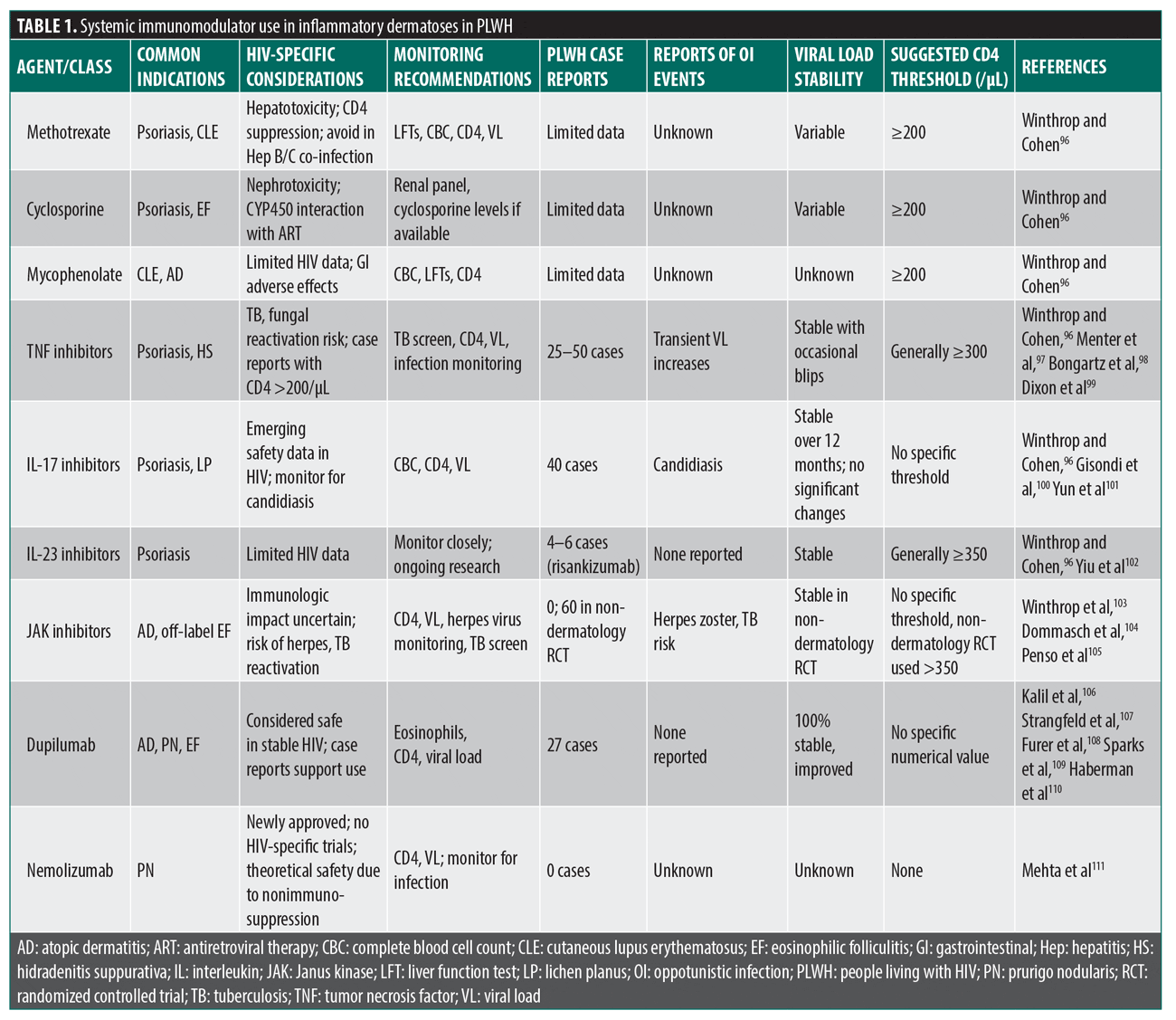
The management of these dermatologic inflammatory conditions in PLWH requires a nuanced approach, balancing disease control with infection risks and ART interactions. Multidisciplinary care involving dermatologists, infectious disease specialists, and pharmacists is critical to optimize outcomes. Table 1 provides a comprehensive list of systemic immunomodulators used for inflammatory dermatoses in PLWH.
Current Protocols and Monitoring
Rationale for use. Despite longstanding concerns regarding the use of immunomodulatory agents in individuals with HIV, growing evidence supports their sensible use in select patients.58 As combination ART has improved immune reconstitution and suppressed viral replication in the majority of PLWH, the paradigm has shifted from avoiding immunosuppression altogether to balancing its risks and benefits.59 Inflammatory skin diseases such as psoriasis, atopic dermatitis, and prurigo nodularis can be debilitating and refractory to topical therapy alone. For patients with controlled HIV, immunomodulators, including systemic agents and targeted biologics, may offer significant improvement in skin-related quality of life and reduction in systemic inflammation, which itself may contribute to HIV-related comorbidities.59 Several small cohort studies and case reports suggest that these therapies can be safely administered in virologically suppressed patients with appropriate screening and monitoring protocols.60
Patient selection (CD4, viral load, ART). Before starting any systemic agent, confirm the patient’s HIV status. For the patient’s CD4+ lymphocyte count, aim for ≥200/μL as a general minimum for any new immunosuppression. Many infectious disease specialists prefer >350/μL before starting biologics,61 but dupilumab has been given to some patients with CD4+ lymphocyte count of 100 to 200/μL in desperate cases, though that is exceptional. If the CD4+ count falls below 200/μL, biologic therapy should generally be deferred.61 Most physicians require an undetectable HIV RNA, typically documented on ≥2 occasions over the prior 3 to 6 months.62 The exact duration is not evidence based, but expert consensus is often 3 months for standard biologics and 6 months for higher-risk therapies, such as JAK inhibitors. ART adherence is critical. Systemic immunosuppression should never be started if the patient is nonadherent to ART. This is because the risk of HIV rebound and new opportunistic infections is too high.63 In summary, select patients who have stable, well-controlled HIV and are maintaining ART during dermatologic therapy are good candidates for immunomodulatory agents.
Baseline workup. Perform a thorough baseline assessment before any new immunosuppressant, including CD4+ count and viral load within 4 weeks of therapy initiation.62
Given the increased susceptibility of PLWH to tuberculosis, screening for latent tuberculosis infection is imperative.61 This is especially important before starting TNF-α inhibitors.61 Both the tuberculin skin test (TST) and IFN-γ release assays (IGRAs) are recommended, with a TST induration of ≥5 mm considered positive in this population.63 A positive result warrants further evaluation to exclude active tuberculosis disease before starting immunosuppressive therapy. Patients should also be tested for hepatitis B surface antigen (HBsAg), hepatitis B surface antibodies, hepatitis B core antibodies (anti-HBc), and hepatitis C antibodies.63 PLWH are at a higher risk for viral hepatitis, and immunosuppressive drugs can reactivate hepatitis B. If HBsAg or anti-HBc are positive, plan antiviral prophylaxis. Any active hepatitis C should be treated.63 Physicians should consider regional risks. For example, PLWH with a CD4+ lymphocyte count <100/μL should be screened for histoplasma antigen and cryptococcal antigen.63
Ensuring that patients receive comprehensive preventive care in accordance with established HIV management guidelines is a cornerstone of treatment. Current recommendations from the Centers for Disease Control and Prevention and the Infectious Diseases Society of America emphasize routine health maintenance beyond immunizations, including regular screening for sexually transmitted infections, tuberculosis, and viral hepatitis; age- and risk-appropriate cancer screening; and evaluation for comorbid conditions. Preventive care also includes mental health and substance use screening, risk-reduction counseling, and ongoing laboratory monitoring as part of longitudinal care. Within this framework, immunizations remain an essential component, with specific considerations for immune status in patients with HIV.61,62 This baseline workup parallels standard HIV care and is mandatory before adding any immunomodulator.
Ongoing monitoring. Once therapy is started, close follow-up is essential. Perform complete blood cell count and liver function tests at least every 3 to 6 months to monitor for drug toxicity on MTX or azathioprine. Recheck CD4 lymphocyte count and HIV viral load every 3 to 6 months to ensure ongoing immune competence.61 If the CD4 lymphocyte count unexpectedly falls or the viral load rises, re-evaluate the medication regimen promptly. If a patient develops any possible opportunistic infection, obtain appropriate workup immediately. Standard prophylaxis for conditions such as Pneumocystis jirovecii pneumonia (PJP), Mycobacterium avium complex (MAC), or toxoplasmosis should be continued based on CD4 lymphocyte count thresholds while on immunosuppression.64 The key is proactive monitoring—do not wait for a severe infection to intervene.
Dosing and Combination Use
Generally, use standard doses of immunosuppressive drugs as used in HIV-negative patients, with some caveats.64 In patients with a CD4+ lymphocyte count between 200 and 350/μL, consider starting at the low end of the dosing range and slowly uptitrating. This might mitigate some infection risk. For example, the standard dosage of adalimumab 40 mg every 2 weeks should be given only if the CD4+ lymphocyte count is robust; if the CD4+ count is near 200/μL, one might space doses to 3 to 4 weeks initially.65
Combining agents is not categorically forbidden but should be rare. If necessary, patients must have a good CD4+ lymphocyte count (>350/μL) and well-suppressed virus and typically use low doses of the agents (eg, MTX 7.5–10 mg/week). Avoid triple immunosuppression altogether. Each additional drug substantially raises infection risk.66 For corticosteroids or MTX used as bridging therapy, plan an early taper. The goal is to use the minimal effective immunosuppression. Once a stable biologic is maintained, reassess whether a dose reduction or drug holiday is possible every 3 to 6 months. Long term, maintain only as much therapy as needed to control disease.67
The primary concern with systemic immunomodulation in HIV is the potential for opportunistic infections.65 The risk varies depending on the agent used and the patient’s immune status. TNF-α inhibitors, for example, have been associated with increased risk of reactivation of tuberculosis and histoplasmosis, while systemic corticosteroids may predispose patients to PJP, candidiasis, and herpes virus reactivation.67 Some key factors to keep in mind include that TNF inhibitors and JAK inhibitors may increase reactivation risk; therefore, screening and prophylaxis (eg, isoniazid) are critical.66 Systemic immunosuppression may predispose patients to candidiasis, histoplasmosis, or cryptococcosis.66 Herpes simplex and zoster infections are more common, especially with corticosteroids or calcineurin inhibitors.63 Skin barrier disruption in dermatoses, compounded by systemic immunosuppression, may increase risk of cellulitis or abscesses.66 Prophylactic strategies, such as trimethoprim-sulfamethoxazole for PJP prevention in those with a CD4+ lymphocyte count <200/μL, should be maintained or initiated depending on individual risk.66
Despite these risks, many PLWH with severe dermatologic disease experience marked clinical improvement with immunomodulatory therapy.67 Another benefit is the reduction in inflammatory burden, which may also contribute to systemic health improvement by reducing chronic immune activation.68 Improved skin integrity and reduced flares, thereby lowering secondary infection risk from open or eczematous lesions, is another positive outcome.68 The overall enhanced quality of life that patients experience strengthens mental health and adherence to HIV treatment, leading to a positive outcome for the patient as well as the patient-provider relationship.68
Recommendations from Existing Dermatology, Infectious Disease, and HIV Guidelines
The current recommendations include initiation of immunomodulatory therapy, which is generally considered safe in patients with well-controlled HIV with a CD4+ lymphocyte count ≥200/μL and undetectable viral load.68 The use of biologic agents also can be considered in select patients, with careful monitoring and coordination with infectious disease clinicians. Prophylactic measures can be implemented against opportunistic infections as indicated by CD4 lymphocyte count thresholds and patient history.69 While existing guidelines provide general recommendations, specific guidance on the use of newer immunomodulatory agents in PLWH is limited. There are no prospective studies that have established a minimum duration of viral suppression prior to immunomodulator initiation. Expert consensus generally recommends ≥3 months of documented undetectable viral load before initiating standard biologics and ≥6 months for higher-risk agents such as JAK inhibitors or combination immunosuppression.69
Proposed treatment algorithm. Experts often conceptualize PLWH into risk tiers. A proposed treatment algorithm follows:
Tier 1: Optimal candidates
Criteria:
- CD4+ lymphocyte count >350/μL
- ≥3 to 6 months of documented viral suppression
- Good ART adherence with no recent opportunistic infections
Approach:
-
- Most systemic therapies are permitted at standard doses. IL-17 or IL-23 inhibitors and dupilumab are preferred. No special prophylaxis is needed. TNF inhibitors may also be used if indicated, but opt for a IL-17 or IL-23 inhibitor first. Continue monitoring CD4 lymphocyte count and viral load every 3 to 4 months for the first year70
Tier 2: Intermediate
Criteria:
- CD4+ lymphocyte count of 200 to349/μL
- ≥6 months of viral suppression
- ART adherent, with no opportunistic infections in the past 6 months
Approach:
- Systemic therapy is still possible but with more caution. IL-17 or IL-23 inhibitors or dupilumab are preferred; avoid TNF inhibitors. Start at a low therapeutic dose and slowly increase as needed.70 Avoid combining immunosuppressants. Patients require a tuberculosis screen. If the CD4+ lymphocyte count is between 150 to 200/μL, add PJP prophylaxis (trimethoprim-sulfamethoxazole).
- Check the CD4+ lymphocyte count every ~3 months69
Tier 3: High risk
Criteria:
- CD4+ lymphocyte count <200/μL
- Any detectable HIV viremia
- Recent or current opportunistic infection
- Poor ART adherence70
Approach:
- Generally, defer systemic immunosuppression. Focus on optimizing ART and topical treatments. If the patient’s skin disease is life-threatening, such as severe erythroderma, consider dupilumab, which is sometimes used when the CD4+ lymphocyte count is <200/μL, provided there is intensive monitoring.69 In such cases, ensure full prophylaxis: continue trimethoprim-sulfamethoxazole, consider azithromycin if the CD4+ cell count is <50/μL, and treat latent infections.70 Reassess in 3 to 6 months; if the viral load is suppressed and the CD4+ lymphocyte count recovers to >200/μL, move the patient to Tier 2.
Prophylactic Regimens
HIV-specific prophylaxis guidelines still apply when patients are immunosuppressed. Prophylactic strategies should be tailored based on CD4+ lymphocyte count and individual risk factors.60 PJP is one opportunistic infection in which prophylaxis with trimethoprim-sulfamethoxazole in patients with a CD4+ cell count <200/μL should be initiated.60 In patients with CD4+ lymphocyte counts <100/μL and positive serology, prophylaxis against Toxoplasma gondii is recommended.60 MAC prophylaxis should be initiated with CD4+ lymphocyte counts <50/μL.69 Isoniazid prophylaxis should be provided in patients with latent tuberculosis infection.63 These prophylaxis measures follow standard HIV opportunistic infection guidelines. Always continue the patient’s HIV prophylaxis (eg, trimethoprim-sulfamethoxazole for CD4 lymphocyte counts <200/μL) even after systemic therapy is started.
Contraindications and Special Precautions
Live vaccines (eg, yellow fever, measles-mumps-rubella, varicella, intranasal influenza) are contraindicated if the patient’s CD4+ lymphocyte count is <200/μL. If a patient is significantly immunosuppressed, avoid live vaccines during therapy.68 If the patient has an active opportunistic infection, delay immunosuppression until the infection is under control.
Many systemic immunomodulators are metabolized via CYP3A4. For example, protease inhibitor ART can raise JAK inhibitor levels dramatically. Always check for interactions between chosen immunomodulators and the patient’s ART regimen, and adjust doses if needed.71 Many systemic agents have contraindications for pregnancy.72
Recent Updates and Emerging Evidence
Case series, retrospective studies, and real-world data on biologic safety. Historically, PLWH were excluded from trials evaluating immunomodulatory agents, especially biologics, due to concerns regarding infectious complications. However, several recent retrospective studies and case series have provided preliminary evidence supporting the safety of select biologics in virologically suppressed patients with adequate CD4+ lymphocyte counts.72 IL-17 inhibitors, including secukinumab and ixekizumab, have emerged as agents in this population. A recent case report documented the successful use of ixekizumab in a PLWH with moderate-to-severe psoriasis, demonstrating marked clinical improvement without compromise to HIV disease control.73 Similarly, a systematic review of 112 studies covering 179 PLWH treated across multiple biologic classes was conducted. TNF-α inhibitors were the most used class, and IL-17 inhibitors represented a significant portion of cases. Nearly all biologic classes demonstrated a favorable safety profile with minimal or minor adverse events. Anti-CD20 inhibitors and TNF-α inhibitors were specifically associated with opportunistic infections. Transient increases in viral load were noted with TNF-α inhibitors. However, it is important to note that there were no instances of virologic failure reported. This review acknowledged that evidence quality was low, restricted to case reports and retrospective reviews, but the overall safety profile was favorable.74
Additional real-world data from Peluso et al75 included a retrospective cohort of 77 PLWH with 110 treatment episodes from 2000 to 2019. The median pretreatment CD4+ lymphocyte count was around 600/μL; 90% of treatment episodes for rheumatologic comorbidities involved concomitant ART; and the most used medications were TNF inhibitors, antimetabolites, and checkpoint inhibitors. There were 0 instances of virologic failure reported, which was defined as progression to >200 copies/mL on 2 consecutive measurements. No infections were attributed to immunomodulators, and follow-up was 1 year later to assess viral load.75
A study by Xu et al76 included a real-world cohort of 36 PLWH with psoriasis on biologics, with follow-up 12 months later. There was no significant change in CD4+ lymphocyte counts or HIV viral load and no difference in infection rates compared to PLWH without psoriasis.
In regard to IL-17 or IL-23 inhibitors over TNF-α inhibitors in PLWH, no head-to-head trials exist specifically in HIV populations; the evidence is extrapolated. In the Xu et al76 systematic review of 179 PLWH taking biologics, TNF-α inhibitors were specifically associated with opportunistic infections, and transient HIV viral load increases extrapolated from general population studies showed lower infection risks. Given that PLWH already face elevated infection risk due to immune dysregulation, the lower infection profile of IL-17 and IL-23 inhibitors provides a theoretical safety advantage. However, the HIV-specific association of TNF inhibitors with opportunistic infections and viral load increases, combined with general population data showing lower infection rates with IL-17 and IL-23 inhibitors, supports preferential use of the newer agents when clinically appropriate.76
A study by Peluso et al75 found that IL-17 inhibitor use was associated with significantly lower tuberculosis risk compared to TNF inhibitors (odds ratio: 0.126; P=0.0457). IL-23 inhibitors demonstrated significantly lower risk than TNF inhibitors for multiple infections, including herpes zoster (hazard ratio [HR]: 0.58), hepatitis B reactivation (HR: 0.24), cytomegalovirus (HR: 0.25), and influenza (HR: 0.52).76
When considering JAK inhibitors, there are no published data on JAK inhibitor safety specifically in PLWH. JAK inhibitors carry higher infection risk than IL-17 or IL-23 inhibitors. Serious infection rates with IL-17 or IL-23 inhibitors are 1.0% to 2.0% compared to 2.0% to 3.2% with JAK inhibitors.75 The evidence regarding their risk compared to TNF inhibitors is mixed. A 2025 meta-analysis of 226,788 patients found no statistically significant difference in overall serious infection risk between JAK inhibitors and TNF antagonists.77 However, another 2025 study found TNF inhibitors were associated with significantly lower serious infection risk than JAK inhibitors, with lower rates of respiratory tract infections, urogenital infections, and sepsis.78 Herpes zoster reactivation is 2 to 3 times more common with JAK inhibitors compared to TNF inhibitors. This is particularly concerning for PLWH, who already have elevated baseline herpes zoster risk due to immune dysregulation.79 Baricitinib specifically showed increased opportunistic infection risk; however, most JAK inhibitors did not raise opportunistic infection risks compared to TNF inhibitors.80 For PLWH with inflammatory dermatoses requiring systemic therapy, IL-17 and IL-23 inhibitors should be strongly preferred over both TNF inhibitors and JAK inhibitors due to lower overall infection risk and no broad immunosuppression of antiviral pathways.80 JAK inhibitors should be reserved only for cases where all other options have failed and only in patients with CD4+ lymphocyte count >500/μL and undetectable viral loads, and this therapy requires close collaboration with infectious disease specialists. Varicella zoster vaccination should be considered prior to initiation if CD4+ lymphocyte count permits.
Overall, these findings suggest that PLWH with CD4+ cell count >300 to 600/ μL were safely treated, supporting current recommendations for CD4+ cell count >200/μL as the minimum threshold. Another takeaway is that IL-17 and IL-23 inhibitors have a more favorable safety profile than TNF-α inhibitors for opportunistic infections.
With careful patient selection and monitoring, combination therapy can be used safely and effectively in immunosuppressed patients. A study of 32 PLWH receiving immunosuppressive therapy, mainly MTX, had good safety outcomes while being followed for over 3.5 years.81 Interestingly, there was an increase in CD4+ cell count during immunosuppressive therapy, and 81% of patients achieved remission of their autoimmune disease.81 Combination therapy may be appropriate if the patient’s CD4+ lymphocyte count is 350 to 500/μL and their viral load is undetectable for >6 months, there is a need to reduce immunogenicity, and a lower dose of MTX is used. Combination therapy should be avoided if the patient’s CD4+ lymphocyte count is <200/μL; if the patient has a detectable viral load, poor adherence to ART, history of opportunistic infections, or active hepatitis infection without antivirals; or if the combination involves 2 biologics or combining JAK inhibitors with other immunosuppressants.82 If combination therapy is pursued, initiate prophylaxis for hepatitis B, as reactivation can occur with immunomodulatory therapy even in patients who are negative for HBsAg and positive for anti-HBc.83
Immune Reconstitution Inflammatory Syndrome (IRIS)
One important consideration before initiating immunomodulatory therapy in patients with HIV is the risk of IRIS, a paradoxical worsening of underlying infections or inflammatory conditions that can occur after the initiation of ART.84 IRIS may complicate the management of dermatologic diseases, particularly in patients with low baseline CD4+ cell counts. Current World Health Organization guidance (2023) emphasizes careful assessment for IRIS risk and underlying opportunistic infections when introducing additional immunomodulatory therapies after ART initiation, recommending a cautious, individualized approach to timing based on clinical stability and risk of inflammatory complications.86
Clinical Trials and Registries
There is a gradual shift in the inclusion of PLWH in dermatologic clinical trials, particularly those investigating biologics. Although most large-scale biologic trials still underrepresent this population, some safety data can be extrapolated. For example, an integrated analysis of 17 clinical trials involving ixekizumab in patients with psoriasis (totaling over 18,000 patient-years of exposure) demonstrated a low incidence of serious infections and no reported cases of tuberculosis reactivation.84 While these trials primarily enrolled HIV-negative individuals, the absence of opportunistic infections provides a reassuring signal that may be generalizable to immunocompetent PLWH.84 Continued efforts to develop HIV-specific biologic safety registries are needed to provide real-world safety and efficacy data.
Future Directions
There remains a significant gap in prospective studies specifically evaluating the use of immunomodulatory therapies in PLWH. Data on the use of newer small-molecule inhibitors, such as JAK inhibitors and IL-23 inhibitors, are lacking. Additionally, no standardized guidelines exist for the long-term monitoring of these patients while on immunomodulators, representing a critical area for future research and consensus development. Large-scale registries and multicenter cohort studies incorporating PLWH with inflammatory dermatoses are essential to refining clinical protocols and informing individualized risk-benefit analyses.
As HIV care continues to evolve and more patients achieve long-term viral suppression, inclusion of this population in clinical research must be prioritized. Future directions should also explore the potential immunologic interplay between chronic HIV infection, systemic inflammation, and biologic immunomodulation, particularly in the context of skin-limited disease vs systemic inflammatory involvement.85
Special Populations
Pediatric patients. Managing inflammatory dermatoses in pediatric patients with HIV presents unique challenges due to the distinct immunologic and developmental characteristics of this population. Children with HIV often exhibit higher viral loads and variable immune recovery following ART, complicating decisions regarding the initiation of systemic immunomodulators.86 Data on the safety and efficacy of biologic agents or traditional immunosuppressants in children living with HIV remain limited.
Topical therapies are typically the first-line treatment for most dermatoses in pediatric patients with HIV.87 However, in severe or refractory cases, systemic therapies may be considered. For instance, a case report detailed the successful use of dupilumab combined with MTX in a 3-year-old boy with severe atopic dermatitis unresponsive to previous treatments.87 The patient showed significant improvement without adverse effects, suggesting that dupilumab may be a viable option in similar cases.87
Current guidelines from the World Health Organization and the US Department of Health and Human Services emphasize the importance of achieving virologic suppression before initiating any immunosuppressive therapy in children.86 Regular monitoring, including complete blood cell counts, liver function tests, CD4+ cell counts, and viral loads, is crucial due to the increased risk of metabolic complications and opportunistic infections in this population.88
Given the paucity of randomized trials in pediatric patients with HIV, systemic immunomodulatory treatments should be comanaged with pediatric infectious disease and dermatology specialists, and ideally, cases should be reported in registries to build future evidence.89
Pregnancy. Pregnancy in women with HIV introduces additional complexities when considering immunomodulatory therapy. Immunologic changes during pregnancy, characterized by a shift toward Th2-mediated immunity, can alter the course of inflammatory dermatoses and HIV viral dynamics.90 Moreover, concerns about teratogenicity, placental transfer, and potential neonatal immunosuppression must be factored into therapeutic decisions.91 Systemic corticosteroids, while commonly used during pregnancy, carry increased risks of gestational diabetes, hypertension, and intrauterine growth restriction.92 Agents such as MTX and MMF are contraindicated due to their teratogenic potential.93 Biologics such as certolizumab pegol, a TNF-α inhibitor, have demonstrated relative safety in pregnancy due to minimal placental transfer, particularly in the first 2 trimesters.94 While data on biologic use in pregnant patients living with HIV are limited, expert consensus generally favors deferring immunomodulatory therapy during pregnancy unless the underlying skin condition poses significant risks to maternal or fetal health.95
Adherence to ART and maintenance of viral suppression remain paramount. Guidelines from the US Department of Health and Human Services recommend close collaboration among dermatology, infectious disease, and maternal-fetal medicine teams when initiating or continuing immunomodulatory treatment during pregnancy.95
Until more robust evidence becomes available, systemic immunomodulatory therapy during pregnancy in women with HIV should be limited to carefully selected cases, balancing maternal disease severity with fetal safety, and guided by multidisciplinary input.95
Conclusion
HIV is no longer a contraindication to immunomodulatory therapy per se but rather a factor requiring caution and strategy. The evidence reviewed here indicates that with the right safeguards, systemic therapies (including biologics) can be safely used in PLWH with severe skin disease. We emphasize that clinical decision-making should be evidence based but individualized. Current guidelines and case series are reassuring, but each patient’s situation is unique. The goal is to control debilitating skin disease and improve quality of life without compromising HIV control. With ongoing research and registry data, our strategies will continue to improve. For now, the key message is clear: do not automatically withhold appropriate dermatologic treatment from a PLWH; rather, manage it with informed caution, rigorous monitoring, and coordination of care.
References
- Samji H, Cescon A, Hogg RS, et al. Closing the gap: increases in life expectancy among treated PLWH individuals in the United States and Canada. PLoS One. 2013;8(12):e81355.
- Lederman MM, Funderburg NT, Sekaly RP, et al. Residual immune dysregulation syndrome in treated HIV infection. Adv Immunol. 2013;119:51–83.
- Kalb RE, Fiorentino DF, Lebwohl MG, et al. Risk of serious infection with biologic and systemic treatment of psoriasis: results from the Psoriasis Longitudinal Assessment and Registry (PSOLAR). JAMA Dermatol. 2015;151(9):961–969.
- Liu T, Li S, Ying S, et al. The IL-23/IL-17 pathway in inflammatory skin diseases: from bench to bedside. Front Immunol. 2020;11:594735.
- Dogra S, Mahajan R. Systemic methotrexate therapy for psoriasis: past, present and future. Clin Exp Dermatol. 2013;38(6):573–588.
- Calabrese LH, Zein N, Vassilopoulos D. Safety of antitumour necrosis factor (anti-TNF) therapy in patients with chronic viral infections: hepatitis C, hepatitis B, and HIV infection. Ann Rheum Dis. 2004;63(Suppl 2):ii18–ii24.
- Deeks SG, Lewin SR, Havlir DV. The end of AIDS: HIV infection as a chronic disease. Lancet. 2013;382(9903):1525–1533.
- Hunt PW. HIV and inflammation: mechanisms and consequences. Curr HIV/AIDS Rep. 2012;9(2):139–147.
- Moir S, Fauci AS. B-cell exhaustion in HIV infection: the role of immune activation. Curr Opin HIV AIDS. 2014;9(5):472–477.
- Cupps TR, Fauci AS. Corticosteroid-mediated immunoregulation in man. Immunol Rev. 1982;65:133–155.
- Fardet L, Petersen I, Nazareth I. Risk of cardiovascular events in people prescribed glucocorticoids with iatrogenic Cushing’s syndrome: cohort study. BMJ. 2012;345:e4928.
- Kanatoula DD, Bodner E, Ghoreschi K, et al. Non-biologic immunosuppressive drugs for inflammatory and autoimmune skin diseases. J Dtsch Dermatol Ges. 2024;22(3):400–421.
- Otto AO, Rivera CG, Zeuli JD, Temesgen Z. Hepatotoxicity of contemporary antiretroviral drugs: a review and evaluation of published clinical data. Cells. 2021;10(5):1263.
- Soriano V, Barreiro P, Núñez M. Management of chronic hepatitis B and C in HIV-coinfected patients. J Antimicrob Chemother. 2006;57(5):815–818.
- Freeman ML, Clagett BM, Moisi D, et al. Methotrexate inhibits T cell proliferation but not inflammatory cytokine expression to modulate immunity in people living with HIV. Front Immunol. 2022;13:924718.
- Kaplan JE, Benson C, Holmes KK, et al; Centers for Disease Control and Prevention, National Institutes of Health, HIV Medicine Association of the Infectious Diseases Society of America. Guidelines for prevention and treatment of opportunistic infections in HIV-infected adults and adolescents: recommendations from CDC, the National Institutes of Health, and the HIV Medicine Association of the Infectious Diseases Society of America. MMWR Recomm Rep. 2009;58(RR-4):1-207.
- Cronstein BN, Aune TM. Methotrexate and its mechanisms of action in inflammatory arthritis. Nat Rev Rheumatol. 2020;16(3):145–154.
- Karran P, Attard N. Thiopurines in current medical practice: molecular mechanisms and contributions to therapy-related cancer. Nat Rev Cancer. 2008;8(1):24–36.
- Allison AC, Eugui EM. Mycophenolate mofetil and its mechanisms of action. Immunopharmacology. 2000;47(2–3):85–118.
- Bang AS, Hakimi M, Tahir P, et al. Biologic therapies in HIV/AIDS patients with inflammatory diseases: a systematic review of the literature. AIDS Patient Care STDS. 2023;37(5):215–242.
- Gong J, Wu W, Qiu L, et al. Interleukin-17A inhibitor secukinumab treatment in HIV-positive psoriasis patient: a case report. Clin Cosmet Investig Dermatol. 2022;15:2949–2956.
- Renault C, Veyrenche N, Mennechet F, et al. Th17 CD4+ T-cell as a preferential target for HIV reservoirs. Front Immunol. 2022;13:822576.
- Alvarez Y, Tuen M, Shen G, et al. Preferential HIV infection of CCR6+ Th17 cells is associated with higher levels of virus receptor expression and lack of CCR5 ligands. J Virol. 2013;87(19):10843–10854.
- Fernandes JR, Berthoud TK, Kumar A, Angel JB. IL-23 signaling in Th17 cells is inhibited by HIV infection and is not restored by HAART: implications for persistent immune activation. PLoS One. 2017;12(11):e0186823.
- Wu S, Xu Y, Yang L, et al. Short-term risk and long-term incidence rate of infection and malignancy with IL-17 and IL-23 inhibitors in adult patients with psoriasis and psoriatic arthritis: a systematic review and meta-analysis. Front Immunol. 2023;14:1294416.
- Yamanaka-Takaichi M, Ghanian S, Katzka DA, et al. Candida infection associated with anti-IL-17 medication: a systematic analysis and review of the literature. Am J Clin Dermatol. 2022;23(4):469–480.
- Davidson L, van den Reek JMPA, Bruno M, et al. Risk of candidiasis associated with interleukin-17 inhibitors: a real-world observational study of multiple independent sources. Lancet Reg Health Eur. 2022;13:100266.
- Schinocca C, Rizzo C, Fasano S, et al. Role of the IL-23/IL-17 pathway in rheumatic diseases: an overview. Front Immunol. 2021;12:637829.
- Edmonds N, Zhao P, Flowers RH. The use of dupilumab in patients with HIV. Int J STD AIDS. 2022;33(14):1165–1173.
- McInnes IB, Byers NL, Higgs RE, et al. Comparison of baricitinib, upadacitinib, and tofacitinib mediated regulation of cytokine signaling in human leukocyte subpopulations. Arthritis Res Ther. 2019;21(1):183.
- Liu MQ, Zhao M, Kong WH, et al. Combination antiretroviral therapy (cART) restores HIV-1 infection-mediated impairment of JAK-STAT signaling pathway. Oncotarget. 2017;8(14):22524–22533.
- Amariles P, Rivera-Cadavid M, Ceballos M. Clinical relevance of drug interactions in people living with human immunodeficiency virus on antiretroviral therapy—update 2022: systematic review. Pharmaceutics. 2023;15(10):2488.
- Cedeno-Laurent F, Gómez-Flores M, Mendez N, et al. New insights into HIV-1-primary skin disorders. J Int AIDS Soc. 2011;14:5.
- Krishnan S, Wilson EM, Sheikh V, et al. Evidence for innate immune system activation in HIV type 1-infected elite controllers. J Infect Dis. 2014;209(6):931–939.
- Zancanaro PCQ, McGirt LY, Mamelak AJ, et al. Cutaneous manifestations of HIV in the era of highly active antiretroviral therapy: an institutional urban clinic experience. J Am Acad Dermatol. 2006;54(4):581–588.
- Chatzikokkinou P, Sotiropoulos K, Katoulis A,et al. Seborrheic dermatitis – an early and common skin manifestation in HIV patients. Acta Dermatovenerol Croat. 2008;16(4):226–230.
- Saunte DML, Gaitanis G, Hay RJ. Malassezia-associated skin diseases, the use of diagnostics and treatment. Front Cell Infect Microbiol. 2020;10:112.
- Dall’Oglio F, Nasca MR, Gerbino C, Micali G. An overview of the diagnosis and management of seborrheic dermatitis. Clin Cosmet Investig Dermatol. 2022;15:1537–1548.
- Patel RV, Weinberg JM. Psoriasis in the patient with human immunodeficiency virus, part 2: review of treatment. Cutis. 2008;82(3):202–210.
- Arcutis Biotherapeutics. FDA Approves Arcutis’ ZORYVE® (roflumilast) Topical Foam, 0.3% for the Treatment of Seborrheic Dermatitis in Individuals Aged 9 Years and Older. Press release. 15 Dec 2023. Accessed 15 Jul 2026. https://www.arcutis.com/fda-approves-arcutis-zoryve-roflumilast-topical-foam-0-3-for-the-treatment-of-seborrheic-dermatitis-in-individuals-aged-9-years-and-older/
- Blauvelt A, Draelos ZD, Stein Gold L, et al. Roflumilast foam 0.3% for adolescent and adult patients with seborrheic dermatitis: a randomized, double-blinded, vehicle-controlled, phase 3 trial. J Am Acad Dermatol. 2024;90(5):986–993.
- Silverberg JI, Boguniewicz M, Quintana FJ, et al. Tapinarof validates the aryl hydrocarbon receptor as a therapeutic target: a clinical review. J Allergy Clin Immunol. 2024;154(1):1–10.
- Winthrop KL. The emerging safety profile of JAK inhibitors in rheumatic disease. Nat Rev Rheumatol. 2017;13(4):234–243.
- Morar N, Willis-Owen SA, Maurer T, Bunker CB. HIV-associated psoriasis: pathogenesis, clinical features, and management. Lancet Infect Dis. 2010;10(7):470–478.
- Rudikoff D. The relationship between HIV infection and atopic dermatitis. Curr Allergy Asthma Rep. 2002;2(4):275–281.
- Lor M, Villa N, Holland V. Safe and effective treatment of atopic dermatitis using dupilumab over 23 months in a patient with HIV. Dermatol Ther. 2020;33(6):e14271.
- Huang AH, Williams KA, Kwatra SG. Prurigo nodularis: epidemiology and clinical features. J Am Acad Dermatol. 2020;83(6):1559–1565.
- Maurer T, Poncelet A, Berger T. Thalidomide treatment for prurigo nodularis in human immunodeficiency virus-infected subjects: efficacy and risk of neuropathy. Arch Dermatol. 2004;140(7):845–849.
- Sardana K, Gupta A, Sinha S. An observational analysis of low-dose thalidomide in recalcitrant prurigo nodularis. Clin Exp Dermatol. 2020;45(1):92–96.
- Husein-ElAhmed H, Steinhoff M. Dupilumab in prurigo nodularis: a systematic review of current evidence and analysis of predictive factors to response. J Dermatolog Treat. 2022;33(3):1547–1553.
- NEMLUVIO. Prescribing information. Galderma Laboratories, L.P; revised Jun 2025. Accessed 15 Jul 2026. https://www.accessdata.fda.gov/drugsatfda_docs/label/2025/761391s001s002lbl.pdf
- Garg T, Sanke S. Inflammatory dermatoses in human immunodeficiency virus. Indian J Sex Transm Dis AIDS. 2017;38(2):113–120.
- Amerio P, Verdolini R, Proietto G, et al. Role of Th2 cytokines, RANTES and eotaxin in AIDS-associated eosinophilic folliculitis. Acta Derm Venereol. 2001;81(2):92–95.
- Eosinophilic folliculitis. In: Bolognia JL, Schaffer JV, Cerroni L, eds. Dermatology. 4th ed. Philadelphia, PA: Elsevier; 2017.
- Shah PM, Dhakre VW. The rare occurrence of cutaneous and mucosal lichen planus in HIV infection. BMJ Case Rep. 2017;2017:bcr-2017222625.
- Vičić M, Hlača N, Kaštelan M, et al. Comprehensive insight into lichen planus immunopathogenesis. Int J Mol Sci. 2023;24(3):3038.
- Romita P, Foti C, Calianno G, Chiricozzi A. Successful treatment with secukinumab in an HIV-positive psoriatic patient after failure of apremilast. Dermatol Ther. 2022;35(8):e15610.
- HIV Medicine Association; Infectious Diseases Society of America. Clinical practice guidance for persons with human immunodeficiency virus: 2020 update. Infectious Diseases Society of America. Accessed 15 May 2025. https://www.idsociety.org/practice-guideline/primary-care-management-of-people-with-hivIDS.
- Shimon SV, Romanelli P. Systematic review of biologic use for psoriasis in HIV-positive individuals from 2018 to 2024. Arch Dermatol Res. 2024;317(1):14.
- US Department of Health and Human Services. Guidelines for the Use of Antiretroviral Agents in Adults and Adolescents with HIV. Updated 27 Feb 2024. Accessed 15 May 2025. https://clinicalinfo.hiv.gov/sites/g/files/mnhszr391/files/guidelines/documents/adult-adolescent-arv/guidelines-adult-adolescent-arv.pdf
- HIV Clinical Guidelines Program. Virologic and Immunologic Monitoring in HIV Care. Accessed 15 May 2025. https://www.hivguidelines.org/guideline/hiv-monitoring
- HIV Clinical Guidelines Program. Adherence to the Continuum of Care. Accessed 15 May 2025. https://clinicalinfo.hiv.gov/en/guidelines/hiv-clinical-guidelines-adult-and-adolescent-arv/adherence-continuum-care
- Centers for Disease Control and Prevention. Updated Recommendations for Prevention and Treatment of Opportunistic Infections in Adults and Adolescents with HIV. 2023. Accessed 15 May 2025. https://stacks.cdc.gov/view/cdc/129042
- Schweitzer L, Miko BA, Pereira MR. Infectious disease prophylaxis during and after immunosuppressive therapy. Kidney Int Rep. 2024;9(8):2337–2352.
- Chin-Hong PV. Infectious and other complications of immunobiologic agents. Top Antivir Med. 2019;26(4):100–103.
- Fernández-Sánchez M, Iglesias MC, Ablanedo-Terrazas Y, et al. Steroids are a risk factor for Kaposi’s sarcoma–immune reconstitution inflammatory syndrome and mortality in HIV infection. AIDS. 2016;30(6):909–914.
- Goldschmidt R, Chu C. HIV infection in adults: initial management. Am Fam Physician. 2021;103(7):407–416.
- European AIDS Clinical Society. EACS Guidelines. Accessed 15 May 2025. https://www.eacsociety.org/guidelines/eacs-guidelines/
- University of Washington HIV Institute. Opportunistic infections prevention core concept. Accessed 15 May 2025. https://www.hiv.uw.edu/pdf/co-occurring-conditions/opportunisticinfections-prevention/core-concept/all
- Panel on Antiretroviral Guidelines for Adults and Adolescents. Guidelines for the use of antiretroviral agents in adults and adolescents with HIV. Office of AIDS Research Advisory Council. Updated 2024. Accessed 21 Apr 2026. https://clinicalinfo.hiv.gov/en/guidelines/adult-and-adolescent-arv
- Guidelines for the Prevention and Treatment of Opportunistic Infections in Adults and Adolescents with HIV: Prophylaxis to Prevent First Episode of Opportunistic Disease. ClinicalInfo HIV.gov. Accessed 15 May 2025. https://clinicalinfo.hiv.gov/en/guidelines/hivclinical-guidelines-adult-and-adolescent-opportunistic-infections/prophylaxis-prevent-first-episode
- Myers B, Thibodeaux Q, Reddy V, et al. Biologic treatment of 4 HIV-positive patients: a case series and literature review. J Psoriasis Psoriatic Arthritis. 2021;6(1):19–26.
- Liu F, Liu Z, Yang R, et al. Ixekizumab: an alternative for HIV-positive psoriasis patients. AIDS Res Ther. 2024;21(1):87.
- World Health Organization. Guidelines for Managing Advanced HIV Disease and Rapid Initiation of Antiretroviral Therapy. WHO; 2023. Accessed 15 Jul 2026. https://www.who.int/publications/i/item/9789241550062
- Peluso MJ, Chen J, Munter S, et al. Outcomes of immunomodulatory and biologic therapy in people living with HIV. AIDS. 2020;34(8):1171-1179.
- Xu J, Gill K, Flora A, et al. The impact of psoriasis biologic therapy on HIV viral load and CD4+ cell counts in HIV-positive individuals: a real-world cohort study. J Eur Acad Dermatol Venereol. Published online 10 Mar 2023.
- Solitano V, Ahuja D, Lee HH, et al. Comparative safety of JAK inhibitors vs TNF antagonists in immune-mediated inflammatory diseases: a systematic review and meta-analysis. JAMA Netw Open. 2025;8(9):e2531204.
- Ota R, Hirata A, Hata T, et al. Serious infection risk with TNF inhibitors vs JAK inhibitors in rheumatoid arthritis: a nationwide claims-based cohort study. Clin Pharmacol Ther. 2026;119(5):1307–1316.
- Uchida T, Iwamoto N, Fukui S, et al. Comparison of risks of cancer, infection, and MACEs associated with JAK inhibitor and TNF inhibitor treatment: a multicentre cohort study. Rheumatology (Oxford). 2023;62(10):3358–3365.
- Ouranos K, Avila DV, Mylona EK, et al. Cumulative incidence and risk of infection in patients with rheumatoid arthritis treated with Janus kinase inhibitors: a systematic review and meta-analysis. PLoS One. 2024;19(7):e0306548.
- Shoor S. Risk of serious infection associated with agents that target T-cell activation and interleukin-17 and interleukin-23 cytokines. Infect Dis Clin North Am. 2020;34(2):179–189.
- Guitton Z, Viget N, Surgers L, et al. Impact and tolerance of immunosuppressive treatments in patients living with HIV with inflammatory or autoimmune diseases. Microorganisms. 2022;10(10):1891.
- Armstrong AW, Bagel J, Van Voorhees AS, et al. Combining biologic therapies with other systemic treatments in psoriasis: evidence-based, best-practice recommendations from the Medical Board of the National Psoriasis Foundation. JAMA Dermatol. 2015;151(4):432–438.
- Griffiths CEM, Gooderham M, Colombel JF, et al. Safety of ixekizumab in adult patients with moderate-to-severe psoriasis: data from 17 clinical trials with over 18,000 patient-years of exposure. Dermatol Ther (Heidelb). 2022;12(6):1431–1446.
- HIV Clinical Guidelines Program. HIV monitoring. Accessed 15 May2025. https://www.hivguidelines.org/guideline/hiv-monitoring/
- World Health Organization. Consolidated guidelines on HIV prevention, testing, treatment, service delivery and monitoring. WHO; 2022. Accessed 15 Jul 2026. https://www.who.int/publications/i/item/9789240031593
- Gottschalk GM. Pediatric HIV/AIDS and the skin: an update. Dermatol Clin. 2006;24(4):531–536, vii.
- US Department of Health and Human Services. Guidelines for the Use of Antiretroviral Agents in Pediatric HIV Infection. 2023. Accessed 15 May 2025. https://clinicalinfo.hiv.gov/sites/g/files/mnhszr391/files/guidelines/documents/pediatric-arv/guidelines-pediatric-arv.pdf
- Davis M, Smith S, Skaggs B, et al. Narrative review of skin and pregnancy: dermatologic manifestations and medication management during pregnancy. Dermatol Ther (Heidelb). 2026;16:2675–2688.
- Mahadevan U, Robinson C, Bernasko N, et al. Inflammatory bowel disease in pregnancy clinical care pathway: a report from the American Gastroenterological Association IBD Parenthood Project Working Group. Gastroenterology. 2019;156(5):1508–1524.
- Bandoli G, Palmsten K, Forbess Smith CJ, Chambers CD. A review of systemic corticosteroid use in pregnancy and the risk of select pregnancy and birth outcomes. Rheum Dis Clin North Am. 2017;43(3):489–502.
- Sammaritano LR, Bermas BL, Chakravarty EE, et al. 2020 American College of Rheumatology guideline for the management of reproductive health in rheumatic and musculoskeletal diseases. Arthritis Rheumatol. 2020;72(4):529–556.
- Mariette X, Förger F, Abraham B, et al. Lack of placental transfer of certolizumab pegol during pregnancy: results from CRIB, a prospective, postmarketing, pharmacokinetic study. Ann Rheum Dis. 2018;77(2):228–233.
- American College of Obstetricians and Gynecologists. ACOG Committee Opinion No. 776: immune modulating therapies in pregnancy and lactation. Obstet Gynecol. 2019;133(4):e287–e295.
- US Department of Health and Human Services. Recommendations for Use of Antiretroviral Drugs During Pregnancy. 2023. Accessed 15 May 2025. https://clinicalinfo.hiv.gov/sites/g/files/mnhszr391/files/guidelines/documents/pediatric-arv/guidelines-pediatric-arv.pdf
- Winthrop KL, Cohen SB. Oral surveillance and JAK inhibitor safety: the theory of relativity. Nat Rev Rheumatol. 2022;18(5):301–304.
- Menter A, Strober BE, Kaplan DH, et al. Joint AAD-NPF guidelines of care for the management of psoriasis with biologics. J Am Acad Dermatol. 2019;80(4):1029–1072.
- Bongartz T, Sutton AJ, Sweeting MJ, et al. Anti–tumor necrosis factor antibody therapy in rheumatoid arthritis and the risk of serious infections and malignancies: systematic review and meta-analysis of randomized controlled trials. JAMA. 2006;295(19):2275–2285.
- Dixon WG, Watson KD, Lunt M, et al; British Society for Rheumatology Biologics Register. Serious infection rates in patients with rheumatoid arthritis receiving anti–tumor necrosis factor therapy: results from the British Society for Rheumatology Biologics Register. Arthritis Rheum. 2006;54(8):2368–2376.
- Gisondi P, Bellinato F, Targher G, et al. Biological disease-modifying antirheumatic drugs may mitigate the risk of psoriatic arthritis in patients with chronic plaque psoriasis. Ann Rheum Dis. 2022;81(1):68–73.
- Yun H, Xie F, Delzell E, et al. Risk of hospitalised infection in rheumatoid arthritis patients receiving biologics following a previous infection while on treatment with anti-TNF therapy. Ann Rheum Dis. 2015;74(6):1065–1071.
- Yiu ZZN, Exton LS, Jabbar-Lopez Z, et al. Risk of serious infections in patients with psoriasis on biologic therapies: a systematic review and meta-analysis. J Invest Dermatol. 2016;136(8):1584–1591.
- Winthrop K, Isaacs J, Calabrese L, et al. Opportunistic infections associated with Janus kinase inhibitor treatment for rheumatoid arthritis: a structured literature review. Semin Arthritis Rheum. 2023;58:152120.
- Dommasch ED, Kim SC, Lee MP, Gagne JJ. Risk of serious infection in patients receiving systemic medications for the treatment of psoriasis. JAMA Dermatol. 2019;155(10):1142–1152.
- Penso L, Dray-Spira R, Weill A, et al. Association between biologic therapies and risk of serious infections in patients with psoriasis. JAMA Dermatol. 2021;157(9):1056–1065.
- Kalil AC, Patterson TF, Mehta AK, et al. Baricitinib plus remdesivir for hospitalized adults with COVID-19. N Engl J Med. 2021;384(9):795–807.
- Strangfeld A, Schäfer M, Gianfrancesco MA, et al. Factors associated with COVID-19–related death in people with rheumatic diseases: results from the COVID-19 Global Rheumatology Alliance physician-reported registry. Ann Rheum Dis. 2021;80(7):930–942.
- Furer V, Rondaan C, Heijstek MW, et al. 2019 update of EULAR recommendations for vaccination in adult patients with autoimmune inflammatory rheumatic diseases. Ann Rheum Dis. 2020;79(1):39–52.
- Sparks JA, Wallace ZS, Seet AM, et al. Associations of baseline use of biologic or targeted 850 synthetic DMARDs with COVID-19 severity in rheumatoid arthritis: results from the COVID-19 Global Rheumatology Alliance physician registry. Ann Rheum Dis. 2021;80(9):1137–1146.
- Haberman R, Axelrad J, Chen A, et al. Covid-19 in immune-mediated inflammatory diseases—case series from New York. N Engl J Med. 2020;383(1):85–88.
- Mehta S, Metko D, Seigel K, et al. Predictors of outcomes in prurigo nodularis in patients living with HIV: a scoping review. J Cutan Med Surg. 2024;28(6):601–602.