
J Clin Aesthet Dermatol. 2026;19(9):12–13.
Two-Dimensional and Three-Dimensional High-Frequency Ultrasound of Seborrheic Keratosis
Dear Editor:
Seborrheic keratosis (SK) is the most prevalent form of human tumor, affecting both men and women equally and increasing in number with age.1 Diagnosis is clinically based on characteristic texture and morphology; however, dermatoscopy and biopsy can assist in ruling out malignancy when the lesion presents with clinical variability and diagnostic uncertainty. High-frequency ultrasound (HFUS) may aid in the noninvasive diagnosis of SK and differentiate it from malignancy and other benign lesions by identifying characteristic sonographic features such as lesion echogenicity, surface architecture, and depth.2 HFUS uses nonionizing acoustic energy, generated through high-frequency sound waves, to produce real-time images of the skin.3 Although it offers excellent resolution, its depth of penetration is limited to approximately 3 to 4 mm at 50 MHz, which restricts its use in evaluating deeper structures such as subcutaneous fat or muscle.3
This report provides sonographic findings of SK using HFUS and introduces a novel three-dimensional (3D) reconstruction created from an orthogonal HFUS image series. Gross images, dermatoscopic images, and 36 sonographic images were taken across 4 different lesions using a 50-MHz probe. Our analysis highlights lesion architecture, margin depth, and unique descriptions.
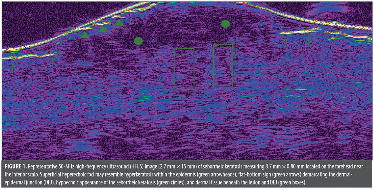
Using 2 sets of orthogonal HFUS image series, a 3D representation of the lesion was created via manual segmentation and plotting along the ultrasound scanning direction in MATLAB (R2025a). Dermatoscopy demonstrated the classic features of SK, including milia-like cysts and comedo-like openings.4 For all lesions under HFUS, the epidermis appears verrucous, resembling the lesion’s irregular surface texture, and contains superficial hyperechoic foci suggestive of hyperkeratosis within the epidermal layer (green arrows; Figure 1). We found that the dermal-epidermal junction (DEJ) consistently demonstrates a flat-bottom sign (green arrows; Figure 1), indicating the lesion is confined within the epidermis. While the DEJ disappears on HFUS within the tumor margins, the DEJ remains beneath the hypoechoic, hyperkeratotic lesion body as is pathognomonic for SKs. The hypoechogenicity of the lesion is consistent with high cellular density within the epidermis. Hyperechoic dermal tissue can be noted immediately beneath the inferior hypoechoic border. We note there are enlarged regions (green boxes; Figure 1) of increased density, possibly resembling sebaceous involvement underlying the lesion.
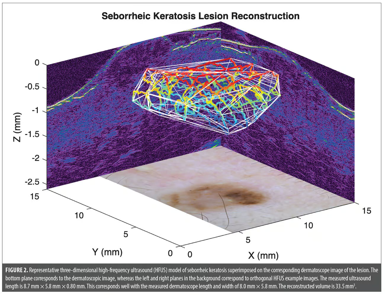
The 3D representation of the SK (Figure 2) illustrates hypoechoic boundaries spanning multiple cross-sections of the lesion. This reconstruction lacking dermal penetration further confirms a well-demarcated lesion confined within the epidermis that clinically exhibits a “stuck-on” appearance and exophytic growth pattern characteristic of SK. The immediate utility of noninvasive 3D probing lies within evaluations of margins, morphology, and consistent interior homogeneity. When superimposed on dermatoscopic features typical of SK, 3D tumor evaluation provides necessary lesion characteristics otherwise typically accessible via biopsy and subsequent histopathologic analysis.
However, the use of HFUS for evaluating benign tumors remains discretionary; while some physicians use it primarily for indeterminate lesions, others prefer incorporating it as part of a comprehensive imaging assessment.2 We demonstrate that HFUS may serve as a diagnostic tool adjunct to dermatoscopy to aid in lesion characterization, reduce the use of invasive procedures, and possibly inform clinical management in the future.
With regard,
Julian Henke, BS; Benazir Merchant, BS; Caroline Serafini, PhD; Devon Barrett, MD; Myer Khan; Zakaria Khan; Jiwoong Jason Jeong; Xiaofeng Yang, PhD; and Mohammed K. Khan MD, PhD
Keywords: Seborrheic keratosis, general dermatology, high frequency ultrasound, 3D ultrasound, clinical research
Affiliations: Mr. Henke and Dr. Yang are with the Emory University School of Medicine, Atlanta, Georgia. Ms. Merchant is with the Medical College of Georgia, Augusta, Georgia. Drs. Serafini and Khan are with the Winship Cancer Center at Emory University, Atlanta, Georgia. Dr. Barrett is with the Department of Dermatology, Emory Unversity School of Medicine, Atlanta, Georgia. Mr. M. Khan is with Georgia Institute of Technology, Atlanta, Georgia. Mr. Z. Khan is with the University of Washington, Seattle, Washington. Mr. Jeong is with Arizona State University, Phoenix, Arizona.
Funding: No funding was provided for this article.
Disclosures: The authors have no relevant conflicts of interest.
References:
- Barthelmann S, Butsch F, Lang BM, et al. Seborrheic keratosis. J Dtsch Dermatol Ges. 2023;21(3):265–277.
- Qin J, Wang J, Zhu Q, et al. Usefulness of high-frequency ultrasound in differentiating basal cell carcinoma from common benign pigmented skin tumors. Skin Res Technol. 2021;27(5):766–773.
- Vergilio MM, Monteiro E Silva SA, Jales RM, Leonardi GR. High-frequency ultrasound as a scientific tool for skin imaging analysis. Exp Dermatol. 2021;30(7):897–910.
- Braun RP, Rabinovitz HS, Krischer J, et al. Dermoscopy of pigmented seborrheic keratosis: a morphological study. Arch Dermatol. 2002;138(12):1556–1560.