
J Clin Aesthet Dermatol. 2026;19(9):73–78.
Albert E. Zhou MD, PhD; Christian Gronbeck, MD; Jenny J. Kim, MD, PhD; Siegrid Yu, MD; Hao Feng, MD, MHS; and Daniel M. Klufas, MD
Drs. Zhou and Kim are with the Department of Dermatology, University of California Los Angeles, Los Angeles, California. Dr. Gronbeck is with Zitelli, Brodland, and Lim Skin Cancer Center, Pittsburgh, Pennsylvania. Dr. Yu is with the Department of Dermatology, University of California San Francisco, San Francisco, California. Drs. Feng and Klufas are with the Department of Dermatology, University of Connecticut School of Medicine, Farmington, Connecticut.
Abstract: BACKGROUND: Mohs micrographic surgery (MMS) unites staged extirpation, comprehensive intraoperative histopathologic assessment, and immediate reconstruction to deliver durable local control with tissue conservation in anatomically critical sites. Contemporary practice, however, is shaped by intersecting forces: stewardship of indications, reimbursement trends, vertical consolidation, expanding use of intraoperative immunohistochemistry (IHC), and the emergence of complementary modalities (superficial radiation therapy; hedgehog and programmed cell death-1 [PD-1] inhibitors) alongside digital tools (teledermatology, artificial intelligence [AI], optical coherence tomography [OCT], reflectance confocal microscopy [RCM]). OBJECTIVE: To provide a narrative synthesis that clarifies where MMS adds the greatest value, delineates the appropriate roles of competing and complementary treatments, and outlines pragmatic steps (clinical, economic, and educational) to preserve high-quality, margin-controlled cutaneous oncology. METHODS: We conducted a structured narrative review of MEDLINE and Embase (January 2000–December 2025) using controlled vocabulary and keywords for MMS; basal cell and cutaneous squamous cell carcinoma; melanoma in situ; radiation therapy; systemic therapy (hedgehog/PD-1); IHC; teledermatology/AI; and optical imaging (OCT/RCM). Eligible studies included randomized trials, prospective cohorts, comparative observational studies, systematic reviews/meta-analyses, and cost or reimbursement analyses reporting local control/recurrence, complications, episode-level cost, or implementation outcomes. CONCLUSION: MMS provides superior or noninferior oncologic control with tissue preservation, same-day repair, and favorable safety in ambulatory settings. Appropriate use criteria should be framed as clinical stewardship, while superficial radiation and systemic therapies are best positioned as complements to MMS when surgery is infeasible or as neoadjuvant/adjuvant strategies. Sustaining the value of MMS will require explicit documentation of surgeon work and practice expense, standardized outcomes and patient-reported measures, equitable workforce strategies, and measured adoption of validated technologies such as OCT/RCM and AI. Payers and policymakers should recognize and preserve the true clinical and economic contribution of MMS. Keywords: Mohs micrographic surgery, basal cell carcinoma, cutaneous squamous cell carcinoma, appropriate use criteria, reimbursement, cost-effectiveness, superficial radiation therapy, immunohistochemistry, teledermatology, artificial intelligence
Introduction
Mohs micrographic surgery (MMS) is a definitive treatment for many forms of skin cancer, combining complete margin control with tissue preservation and excellent cosmetic outcomes. As skin cancer incidence climbs with an aging population and the therapeutic landscape broadens, the specialty faces intersecting pressures. Herein, we highlight several forces shaping MMS practice, including the evaluation of clinical stewardship, emergence of systemic therapies, digital innovation, and new workforce and economic realities with implications for practice, policy, and training.
Utilization and Appropriate Use
Use of MMS increased substantially from the mid-1990s through 2009,1,2 prompting the development of the 2012 Appropriate Use Criteria (AUC) from the AAD/ACMS/ASDSA/ASMS Task Force.3 The AUC provided a unified framework that rates more than 270 tumor-site clinical scenarios by appropriateness and was designed as a stewardship tool that concentrates MMS where comprehensive margin control most improves outcomes—high-risk histology, head and neck subsites, and recurrent disease—while complementing, not replacing, clinician judgment. Framed correctly, the AUC supports consistent, evidence-concordant selection without fully functioning as a cost-containment proxy.2 The core challenge is preserving the value of MMS while employing alternatives (ie, wide local excision, electrodesiccation and curettage, topical therapy) when outcomes are comparable in truly low-risk disease.
Clinical Effectiveness, Safety, and Outcomes
Evidence continues to support the high-volume, high-impact role of MMS. Across randomized and long-term prospective studies, MMS provides lower or at least noninferior recurrence than excision for both basal cell carcinoma (BCC) and high-risk cutaneous squamous cell carcinoma (cSCC).4–8 MMS is also increasingly used for melanoma in situ (MIS)/lentigo maligna and thinly invasive melanomas on the head and neck, where standard 5-mm fixed margins are often inadequate and wider excision risks functional or cosmetic impairment.9,10
In a multicenter cohort comprising >20,000 MMS cases, adverse events occurred in <1%, with complications being exceedingly rare,11 thus illustrating its utility in an outpatient setting. Among Medicare beneficiaries in 2009, approximately 558,447 MMS cases were performed, with a mean of 1.75 stages per tumor, and most patients received same-day reconstruction.12 Taken together, these data justify MMS as the preferred technique in defined high-risk scenarios.
Competing and Complementary Therapeutic Modalities
Systemic and targeted therapies for cutaneous malignancies have reshaped the therapeutic landscape (Table 1). Hedgehog inhibitors (vismodegib, sonidegib)13 and programmed cell death-1 (PD-1) blockade (cemiplimab, pembrolizumab)13 expand options for advanced or unresectable disease and can be deployed neoadjuvantly to facilitate removal of complex tumors, with both durable and impressive response rates.14,15 Yet, therapy-induced fibrosis or regression can complicate intraoperative histologic interpretation, underscoring the need for close collaboration among surgeons, oncologists, and dermatopathologists.
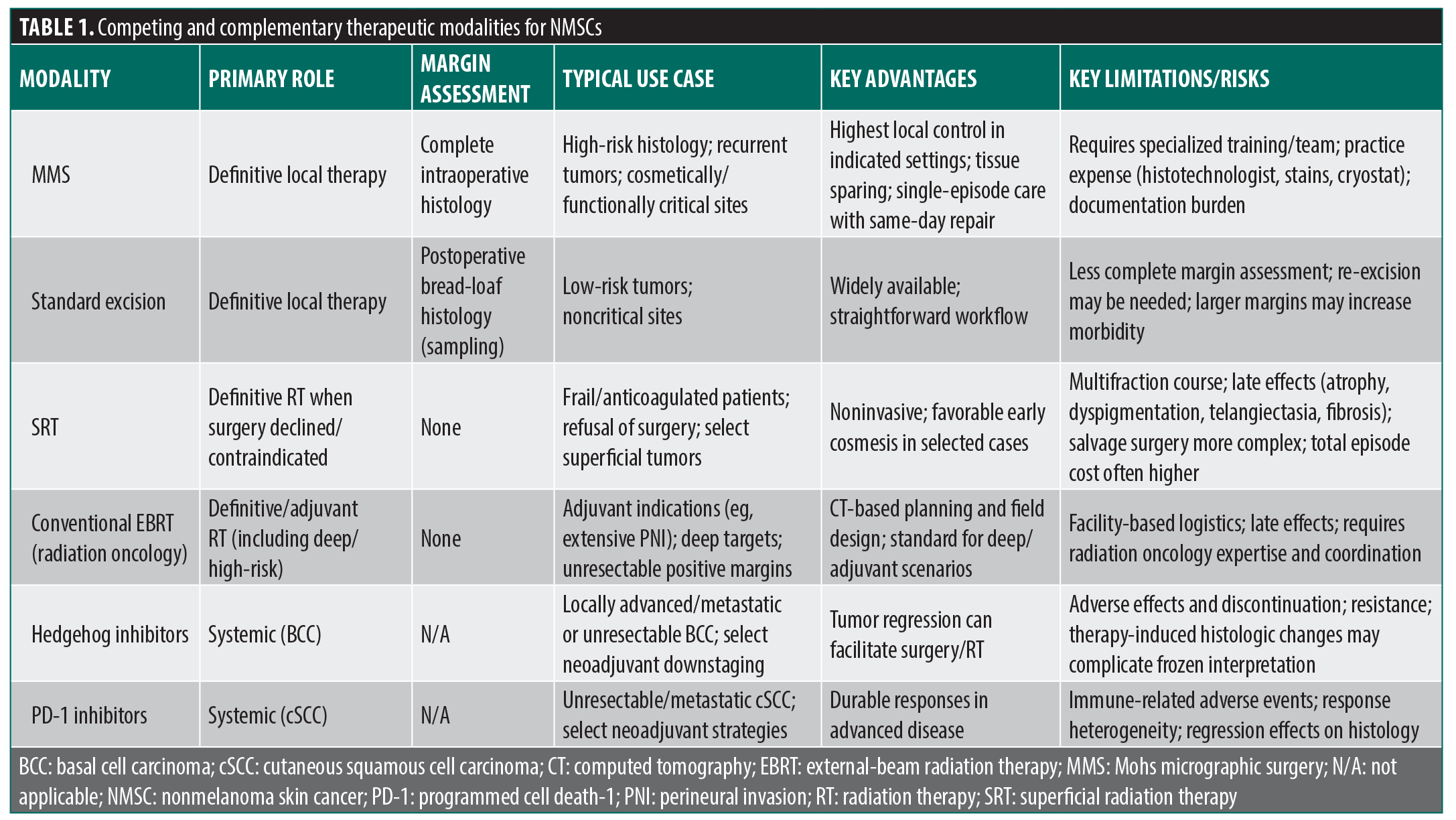
The use of superficial radiation therapy (SRT), including electronic brachytherapy and “image-guided” variants, in the treatment of keratinocyte carcinomas has risen dramatically.16 While its use is appropriate for poor surgical candidates or patients who decline surgery,16,17 unlike MMS, SRT lacks histologic margin control, generally demonstrates higher 5-year recurrence rates than MMS in high-risk facial tumors,18 requires multifraction courses, and can carry delayed adverse effects (eg, atrophy, dyspigmentation, telangiectasia, fibrosis). Medicare analyses have also shown higher total spending for SRT than for MMS, with image guidance adding further expense.16,17 When definitive radiation is indicated, conventional external-beam radiation therapy delivered by board-certified radiation oncologists remains the standard for deeper or high-risk disease (eg, named-nerve or large-caliber perineural invasion, postoperative adjuvant indications), whereas SRT may be a modality suited for shallower targets. Accordingly, MMS remains as a first‑line option when surgery is feasible and indicated; SRT and systemic targeted therapy serve selective definitive, neoadjuvant, or adjuvant roles.
Emerging Use of Biomarkers
Circulating tumor DNA (ctDNA) is emerging as a surveillance and molecular residual disease (MRD) biomarker in cutaneous oncology. In resectable stage III melanoma, serial ctDNA dynamics independently predict recurrence and complement imaging,19 while a prospective multicenter study in Merkel cell carcinoma showed ctDNA detects MRD and anticipates clinical relapse, supporting risk-adapted surveillance pathways.20 Early cSCC reports demonstrate feasibility and signal sensitivity in advanced disease,21,22 and ongoing trials are evaluating postoperative ctDNA to detect residual disease—use cases that may better stratify and triage for very-high-risk Mohs cohorts and open multidisciplinary discussions surrounding the need for surveillance imaging and adjuvant therapy.
Gene expression profiles (GEPs) have also refined preoperative risk stratification. In melanoma, the validated 31-gene assay provides independent prognostic information beyond clinicopathologic staging, whereas in cSCC, the validated 40-gene assay independently predicts nodal/metastatic events and can augment staging to guide selective imaging, nodal evaluation, and adjuvant radiation decisions when integrated with clinicopathologic risk.23,24 The National Comprehensive Cancer Network (NCCN) Cutaneous Melanoma Guidelines now recognize the Merlin CP-GEP assay as an appropriate molecular test for shared decision-making regarding Sentinel Lymph Node Biopsy in select T1b and T2a melanoma patients. As precision medicine continues to evolve, the future of MMS will not only involve greater interdisciplinary coordination and decision-making but also emphasize the importance of Mohs surgeons in serving as experts in cutaneous oncology.
Immunohistochemistry (IHC) in Mohs
Selective use of IHC (MART-1, SOX10, PRAME for melanoma25; CD34 for dermatofibrosarcoma protuberans; cytokeratins/p63 for poorly differentiated keratinocyte carcinomas and adnexal neoplasms26) improves diagnostic sensitivity when hematosylin-eosin is equivocal, thereby preventing undertreatment and preserving tissue in constrained sites. IHC, however, prolongs intraoperative time, adds appreciable cost, requires specialized platforms and protocols that affect laboratory flow, and may demand dermatopathologist-level interpretation. Adoption is expanding,27 and therefore, fellowship training should not only consider defining minimum IHC training, competencies, and case-log expectations but also committees to formalize panel selection and indications, stain interpretation and common artifacts, laboratory workflows and quality control, documentation, billing, and compliance. Ongoing research on outcomes can quantify the impact of IHC on recurrence, tissue preservation, and reconstructive complexity and link these benefits to the work and expense (added histotechnologist time, reagents, equipment depreciation) so that payment and policy accurately reflect the true demands of modern MMS.
Value, Payment Headwinds, and Practice Economics
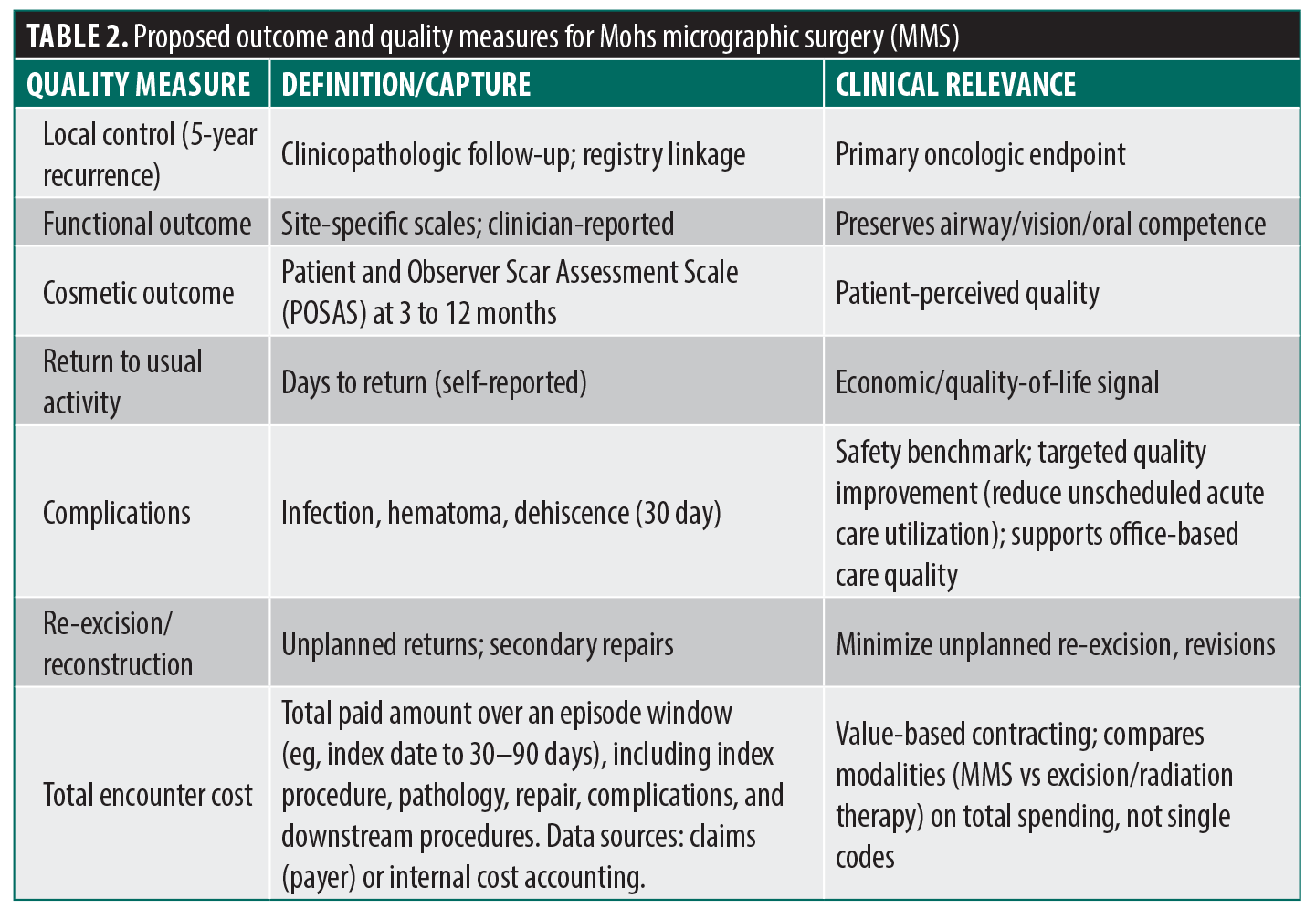
MMS is cost effective—and often cost saving—once recurrence, re-excision, and reconstruction factors are incorporated.28–32 Ambulatory site-of-service concentrates value by enabling same-day repair without facility or anesthesia fees and minimizing indirect costs (travel, time away from work/caregiving).33 Despite expanding use in an aging population,2 inflation-adjusted reimbursement has declined steadily by more than 14% for stages and 15% to 20% for flaps/grafts since 2007.34 Meanwhile, clinician work (frozen-section interpretation, staged mapping, on-table reconstruction) and practice expense (eg, consumables, histotechnologists, medical support staff, stains, cryostats, microscopes) remain substantial. Recent fee schedule rulemaking has prioritized “efficiency adjustments,” and continued economic compression will accelerate practice consolidation and procedural diversification. Safety-net and small practices may become incentivized to chase volume or shift payer mix. Therefore, payment should reflect staff, supplies, and equipment that enable safe and effective MMS. To align with value-based care models (Table 2),35 the field should standardize practical outcome sets such as recurrence, function, cosmesis, return-to-activity, and patient-reported outcomes—captured by low-friction methods (eg, short message system [SMS]) to inform quality improvement and payer dialogue.
Consolidation and Private Equity (PE)
Dermatology has been a leading target for PE acquisitions over the past decade,36 which has been associated with increased healthcare spending and higher encounter volumes.37 For MMS, consolidation and scale can provide negotiating leverage, capital for technology, and administrative relief36 but impose productivity targets, downstream service steering, or narrower networks that may not always align with AUC stewardship as well as patient or community needs. Mohs surgeons will need to weigh the impact of these effects at the expense of autonomy over case selection, scheduling/patient flow, and participation in teaching/tumor boards, which are core to quality and providing ideal patient care. Regulations, additional governance safeguards, and transparent outcomes may be needed to better align financial incentives with the preservation of clinician autonomy.38
Digital Innovation: Telemedicine, Artificial Intelligence (AI), and Mixed Reality
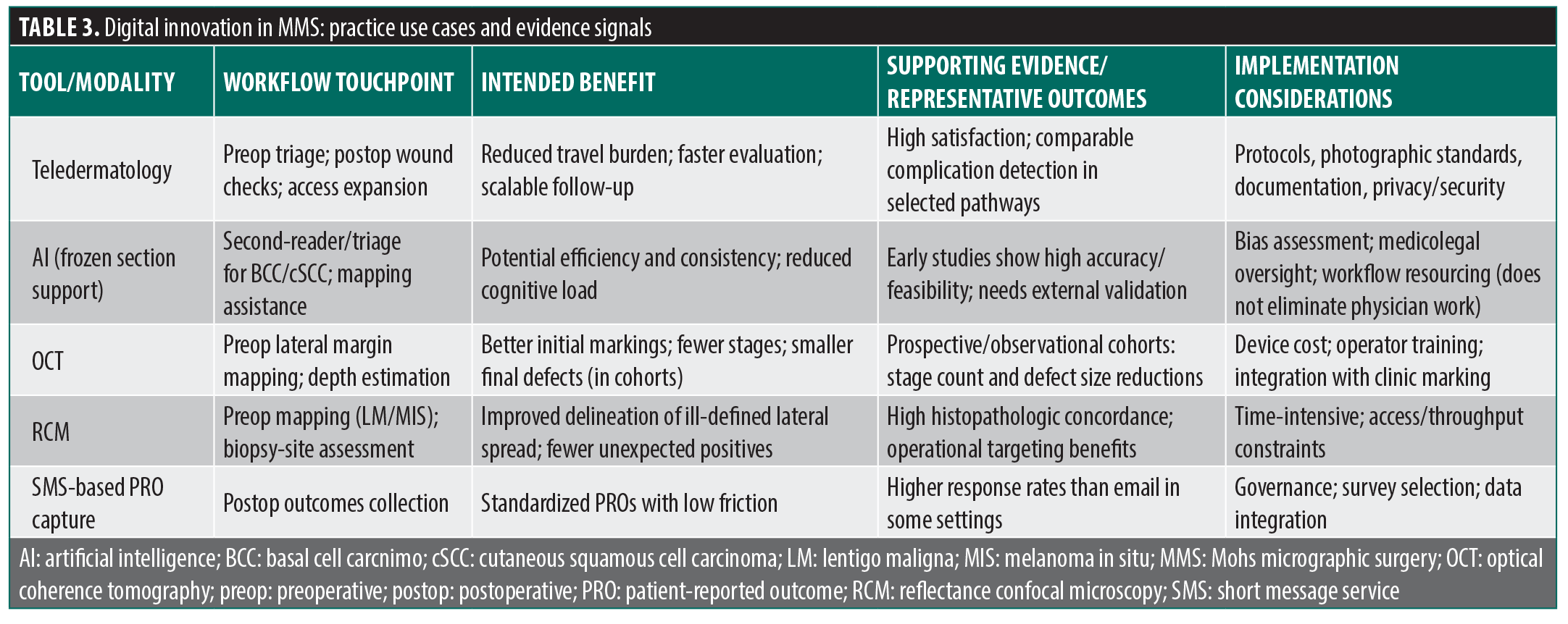
Teledermatology has found meaningful application in the perioperative continuum of MMS, including preoperative triage to postoperative wound surveillance, with strong patient satisfaction, safety, and comparable complication detection rates.39,40 Virtual touchpoints reduce travel barriers, particularly for older or mobility-limited populations. Beyond telehealth, AI and extended reality platforms are poised to redefine surgical training and scheduling, workflow, and patient engagement (Table 3).41–44 Early AI models show promise as second readers for frozen sections and have demonstrated high accuracy in detecting keratinocyte carcinoma and delineating tumor margins on histopathology,45–48 suggesting the potential for efficiency gains and supporting intraoperative decision-making. Preoperative optical imaging, whether by optical coherence tomography or reflectance confocal microscopy, can also help delineate lateral tumor margins and subclinical extension with concordance to histopathology, thereby reducing Mohs stages and better estimating defect size.49,50 Virtual and augmented reality (VR/AR) technologies are emerging as immersive training tools and intraoperative adjuncts, with pilot studies demonstrating growing applications in 3-dimensional reconstructive planning.51 In any scenario, digital integration must be evidence based and patient centered to enhance, rather than supplant, surgical judgment and humanism.
Access and Workforce Distribution
Geographic and payer-related disparities persist. Medicaid beneficiaries often travel farther or longer to reach an in-network Mohs surgeon,52 and county-level analyses identified pronounced access gradients.53 Forecasts predict worsening rural workforce shortages, with demand that will continue to outstrip supply in nonmetropolitan regions even under scenarios of increased overall dermatologist numbers.54 Delayed diagnosis and treatment can lead to enlarged tumors that increase reconstructive complexity and costs.55 Policies that broadly devalue office-based MMS would predictably widen inequities by rendering Medicaid and rural practice less viable. Solutions include (1) payer contracting strategies that expand Medicaid participation where feasible, (2) targeted outreach clinics or rotating rural sessions, (3) training pipelines that preferentially recruit and support trainees with rural ties, and (4) tele-enabled triage and postoperative follow-up to reduce travel burden.
The next generation of Mohs surgeons must navigate evolving tumor biology, systemic therapy paradigms, and digital integration. Furthermore, rising case complexity, expanding reconstruction repertoire, integration of IHC, and perioperative medical optimization will require comprehensive fellowship training.56 Fellowship programs, accrediting bodies, and societies can facilitate the growth of the profession by (1) exposing trainees to rural rotations and tele-supported practice models; (2) building curricular elements on health systems science (contracts, payer policy, AUC-based stewardship); (3) promoting research and quality improvement projects that center on access, appropriateness, and long-term outcomes rather than solely short-term volume metrics; (4) incorporating multidisciplinary oncologic, radiation oncology, and surgical training into fellowship curricula; and (5) promoting the responsible adoption of emerging technologies.
Conclusion
Mohs micrographic surgery stands at a defining juncture: anchored in its unparalleled precision yet surrounded by rapid change. As reimbursement declines and systemic therapies expand, the field must reaffirm its delivery of curative, tissue-sparing outcomes and surgical artistry. AI-assisted histology and AR visualization offer opportunities to enhance efficiency, education, and patient experience, but they must be integrated thoughtfully to preserve the patient-physician connection at the heart of MMS. The future of Mohs surgery will rely on embracing collaboration across specialties, technologies, and geographies.
References
- Asgari MM, Olson J, Alam M. Needs assessment for Mohs micrographic surgery. Dermatol Clin. 2012;30(1):167–175.
- Siddiqui FS, Leavitt A. Mohs micrographic surgery appropriate use criteria (AUC) guidelines. In: StatPearls [Internet]. StatPearls Publishing; 2026. Updated 5 May 2024. Accessed 9 Dec 2025. http://www.ncbi.nlm.nih.gov/books/NBK603719/
- Connolly SM, Baker DR, Coldiron FM, et al; Ad Hoc Task Force. AAD/ACMS/ASDSA/ASMS 2012 appropriate use criteria for Mohs micrographic surgery: a report of the American Academy of Dermatology, American College of Mohs Surgery, American Society for Dermatologic Surgery Association, and the American Society for Mohs Surgery. J Am Acad Dermatol. 2012;67(4):531–550.
- Zürcher S, Martignoni Z, Hunger RE, et al. Mohs micrographic surgery for cutaneous squamous cell carcinoma. Cancers (Basel). 2024;16(13):2394.
- Wang DM, Vestita M, Murad FG, et al. Mohs surgery vs wide local excision in primary high-stage cutaneous squamous cell carcinoma. JAMA Dermatol. 2025;161(5):508–514.
- Mosterd K, Krekels GAM, Nieman FH, et al. Surgical excision versus Mohs’ micrographic surgery for primary and recurrent basal-cell carcinoma of the face: a prospective randomised controlled trial with 5-years’ follow-up. Lancet Oncol. 2008;9(12):1149–1156.
- Alsaif A, Hayre A, Karam M, et al. Mohs micrographic surgery versus standard excision for basal cell carcinoma in the head and neck: systematic review and meta-analysis. Cureus. 2021;13(11):e19981.
- Johnson TM, Smith NR. Mohs surgery versus standard local excision for basal cell carcinoma, squamous cell carcinoma, and melanoma skin cancer. Facial Plast Surg. 2020;36(2):133–140.
- Verma K, Lewis DJ, Siddiqui FS, Dane A. Mohs micrographic surgery management of melanoma and melanoma in situ. In: StatPearls [Internet]. StatPearls Publishing; 2026. Updated 28 Aug 2024. Accessed 9 Dec 2025. https://www.ncbi.nlm.nih.gov/sites/books/NBK606123/
- Ellison PM, Zitelli JA, Brodland DG. Mohs micrographic surgery for melanoma: a prospective multicenter study. J Am Acad Dermatol. 2019;81(3):767–774.
- Alam M, Ibrahim O, Nodzenski M, et al. Adverse events associated with mohs micrographic surgery: multicenter prospective cohort study of 20,821 cases at 23 centers. JAMA Dermatol. 2013;149(12):1378–1385.
- Johnstone C, Joiner KA, Pierce J, Krouse RS. Mohs micrographic surgery volume and payment patterns among dermatologists in the Medicare population, 2013. Am J Clin Oncol. 2018;41(12):1199–1203.
- Tvedten E, Jennings T, Alam M, et al. Dermatologic oncology: the past 50 years. Dermatol Surg. 2025;51(10):919–929.
- Bertrand N, Guerreschi P, Basset-Seguin N, et al. Vismodegib in neoadjuvant treatment of locally advanced basal cell carcinoma: first results of a multicenter, open-label, phase 2 trial (VISMONEO study): neoadjuvant vismodegib in locally advanced basal cell carcinoma. EClinicalMedicine. 2021;35:100844.
- Gross ND, Miller DM, Khushalani NI, et al. Neoadjuvant cemiplimab for stage II to IV cutaneous squamous-cell carcinoma. N Engl J Med. 2022;387(17):1557–1568.
- Gronbeck C, Jain NP, Zhou AE, Feng H. Volume and distribution of radiotherapy performed by dermatologists from 2016 to 2021: a national Medicare trend analysis. J Am Acad Dermatol. 2024;91(2):341–344.
- Gronbeck C, Sloan B, Feng H. Image guidance contributes substantial cost and rarely informs dosimetry management in superficial radiotherapy: a national Medicare analysis. J Am Acad Dermatol. 2025;93(3):795–797.
- Patel JR, Engels E, Chinchilli E, Lambert Smith F. Superficial radiation therapy versus Mohs micrographic surgery: a systematic review and meta-analysis. Dermatol Surg. 2026;52(6):521–524.
- Marchisio S, Ricci AA, Roccuzzo G, et al. Monitoring circulating tumor DNA liquid biopsy in stage III BRAF-mutant melanoma patients undergoing adjuvant treatment. J Transl Med. 2024;22(1):1074.
- Park SJ, Kannan A, Harris JP, et al. Circulating tumor DNA as a predictive biomarker in Merkel cell carcinoma. J Am Acad Dermatol. 2022;87(5):1209–1211.
- Geidel G, Heidrich I, Kött J, et al. Emerging precision diagnostics in advanced cutaneous squamous cell carcinoma. NPJ Precis Oncol. 2022;6(1):17.
- Kim EY, Ruiz ES, Hanna GJ, et al. Sensitivity of personalized circulating tumor DNA assay in advanced cutaneous squamous cell carcinoma. J Am Acad Dermatol. 2024;90(2):427–429.
- Mehrmal S, Tan MG, Arron ST, et al. Gene expression profiling (GEP) in dermatology, part 1: introduction, development, benefits, limitations, and future directions of GEP. J Am Acad Dermatol. 2025:S0190-9622(25)03081-6.
- Mehrmal S, Tan MG, Arron ST, et al. Gene expression profiling (GEP) in dermatology, part 2: clinical applications of GEP in dermatology. J Am Acad Dermatol. 2025;S0190-9622(25)03084-1.
- Burshtein J, Marson J, Shah M, et al. Mohs micrographic surgery for melanoma. Dermatol Clin. 2025;43(3):473–482.
- Stranahan D, Cherpelis BS, Glass FL, et al. Immunohistochemical stains in Mohs surgery: a review. Dermatol Surg. 2009;35(7):1023–1034.
- Gronbeck C, Feng H, Knackstedt T. Growing adoption of immunohistochemistry by Mohs micrographic surgeons: a national Medicare trend analysis. Dermatol Surg. 2024;50(9):809–813.
- Sampath AJ, Paci K, Carrasquillo OY, et al. Retrospective analysis shows the cost of Mohs surgery decreases when adjusted for medical inflation. J Am Acad Dermatol. 2023;89(5):1001–1006.
- Cook J, Zitelli JA. Mohs micrographic surgery: a cost analysis. J Am Acad Dermatol. 1998;39(5 Pt 1):698–703.
- Wilson LS, Pregenzer M, Basu R, et al. Fee comparisons of treatments for nonmelanoma skin cancer in a private practice academic setting. Dermatol Surg. 2012;38(4):570–584.
- Ravitskiy L, Brodland DG, Zitelli JA. Cost analysis: Mohs micrographic surgery. Dermatol Surg. 2012;38(4):585–594.
- Seidler AM, Bramlette TB, Washington CV, et al. Mohs versus traditional surgical excision for facial and auricular nonmelanoma skin cancer: an analysis of cost-effectiveness. Dermatol Surg. 2009;35(11):1776–1787.
- Zhou AE, Klufas T, Gronbeck C, Feng H. Sustained shift toward office-based care for dermatologic procedures: a cross-sectional analysis of Medicare data from 2014 to 2023. Dermatol Surg. 2026;52(6):612–614.
- Mazmudar RS, Sheth A, Tripathi R, et al. Inflation-adjusted trends in Medicare reimbursement for common dermatologic procedures, 2007-2021. JAMA Dermatol. 2021;157(11):1–4.
- Rooke-Ley H, Song Z, Zhu JM. Value-based payment and vanishing small independent practices. JAMA. 2024;332(11):871–872.
- McTighe SP, Harvey D. Private equity in Mohs micrographic surgery: pearls and pitfalls. Dermatol Surg. 2026;52(6):614–615.
- Singh Y, Song Z, Polsky D, et al. Association of private equity acquisition of physician practices with changes in health care spending and utilization. JAMA Health Forum. 2022;3(9):e222886.
- Tsai TC, Meyer GS, Blumenthal D. Regulating private equity in health care — the Massachusetts Model. N Engl J Med. 2025;393(8):731–733.
- Algarin YA, Jaalouk D, Pulumati A, Nouri K. The role of teledermatology in Mohs micrographic surgery: a review. Arch Dermatol Res. 2024;316(5):119.
- Werbel T, Farahbakhsh N, Konda S. Store-and-forward teledermatology wound checks following Mohs surgery: a pilot study. Telemed Rep. 2024;5(1):256–262.
- Mirza FN, Haq Z, Abdi P, et al. Artificial intelligence for Mohs and dermatologic surgery: a systematic review and meta-analysis. Dermatol Surg. 2024;50(9):799–806.
- Fardos M, Miller P, Ward J, Miller R. Applications, ethical considerations, and patient perspectives on artificial intelligence in dermatologic surgery: focus on Mohs surgery. Dermatol Surg. 2025;51(10):930–934.
- Jeha GM, Qiblawi S, Jairath N, et al. ChatGPT and generative artificial intelligence in Mohs surgery: a new frontier of innovation. J Invest Dermatol. 2023;143(11):2105–2107.
- Rios-Duarte JA, Hardway HD, Vidal NY. The CLASS project: a proof-of-concept machine learning-driven complexity level algorithm for surgical scheduling in Mohs micrographic surgery. Dermatol Surg. 2026;52(6):525–530.
- Levy JJ, Davis MJ, Chacko RS, et al. Intraoperative margin assessment for basal cell carcinoma with deep learning and histologic tumor mapping to surgical site. NPJ Precis Oncol. 2024;8(1):2.
- Sohn GK, Sohn JH, Yeh J, et al. A deep learning algorithm to detect the presence of basal cell carcinoma on Mohs micrographic surgery frozen sections. J Am Acad Dermatol. 2021;84(5):1437–1438.
- Tan E, Lim S, Lamont D, et al. Development and validation of a deep learning model for improving detection of nonmelanoma skin cancers treated with Mohs micrographic surgery. JAAD Int. 2023;14:39–47.
- Davis MJ, Srinivasan G, Chacko R, et al. A deep learning algorithm to detect cutaneous squamous cell carcinoma on frozen sections in Mohs micrographic surgery: a retrospective assessment. Exp Dermatol. 2024;33(1):e14949.
- Paradisi A, Cornacchia L, Cappilli S, et al. Preoperative evaluation of high-risk basal cell carcinoma with line-field confocal optical coherence tomography (LC-OCT) reduces Mohs micrographic surgery stage number: a case-control study. EJC Skin Cancer. 2024;2:100015.
- Yélamos O, Cordova M, Blank N, et al. Correlation of handheld reflectance confocal microscopy with radial video mosaicing for margin mapping of lentigo maligna and lentigo maligna melanoma. JAMA Dermatol. 2017;153(12):1278–1284.
- Wang Q, Cutting CB, Sifakis E. Computer-based simulation of facial flap and cleft lip reconstruction using multiresolution physics. Plast Reconstr Surg Glob Open. 2025;13(6):e6820.
- Gronbeck C, Kodumudi V, Brodell RT, et al. Dermatology workforce in the United States – part I: overview, transformations, and implications. J Am Acad Dermatol. 2023;89(1):1–14.
- Beltrami EJ, Masison J, Gronbeck C, Feng H. Comparison of travel distance and time to in-network Mohs micrographic surgeons between Medicaid and Medicare beneficiaries. Dermatol Surg. 2023;49(2):200–201.
- Balboul S, Gronbeck C, Feng H. Dermatology workforce projections in the United States, 2021 to 2036. Arch Dermatol Res. 2024;316(5):192.
- Adamson AS, Zhou L, Baggett CD, et al. Association of delays in surgery for melanoma with insurance type. JAMA Dermatol. 2017;153(11):1106–1113.
- Zhou AE, Ravi S, Kovacs L, et al. Micrographic surgery and dermatologic oncology fellowship selection: an overview of the current landscape and considerations for prospective applicants. Dermatol Surg. Published online 29 Oct 2025.