
J Clin Aesthet Dermatol. 2026;19(8):57–66.
Alexandra DeVries, BS; Alec Lawson, BA; Aysham Chaudry, DO; Wilhelmina Lam, DO; and Mark S. Nestor, MD, PhD
All authors are with Center for Clinical and Cosmetic Research, Aventura, Florida. Ms. DeVries is additionally with Rocky Vista University College of Osteopathic Medicine, Parker, Colorado. Mr. Lawson is additionally with Arizona College of Osteopathic Medicine, Midwestern University, Glendale, Arizona. Dr. Nestor is additionally with the Department of Dermatology and Cutaneous Surgery and the Department of Surgery, Division of Plastic Surgery, University of Miami Miller School of Medicine, Miami, Florida.
FUNDING: No funding was provided for this article.
DISCLOSURES: Dr. Nestor is a consultant for Primus Pharmaceuticals. The remaining authors have no relevant conflicts of interest.
Abstract: BACKGROUND: Curcumin (CUR), the principal polyphenol in Curcuma longa, has gained increasing interest in dermatology because of its anti-oxidant, anti-inflammatory, antimicrobial, photoprotective, and regenerative properties. Despite versatile biological activity, its clinical use has been limited by poor bioavailability and significant product variability across formulations. METHODS: A targeted PubMed search was conducted using a combination of the following keywords: “curcumin,” “turmeric,” and “dermatology.” Only articles in English and relevant to this review were included. Additional articles were included through citation tracking and the authors’ clinical expertise. RESULTS: The search yielded 375 results, and 94 articles and 2 webpages were included based on the inclusion and exclusion criteria. DISCUSSION: CUR modulates multiple intracellular pathways, resulting in reduced pro-inflammatory cytokine expression, improved oxidative balance, and enhanced tissue repair. Despite an extremely favorable safety profile, the clinical use of CUR remains limited by poor bioavailability; however, newer formulations, including US Food and Drug Administration-recognized medical foods, have substantially improved absorption and product consistency. Clinically, CUR has shown immunomodulatory and anti-inflammatory effects applicable to cutaneous conditions including psoriasis, atopic dermatitis, and photodamage and has exhibited antimicrobial activity in acne. Aesthetically, CUR reduces melanogenesis and provides firming effects by limiting oxidative stress and matrix metalloproteinase activity. Regeneratively, it modulates key longevity-associated pathways involved in cellular senescence, wound healing, and anti-aging. CONCLUSION: CUR represents a safe and multifaceted agent with clinical, aesthetic, and regenerative dermatologic benefits. Emerging medical food formulations offer improved bioavailability and product consistency, though larger clinical trials are needed to define optimal dosing and long-term efficacy. KEYWORDS: Curcumin, psoriasis, atopic dermatitis, photodamage, hyperpigmentation, anti-aging, wound healing
Introduction
Natural compounds have been an integral part of traditional medical systems such as ayurveda and traditional Chinese medicine for centuries, and they have gained increasing popularity in Western medicine.1 These therapies are valued for their use of natural ingredients to provide both therapeutic and regenerative benefits. Among these natural agents, the use of turmeric (Curcuma longa) has dated back over 4,000 years, and its principal bioactive component, curcumin (CUR), has garnered attention as a versatile ingredient with therapeutic potential across a broad range of conditions, including those in dermatology.1
CUR is a polyphenol compound with diverse biological targets and has demonstrated anti-inflammatory, antimicrobial, antimutagenic, anti-oxidant, anti-allergic, chemopreventive, chemotherapeutic, and regenerative effects.2,3 These properties are of considerable interest in dermatology, where inflammatory dysregulation, oxidative stress, and allergic mechanisms underlie a wide range of conditions. Mechanistically, CUR also influences multiple molecular signaling pathways, including sirtuin 1 (SIRT1) promotion and mammalian target of rapamycin (mTOR) inhibition, that play a key role in regulating cellular senescence and aging.4 Through its pleiotropic effects, CUR represents a promising agent for therapeutic, aesthetic, and regenerative dermatologic applications.
Despite its broad biological potential, the clinical translation of CUR has been challenged by its inherently poor bioavailability and product inconsistencies that arising from its formulations as unregulated supplements. Many CUR supplements have relatively poor absorption, rapid metabolism, fast systemic elimination, chemical instability, and low aqueous solubility.5,6 Additionally, most CUR formulations are sold as dietary supplements, whereby dosing and purity may vary from batch to batch. To address these challenges, numerous formulation strategies have been developed to enhance the bioavailability of CUR, and the development of pharmaceutical-grade medical foods has the potential to resolve dose and batch-to-batch consistency. These advances have enabled more consistent delivery and enhanced therapeutic effects, allowing CUR to be used effectively in clinical, aesthetic, and regenerative settings as an evidence-based, natural, and versatile therapeutic agent.
Methods
A targeted search was conducted on PubMed using combinations of the keywords “curcumin” and “turmeric” with “dermatology” to examine the effects of CUR on a variety of systemic and cutaneous conditions. Articles were screened, and only articles in the English language and relevant to our study were included. Duplicate articles were excluded. Additional sources were identified through citation tracking and the authors’ clinical expertise.
Results and Discussion
The search yielded 375 articles; 94 articles and 2 webpages were included based on inclusion and exclusion criteria.
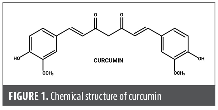
Structure and biological activity. Curcuminoids represent a family of turmeric-derived, structurally related, phenolic compounds predominantly consisting of CUR, desmethoxycurcumin, and bisdemethoxycurcumin, which collectively constitute approximately 2% to 5% of the turmeric rhizome by weight.7 CUR is a lipophilic, low-molecular weight polyphenol and is the principal bioactive component of turmeric.8 It possesses a bis-α,β-unsaturated β-diketone structure, shown in Figure 1, that confers both its characteristic yellow pigmentation and enables interaction with multiple diverse molecular targets.7
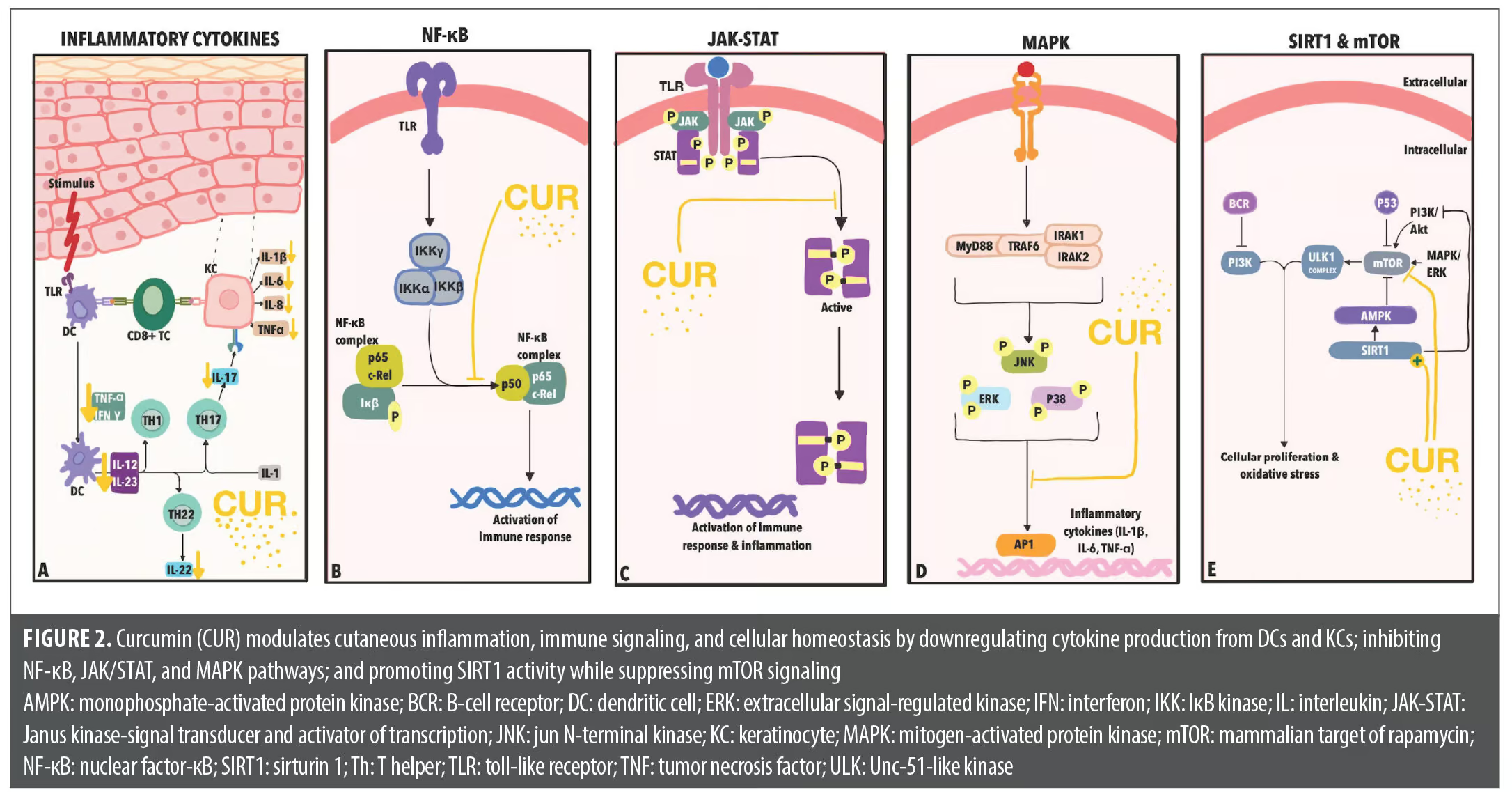
CUR diffuses through cell membranes due to its small size and lipophilic structure, which may explain its wide-ranging effects on a multitude of cellular mediators implicated in cutaneous pathology, as seen in Figure 2.8 It has demonstrated the ability to downregulate numerous inflammatory mediators, including tumor necrosis factor (TNF)-α and interleukin (IL)-12 released from dendritic cells, IL-22 and IL-17 from T cells, and IL-1β, IL-6, IL-8, and TNF-α from keratinocytes.9,10 CUR also inhibits activation of the Janus kinase (JAK) and mitogen-activated protein kinase (MAPK) signaling pathways.9 Both pathways modulate gene expression and promote inflammation, cellular proliferation, and stress responses.11,12 CUR’s effects on the JAK and MAPK pathways lead to decreased levels of inflammatory cytokines IL-1, IL-6, and TNF-α.9 Reduction in TNF-α, along with inhibition of IκB kinase (IKK) and p65 activation, is associated with antineoplastic effects through suppression of nuclear factor-κB (NF-κB) signaling, thereby reducing downstream proproliferative and anti-apoptotic pathways.9,13
In addition, CUR has been shown to modulate the SIRT1 and mTOR pathways. When activated, the mTOR pathway leads to anabolic processes including cell growth and proliferation, which result in oxidative stress on cells.14 SIRT1 also plays a central role in numerous processes linked to cellular homeostasis and longevity, and has an inhibitory effect on mTOR.15,16 By inhibiting mTOR and promoting SIRT1, CUR may provide protection against oxidative damage, attenuation of inflammatory signaling, increased resistance to cellular stress, and promotion of tissue regeneration and wound healing.17–19
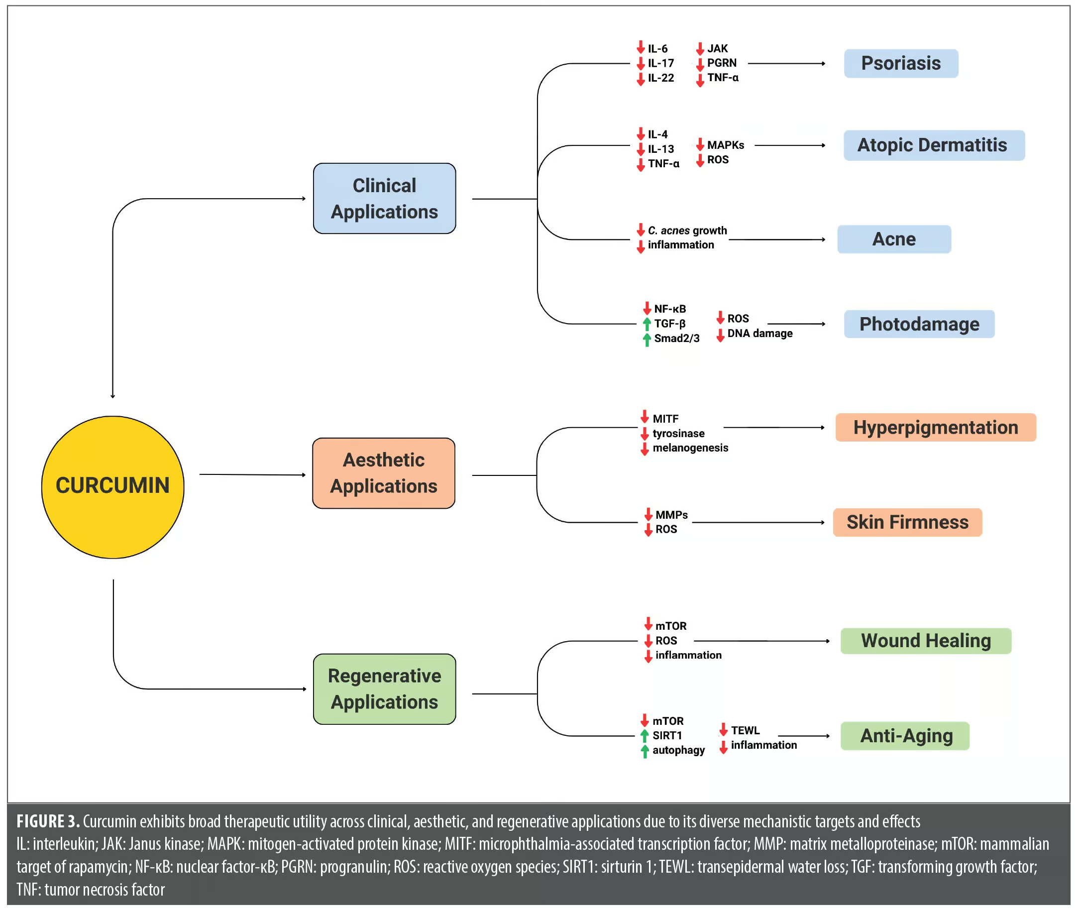
Modulation of these diverse mechanistic targets enables CUR to act on multiple pathogenic pathways relevant to dermatologic disease. Through its combined effects on inflammatory signaling, oxidative stress, cellular proliferation, and tissue regeneration, CUR demonstrates therapeutic potential across a broad spectrum of cutaneous conditions, as illustrated in Figure 3.
Safety. CUR has demonstrated a highly favorable safety profile as evidenced by numerous preclinical and clinical studies. Certain formulations of CUR are recognized as “Generally Recognized as Safe” (GRAS) by the United States Federal Drug Administration (FDA), a designation based on a review of accepted scientific data.20,21 A systematic review and meta-analysis on the use of CUR for arthritis reviewed 29 randomized controlled trials (RCTs) with doses ranging from 120 to 1,500 mg for a duration of 4 to 36 weeks. Based on the trials that reported adverse events, CUR did not increase their occurrence.9 Studies have also shown that CUR exhibits no significant subchronic toxic effects in animal studies and shows no evidence of mutagenic or teratogenic potential.9 Dose-escalation studies have demonstrated that CUR is safe at doses up to 12 g/day for periods of up to 3 months.20 More recently, isolated case reports have described liver injury associated with the use of turmeric supplements; however, the influence of CUR alone relative to other hepatotoxic exposures or contaminants in these cases is unclear.22
Bioavailability. Despite promising findings in clinical and preclinical trials, the clinical translation of CUR is inhibited by poor oral bioavailability. The biochemical structure of CUR exhibits poor aqueous solubility (approximately 11 ng/mL) and is classified as a Biopharmaceutics Classification System Class IV substance, characterized by low solubility and low permeability.23 Along with its poor aqueous solubility, CUR exhibits chemical instability at physiologic pH, resulting in limited intestinal absorption and difficulty achieving therapeutic plasma concentrations.23 These limitations of unformulated CUR, combined with its high plasma protein binding and rapid systemic clearance (half-life of <1 hour), have historically hindered the clinical use of CUR.23
Because of the bioavailability challenges that unformulated CUR presents, there have been recent efforts to develop formulations that enhance its absorption, extend its circulation time, and improve its delivery to tissues.23 Additionally, the optimal dose of CUR required to achieve meaningful biological effects remains equivocal, and excessively high doses may not necessarily translate to increased therapeutic efficacy.
One formulation combined CUR with essential oils derived from turmeric rhizomes.6 A clinical trial assessing this formulation in human participants found that its bioavailability was 6.93-fold greater compared to unformulated CUR, measured by the mean plasma concentration of CUR over an 8-hour period, and that it was tolerated well by study participants.6 The mechanism behind this enhanced bioavailability is likely multifactorial, with proposed explanations including increased extra-intestinal absorption mediated by turmeric essential oils and prolonged circulation that may alter enzymatic metabolism of the compound.6
Curcumin as a medical food. CUR is widely available as an over-the-counter supplement, which introduces risks and additional limitations. Supplements do not require oversight by the FDA or healthcare providers and may exhibit variable product quality and unreliable and inconsistent dosing.24 One study examined 125 CUR supplement preparations from the United States, United Kingdom, India, Australia, and Germany to assess their compliance with evidence-based recommendations. The findings revealed significant regulatory and labeling inconsistencies, with 34% of products failing to disclose their active curcuminoid content.24
Medical foods offer a regulated approach to using natural substances in disease management, with improved consistency in dosing and product quality. They are defined by the FDA as foods formulated to be consumed or administered enterally under the supervision of a physician, and intended for the specific dietary management of a disease or condition with distinctive nutritional requirements, established by medical evaluation.25 Medical foods are regulated under the Orphan Drug Act, and are distinct from both conventional foods and dietary supplements.25 Medical food manufacturers are required by law to adhere to “current good manufacturing practice,” and are subject to FDA oversight.25 Ingredients in medical foods must either be certified by the FDA as GRAS or have food and color additives used in accordance with FDA additive regulations. Importantly, medical foods must be used under medical supervision and are not intended for use by the general population without a diagnosed condition.26
Certain formulations of CUR, such as the medical food Rheumate® (RM; a proprietary multi-ingredient combination of L-5-methyltetrahydrofolate, methylcobalamin, and a curcuminoid-turmerone complex; Primus Pharmaceuticals, Inc.), are recognized by the US FDA as a medical food. RM contains L-5-methyltetrahydrofolate, methylcobalamin, and a curcuminoid-turmerone complex standardized with at least 95% curcuminoids. This complex has been reported to be more bioavailable than unmodified CUR.27 This formulation offers a consistent and reproducible form of highly bioavailable CUR. Safety data indicate that RM is generally well tolerated, with a favorable adverse effect profile even at high doses. With FDA regulation and healthcare provider oversight, RM may offer the benefits of CUR with reliable and consistent dosing.
Role in clinical dermatology. Psoriasis. Psoriasis is a chronic inflammatory disorder involving disruption of the innate and adaptive immune response that results in persistent and uncontrolled cutaneous inflammation.28 Preclinical studies have found that CUR reduces epidermal hyperplasia, suppresses keratinocyte proliferation, and decreases the expression of inflammatory mediators in both murine models and human cell lines.9,29 CUR’s ability to inhibit T-cell proliferation and cytokine secretion, particularly IL-17 and IL-22, is central to its immunomodulatory effect in the setting of psoriasis.29,30 Another hypothesized mechanism includes progranulin (PGRN), a cell-derived growth factor implicated in inflammatory regulation.19,31 Studies have shown that PGRN expression is significantly upregulated in keratinocytes from psoriatic lesions compared to perilesional and normal skin.31,32 In psoriatic murine models, intragastric administration of CUR for 3 weeks was found to suppress the overexpression of PGRN in keratinocytes.19,33 A similar study showed a decrease in pro-inflammatory cytokines, including IL-6, IL-17A, IL-22, IL-23, TNF-α, and transforming growth factor (TGF)-β1 and an improved regulation of the gut microbiota.34
Clinical trials have reinforced these effects by demonstrating that both oral and topical CUR formulations, particularly those with higher bioavailability, can significantly improve clinical outcomes in psoriasis. In a 16-week double-blind RCT, oral CUR (2 g/day in a lecithin-based delivery system) combined with topical corticosteroids led to a ≥50% improvement in Psoriasis Area and Severity Index (PASI-50) in 92% of patients compared to 54% of patients in the placebo plus corticosteroid group.35,36 Notably, 48% achieved PASI-75 and 12% achieved complete clearance, with significant reductions in serum IL-22.35,36 The augmentation of topical corticosteroids with CUR formulations may therefore achieve greater clinical improvement compared to corticosteroids alone.
CUR has also demonstrated efficacy in several other chronic inflammatory conditions, such as ulcerative colitis (UC) and rheumatoid arthritis (RA), which have pathogenic mechanisms that closely mirror that of psoriasis. UC, RA, and psoriasis collectively involve elevated and dysregulated cytokine, T-cell, and JAK-signal transducer and activator of transcription (STAT) signaling.37,38 This mechanistic overlap helps explain why they respond to many of the same targeted therapies. Preclinical studies likewise demonstrated that CUR can modulate these shared inflammatory pathways. In experimental models of inflammatory bowel disease (IBD), CUR suppressed dendritic cell activation by inhibiting JAK, STAT3, and STAT6 phosphorylation and by upregulating negative regulators such as SOCS1 and SOCS3.39 These findings are supported by clinical evidence in both UC and RA. An RCT of CUR plus mesalamine for mild-to-moderate UC reported clinical remission in 53.8% and endoscopic remission in 38% of patients at Week 4, whereas no patients receiving mesalamine plus placebo achieved remission.40 In RA, a meta-analysis of 10 RCTs found significant reductions in tender and swollen joint counts among patients taking CUR compared to placebo.41 One RCT reported that 46.4% of participants taking CUR experienced reductions in joint pain, swelling, stiffness, and fatigue, with greater benefit in those taking more than 200 mg/day.42 Given that the inflammatory pathways targeted by CUR in UC and RA are also central to psoriasis, it is highly plausible that CUR may have similar therapeutic benefits in psoriasis.
Atopic dermatitis. Atopic dermatitis (AD) is characterized by dryness, pruritus, and a localized erythematous rash due to immune dysregulation and skin barrier disruption.43,44 In line with its demonstrated anti-inflammatory effects, CUR has shown potential benefit in treating AD. In preclinical studies of AD-induced mice, a 7-day course of CUR resulted in downregulation of the expression of T helper 2 cell–promoting cytokines and normalization of the epidermal thickness.45 Additionally, CUR was shown to exert anti-allergic effects in a study assessing CUR on mast cell activation.46 CUR, at a concentration of 3 µmol/L, was found to suppress mast cell degranulation and the secretion of TNF-α and IL-4. Comparable concentrations of CUR have also been shown to inhibit mast cell activation by blocking Syk-dependent phosphorylations of the linker for activation of T cells protein. Additionally, the activating phosphorylations of Akt and various MAPKs, which are vital for the production of inflammatory cytokines, were also suppressed.46 Kong et al47 reported that CUR diminished the production of reactive oxygen species (ROS) resulting from immunoglobulin E-mediated or A23187-induced cell degranulation. It also lowered the expression of several pro-inflammatory cytokines, including IL-4 and IL-13.47
Along with preclinical evidence, a clinical trial assessing the effect of an oral CUR formulation on the symptoms and recurrence rates of AD found that those taking oral CUR plus using standard therapy (moisturizers and ceramide creams) for 90 days had a significant reduction in skin breaks when compared to the use of standard therapy alone.48 The study also found that the CUR group showed a more significant improvement of AD symptoms including pruritus, dryness, eczema, edema, and loss of sensitivity compared to the control.48 This study was limited by an unclear proportion of curcuminoids in the oral CUR formulation. Further clinical trials are warranted to determine CUR’s efficacy in improving both clinical outcomes and patient-reported symptoms in AD as well as establishing clear dosing standards.
Acne. Acne is a multifactorial condition involving excess sebum secretion, bacterial colonization, inflammation, and hyperproliferation of follicular keratinocytes.2 The commonly implicated bacterium, Cutibacterium acnes, preferentially colonizes in sebaceous follicles and triggers an inflammatory response, leading to acne lesions.49 The anti-inflammatory and antimicrobial properties of CUR support its use in the treatment of acne. CUR has been found to reduce inflammation in the context of acne vulgaris by targeting the NF-κB pathway and suppressing release of inflammatory cytokines including IL-6 and TNF-α.50 One study assessing the antimicrobial effects of CUR on C. acnes growth in porcine skin found a more significant reduction in bacterial growth when compared to various lengths of antibacterial free fatty acids including decanoic, lauric, myristic, and palmitic acids.51 Notably, the same study also found that 23.7 μg/mL of CUR resulted in a 50% inhibition of C. acnes growth, while 500 μg/mL of the anti-acne medication, azelaic acid, only resulted in a 35% inhibition of growth.51
CUR may also be used as an adjunctive therapy in patients with acne. In a split-face randomized study, the effect of photodynamic therapy (PDT) pretreated with a 20-minute CUR mask (CUR-PDT) was compared to PDT monotherapy administered twice weekly for 2 weeks.52 At the 2-week follow up, CUR-PDT produced significantly greater overall lesion clearance than PDT monotherapy. This benefit was driven primarily by inflammatory lesions, which showed markedly higher clearance with CUR-PDT.52 While this suggests CUR’s potential use as a combination therapy for acne, this study was limited by a small sample size and a short follow-up period, emphasizing the need for further large-scale clinical trials to determine the efficacy of CUR in the treatment of acne.
Photodamage. Photodamage remains a persistent concern in dermatology, but current treatment strategies are limited. CUR has demonstrated protective and reparative effects against photodamage in preclinical models. Topical and oral CUR reduced UV-induced oxidative stress, inflammation, apoptosis, and collagen degradation in skin through the modulation of antioxidant pathways, inhibition of matrix metalloproteinases (MMPs), and suppression of proinflammatory cytokines.18,53 CUR reduced the accumulation of ROS and restored anti-oxidant activity in human dermal fibroblasts.17 CUR decreased NF-κB activity and cleaved caspase‑3, glucose‑regulated protein 78, and C/EBP‑homologous protein expression while upregulating the expression of Bcl‑2, reducing UV-A–induced damage. CUR regulated collagen metabolism through the inhibition of MMPs and Smad7 and upregulation of TGF‑β and Smad2/3.17 These effects were demonstrated in murine models, with CUR pretreatment significantly decreasing erythema, induration, and scale and the number of sunburn cells compared to control.53 Additionally, treatment of human keratinocyte cells significantly reduced UV-B–induced lactate dehydrogenase release, intracellular ROS production and DNA damage, and activating DNA repair enzymes; this was associated with nuclear factor erythroid 2-related factor 2-dependent antioxidant activity.54
These findings are reflected in emerging clinical trials. In a split-face RCT, topical application of CUR in a gel formulation significantly improved markers of photoaging in women aged 34 to 67 years after 4 weeks compared to placebo.55 Moreover, phosphorylase kinase (PhK) promotes photocarcinogenesis in photodamaged skin through the activation of NF-κB and inhibition of photodamaged cell apoptosis. CUR acts as a selective and noncompetitive inhibitor of PhK and targets multiple signaling pathways that are PhK dependent, thus promoting the photodamage repair process including solar elastosis, pigmentary changes, and precancerous lesions.55
CUR may be a promising photoprotective agent with multiple preclinical trials and some clinical evidence supporting its use against photodamage; however, large-scale clinical trials are necessary to establish its efficacy.
Other dermatologic applications. The broad molecular activity of CUR suggests its utility across a wide range of additional dermatologic conditions. Its immunoregulatory effects may provide benefit in autoimmune conditions such as vitiligo and systemic lupus erythematosus (SLE). An RCT of individuals with vitiligo found that a CUR derivative applied topically combined with narrowband UV-B phototherapy twice weekly for 12 weeks produced slightly greater repigmentation compared to phototherapy alone.56 Similarly, another RCT demonstrated that turmeric cream applied over 4 months significantly reduced the size and visibility of vitiligo lesions compared to placebo.57 Evidence of CUR use in SLE is limited but suggests it may play an immunomodulatory role in the disease. In an RCT involving 62 patients with SLE, daily oral administration of CUR for 10 weeks significantly reduced serum levels of anti-dsDNA antibodies and the pro-inflammatory cytokine IL-6 compared to placebo.58 While other studies evaluating inflammatory markers and SLE Disease Activity Index (SLEDAI) scores demonstrated no significant difference between CUR and placebo, there is a lack of trials monitoring clinical symptomatic outcomes in patients.59
CUR has also demonstrated antifibrotic effects relevant to conditions characterized by dysregulated fibroblast activity, such as scleroderma. In a preclinical study, fibroblasts subjected to bleomycin-induced increases in extracellular matrix (ECM) production and treated with varying concentrations of CUR exhibited reductions in total soluble collagen, procollagen I, and fibronectin levels after 24 hours.60 CUR additionally suppressed TGF-β1 via inhibition of the TGF-β1/Smad2 signaling pathway, demonstrating its antifibrotic potential.60
In addition to its immunomodulatory and antifibrotic actions, CUR exhibits antineoplastic properties that have prompted investigation into its potential role in cutaneous malignancies such as melanoma. Preclinical studies suggested that CUR reduces melanoma cell viability and promotes apoptosis in a dose-dependent manner.61 These effects are associated with modulation of apoptotic signaling, including increased expression of Bax, suppression of anti-apoptotic proteins (Mcl-1 and Bcl-2), and inhibition of constitutively active NF-κB and IKK pathways.60,61
Overall, CUR has shown potential in the management of a variety of dermatologic conditions; however, well-designed RCTs evaluating clinically meaningful outcomes are needed to better define its role in clinical practice.
Role in aesthetic dermatology. Hyperpigmentation. There has been a growing interest in nutraceuticals and botanical agents for the treatment of hyperpigmentation.62 CUR in particular has demonstrated antimelanogenic effects in animal models and in vitro.63 Some chemically modified forms of CUR were found to inhibit melanogenesis through significant suppression of tyrosinase activity, while other forms significantly decreased tyrosinase protein levels in human melanocyte cells.63 Moreover, CUR led to decreased levels of melanogenic paracrine mediators in human keratinocytes.63 In another study, CUR inhibited microphthalmia-associated transcription factor and tyrosinase activity.35 Additionally, multiple studies have shown that CUR decreases melanogenesis in α-melanocyte-stimulating hormone (α-MSH)–stimulated cells through the activation of signaling pathways, such as MAPK/extracellular signal-regulated kinase and phosphatidylinositol 3-kinase (PI3K)/Akt.64,65
Systematic and narrative reviews have highlighted the anti-oxidant and depigmenting activity of CUR but emphasized limitations such as poor bioavailability and lack of large clinical trials in humans.2,19,66 Notably, CUR delivery using permeation enhancer nanovesicles in a leporine skin model with sunlight-induced hyperpigmentation had a significant reduction in pigment compared to CUR in a methanol solution, which reflected the influence of formulation and mode of delivery on CUR’s treatment efficacy.67 In a preclinical study in which melanogenesis was induced in murine cells by treatment with 200 nM of α-MSH, CUR at 10 μM reduced both extracellular and intracellular melanin levels compared to control.68 At this dose, CUR was also found to inhibit the increased tyrosinase activity induced by α-MSH.68 CUR’s ability to reduce melanin synthesis and tyrosinase activity, with novel formulations increasing its absorption and efficacy, poses it as a potential dermatologic agent for hyperpigmentation.
Skin firmness/wrinkles. Skin wrinkles arise from the progressive degradation and remodeling of the ECM, particularly collagen and elastin, and are a common concern amongst dermatologic patients.69 Although extrinsic factors, including UV exposure, can accelerate wrinkle formation, intrinsic chronologic aging also contributes.69 The loss of collagen and elastin in wrinkled skin results from reduced fibroblast activity, oxidative stress, and activation of MMPs.69 CUR has been shown to influence many of these processes and may reduce the development of fine lines and wrinkles.69
ROS contribute to the formation of wrinkles by oxidizing collagen, elastin, and DNA.70 Preclinical studies have demonstrated CUR’s ability to alleviate oxidative stress by upregulating antioxidant enzymes. In a study using human dermal fibroblasts, cells treated with a CUR-loaded nanocarrier revealed upregulation of superoxide dismutase 1, heme oxygenase-1, and glutathione peroxidase.18 Another study using UV-B–exposed mice found that those receiving 1,000 mg/kg of oral CUR demonstrated significantly fewer wrinkles than controls, likely due to suppression of MMP-2 expression and therefore the preservation of collagen.71
Clinical evidence also supports the skin firming benefits of CUR. In a trial of 60 healthy participants, a 4-week regimen of an oral curcuminoid supplement (70 mg daily) plus a topical CUR cream (0.02%) was compared to topical CUR alone.72 Both groups showed improvements in wrinkle appearance and skin firmness relative to controls, but the combined oral and topical therapy resulted in greater antiwrinkle effects, suggesting that CUR is effective when applied topically and its efficacy is further enhanced when combined with oral supplementation.72 A split-face RCT in 25 women similarly found that a CUR-containing topical formulation resulted in a significantly reduced ultrasound propagation speed compared to the control, suggesting an increase in skin firmness. Participant self-evaluations of the treatment effect were also significantly better for the CUR-treated side.4 Further clinical trials are needed to confirm the efficacy and long-term benefits of CUR for wrinkle reduction and prevention.
Collectively, CUR’s emerging applications in aesthetic dermatology suggest that its use may continue to expand across a wider range of aesthetic concerns.
Regenerative. Effects on regenerative pathways. CUR’s regenerative potential is largely mediated through its modulation of key cellular pathways involved in senescence and tissue repair. Senescence and SIRT signaling have emerged as major areas of active research in dermatology due to their central roles in cutaneous aging and regeneration. Two pathways of particular relevance are mTOR and SIRT1, which together orchestrate cellular metabolism, autophagy, senescence, and stress responses critical for tissue regeneration.73–75 Chronic or dysregulated mTOR activation is associated with malignancy, metabolic and autoimmune disease, and aging.76 Constitutive activation of this pathway is a key driver of senescent cells, which permanently exit the cell cycle yet remain metabolically active and develop a senescence-associated secretory phenotype (SASP).77 The increased metabolic demand and SASP expression lead to oxidative stress, release of pro-inflammatory mediators, impaired tissue healing, and increased susceptibility to age-related disease.78
In contrast, SIRT1, a nicotinamide adenine dinucleotide positive-dependent deacetylase that influences stress responses, DNA repair, and inflammation, negatively regulates mTOR signaling and suppresses the expression of SASP factors, promoting cellular rejuvenation and longevity.16,79,80 In the skin, SIRT1 plays an important role in the preservation of fibroblast function as well as maintaining genomic integrity.69,80 Its activity is associated not only with improved overall health span and systemic longevity but also with enhanced skin-specific regenerative and anti-aging effects.81
Evidence supporting CUR’s ability to modulate both mTOR and SIRT1 highlights its potential in regenerative applications.82,83 CUR directly inhibits mTOR complex 1 by disrupting the mTOR-raptor complex and reducing phosphorylation of downstream effectors.82,83 In addition, CUR promotes SIRT1 activity, which further suppresses mTOR, shifts cells toward autophagy, reduces senescence and oxidative stress, and enhances tissue repair.74,84–86 This coordinated regulation is mediated in part through the activation of adenosine monophosphate-activated protein kinase (AMPK), linking SIRT1 activity to downstream mTOR inhibition. This SIRT1/AMPK/mTOR axis is critical for maintaining stem cell function and promoting regeneration.84
Wound healing. Tissue regeneration is a crucial component of the wound healing process, and preclinical evidence supports the potential of CUR in enhancing reparative processes. To evaluate CUR’s impact on wound healing, 1 study applied a mucoadhesive C. longa formulation to full-thickness wounds in Wistar rats and compared outcomes to untreated controls. Twice-daily application significantly reduced the wound area on Days 3 and 10 and accelerated re-epithelialization by Day 10.87 CUR-treated wounds showed elevated levels of phospho-Akt and phospho-ribosomal protein S6 (a downstream target of mTOR) at Day 10, followed by a decline in these markers by Day 21.87 This pattern suggests that CUR enhanced healing and tissue regeneration by dynamically modulating the Akt/mTOR pathway, activating it during the proliferative phase to promote tissue regeneration and then suppressing it during the remodeling phase to support proper wound maturation.87 These effects may in part be mediated by CUR-induced SIRT1 activation, as SIRT1 has been shown to suppress inflammation and enhance fibroblast activity during healing.88,89
Another study in murine models of full-thickness excision wounds found that topical CUR enhanced cellular proliferation and collagen production, reflected by elevated DNA, total protein, and type III collagen content relative to untreated wounds.90 The CUR-treated wounds also healed more rapidly, evidenced by faster rates of epithelization, greater wound contraction, and improved tensile strength.90 Mechanistically, this study showed a shift toward anti-oxidative activity through reduced lipid peroxides and increased superoxide dismutase, catalase, and glutathione peroxidase activity in CUR-treated wounds.90 Collectively, these findings indicate that CUR facilitated cutaneous wound repair through coordinated anti-inflammatory, anti-oxidant, and regenerative mechanisms across multiple phases of healing.
Anti-aging. Aging skin is one of the most visible and universally addressed concerns in dermatology, with both intrinsic aging and environmental factors driving cutaneous changes over time. As skin ages, it undergoes progressive structural and functional decline. Histologically, aged skin shows epidermal and subcutaneous atrophy, blunting of the dermal-epidermal junction, degeneration of collagen and elastic fibers, disrupted epidermal barrier function, and reduced microvascularity.69 These structural changes within the skin give rise to the visible signs of skin aging, including wrinkles, roughened skin texture, decreased elasticity, and areas of hyperpigmentation.69
A central component of cutaneous aging is loss of fibroblast function. SIRT1 expression correlates with fibroblast number and proliferative capacity.69 In a study of skin biopsies from women aged 20 to 67 years, the level of SIRT1 in dermal fibroblasts demonstrated an age-dependent decrease, indicating that SIRT1 plays an important role in preserving fibroblast function.69 CUR’s ability to upregulate SIRT1, as well as directly inhibit MMPs, promotes collagen synthesis from fibroblasts within the dermis and has anti-aging effects.69 Its inhibition of mTOR further enhances this activity. At doses ranging from 20 to 25 µM, CUR has been shown to promote cytoprotective autophagy by inhibiting the mTOR-AKT axis, ultimately organizing cellular repair and mitigating age-related DNA damage and dysregulated cell proliferation.18
In addition to its intracellular and fibroblast-specific effects, CUR also enhances skin texture and barrier function. In a 4-week study, combined oral CUR plus a CUR-containing topical cream resulted in reduced transepidermal water loss (TEWL), improved skin hydration, increased dermal density, and improved face roughness compared to placebo.72 The reduction in TEWL suggests a reinforcement of the skin barrier following CUR therapy, resulting in improved skin hydration and addressing a key component of skin aging.72 CUR has additionally been shown to enhance skin barrier integrity by promoting structural proteins and tight junctions.91 In an 8-week RCT of 47 participants, oral CUR increased hyaluronan production and decreased pro-inflammatory cytokines (IL-1β and TNF-α) compared to placebo, highlighting its role in maintaining dermal hydration and reducing chronic underlying inflammation associated with skin aging.92
Through its combined regulation of inflammatory pathways, fibroblast activity, ECM remodeling, autophagy, and barrier function, CUR demonstrates a multifaceted regenerative potential.
Limitations
Critical evaluation of existing studies reveals notable methodological limitations that compromise the strength of evidence supporting oral CUR’s effects on cutaneous conditions.93 The major limitation is inconsistent dosing and bioavailability due to variability in CUR supplement formulations, which complicates the interpretation of efficacy outcomes. This limitation is common among natural compounds, as studied products often vary in formulation and dosing across studies.94,95 In the absence of standardized, reproducible, medical-grade products, meaningful comparison of outcomes across studies remains challenging. Additional limitations include small sample sizes, lack of adequate randomization or blinding, and reliance on subjective self-reporting rather than validated objective assessments, further hindering comparison across studies.93 In several reports, measurement conditions such as room temperature, humidity, and anatomical site are not standardized or clearly described.96 Collectively, these methodological and dosing inconsistencies hinder definitive conclusions regarding CUR’s efficacy for skin conditions and aesthetics. This emphasizes the need for consistent dosing methods and controlled clinical trials adhering to standardized reporting and dosing protocols.
Conclusion
Overall, CUR has accumulated substantial evidence supporting its use in dermatology, with demonstrated efficacy in numerous clinical, aesthetic, and regenerative applications. While there are robust preclinical data and emerging clinical data that support its value as a therapy, the widespread adoption of CUR remains limited due to the challenge of improving its bioavailability. Medical food formulations of CUR, such as RM, may offer an effective means of improving its low bioavailability while delivering a reliable, dose-consistent formulation. Continued research with larger RCTs and longer-term evaluations are warranted to better define CUR’s role in clinical practice.
References
- Sharifi-Rad J, Rayess YE, Rizk AA, et al. Turmeric and its major compound curcumin on health: bioactive effects and safety profiles for food, pharmaceutical, biotechnological and medicinal applications. Front Pharmacol. 2020;11:01021.
- Panahi Y, Fazlolahzadeh O, Atkin SL, et al. Evidence of curcumin and curcumin analogue effects in skin diseases: a narrative review. J Cell Physiol. 2019;234(2):1165–1178.
- Perrone D, Ardito F, Giannatempo G, et al. Biological and therapeutic activities, and anticancer properties of curcumin. Exp Ther Med. 2015;10(5):1615–1623.
- Sommerfeld B. Randomised, placebo-controlled, double-blind, split-face study on the clinical efficacy of Tricutan on skin firmness. Phytomedicine. 2007;14(11):711–715.
- Pan-On S, Dilokthornsakul P, Tiyaboonchai W. Trends in advanced oral drug delivery system for curcumin: a systematic review. J Control Release. 2022;348:335–345.
- Hegde M, Girisa S, BharathwajChetty B, et al. Curcumin formulations for better bioavailability: what we learned from clinical trials thus far? ACS Omega. 2023;8(12):10713–10746.
- Aggarwal BB, Harikumar KB. Potential therapeutic effects of curcumin, the anti-inflammatory agent, against neurodegenerative, cardiovascular, pulmonary, metabolic, autoimmune and neoplastic diseases. Int J Biochem Cell Biol. 2009;41(1):40–59.
- El-Saadony MT, Saad AM, Mohammed DM, et al. Curcumin, an active component of turmeric: biological activities, nutritional aspects, immunological, bioavailability, and human health benefits – a comprehensive review. Front Immunol. 2025;16:1603018.
- Zhang S, Wang J, Liu L, et al. Efficacy and safety of curcumin in psoriasis: preclinical and clinical evidence and possible mechanisms. Front Pharmacol. 2022;13:903160.
- Jahid M, Khan KU, Rehan-Ul-Haq, Ahmed RS. Overview of rheumatoid arthritis and scientific understanding of the disease. Mediterr J Rheumatol. 2023;34(3):284–291.
- Chakraborty J, Chakraborty S, Chakraborty S, Narayan MN. Entanglement of MAPK pathways with gene expression and its omnipresence in the etiology for cancer and neurodegenerative disorders. Biochim Biophys Acta Gene Regul Mech. 2023;1866(4):194988.
- Hu Q, Bian Q, Rong D, et al. JAK/STAT pathway: extracellular signals, diseases, immunity, and therapeutic regimens. Front Bioeng Biotechnol. 2023;11:1110765.
- Liu M, Wang J, Song Z, Pei Y. Regulation mechanism of curcumin mediated inflammatory pathway and its clinical application: a review. Front Pharmacol. 2025;16:1642248.
- Laplante M, Sabatini DM. mTOR signaling at a glance. J Cell Sci. 2009;122(Pt 20):3589–3594.
- Panwar V, Singh A, Bhatt M, et al. Multifaceted role of mTOR (mammalian target of rapamycin) signaling pathway in human health and disease. Signal Transduct Target Ther. 2023;8(1):375.
- Ghosh HS, McBurney M, Robbins PD. SIRT1 negatively regulates the mammalian target of rapamycin. PLoS One. 2010;5(2):e9199.
- Liu X, Zhang R, Shi H, et al. Protective effect of curcumin against ultraviolet A irradiation‑induced photoaging in human dermal fibroblasts. Mol Med Rep. 2018;17(5):7227–7237.
- Nie Y, Li Y. Curcumin: a potential anti-photoaging agent. Front Pharmacol. 2025;16:1559032.
- Kasprzak-Drozd K, Niziński P, Hawrył A, et al. Potential of curcumin in the management of skin diseases. Int J Mol Sci. 2024;25(7):3617.
- Gupta SC, Patchva S, Aggarwal BB. Therapeutic roles of curcumin: lessons learned from clinical trials. AAPS J. 2012;15(1):195–218.
- Generally Recognized as Safe (GRAS). US Food and Drug Administration. 17 Oct 2023. Accessed 21 Oct 2025. https://www.fda.gov/food/food-ingredients-packaging/generally-recognized-safe-gras
- Turmeric. In: LiverTox: Clinical and Research Information on Drug-Induced Liver Injury. National Institute of Diabetes and Digestive and Kidney Diseases; 2012. Updated 16 June 2025. Accessed 17 Nov 2025. http://www.ncbi.nlm.nih.gov/books/NBK548561/
- Anand P, Kunnumakkara AB, Newman RA, Aggarwal BB. Bioavailability of curcumin: problems and promises. Mol Pharm. 2007;4(6):807–818.
- Rahim-Mahdy H, Seifert R. A market and risk assessment of 125 turmeric supplements available in Australia, Germany, India, UK, and USA. Naunyn Schmiedebergs Arch Pharmacol. 2026;399(1):1315–1346.
- Guidance for Industry: Frequently Asked Questions About Medical Foods – Third Edition. 1 Oct 2024. Accessed 11 Nov 2025. https://www.fda.gov/regulatory-information/search-fda-guidance-documents/guidance-industry-frequently-asked-questions-about-medical-foods-third-edition
- Markowitz JS, Gurley PC, Gurley BJ. Medical foods-a closer look at the menu: a brief review and commentary. Clin Ther. 2020;42(7):1416–1423.
- Antony B, Merina B, Iyer VS, et al. A pilot cross-over study to evaluate human oral bioavailability of BCM-95CG (Biocurcumax), a novel bioenhanced preparation of curcumin. Indian J Pharm Sci. 2008;70(4):445–449.
- Rendon A, Schäkel K. Psoriasis pathogenesis and treatment. Int J Mol Sci. 2019;20(6):1475.
- Kumar B, Sahoo PK. Augmented transdermal delivery of curcumin for the effective management of plaque psoriasis – design, formulation, characterisation, and in vivo studies. AAPS PharmSciTech. 2023;24(5):134.
- Kang D, Li B, Luo L, et al. Curcumin shows excellent therapeutic effect on psoriasis in mouse model. Biochimie. 2016;123:73–80.
- Farag AGA, Shoaib MA, Samaka RM, et al. Progranulin and beta-catenin in psoriasis: an immunohistochemical study. J Cosmet Dermatol. 2019;18(6):2019–2026.
- Huang K, Chen A, Zhang X, et al. Progranulin is preferentially expressed in patients with psoriasis vulgaris and protects mice from psoriasis-like skin inflammation. Immunology. 2015;145(2):279–287.
- Zhou T, Zhang S, Zhou Y, et al. Curcumin alleviates imiquimod-induced psoriasis in progranulin-knockout mice. Eur J Pharmacol. 2021;909:174431.
- Cai Z, Wang W, Zhang Y, Zeng Y. Curcumin alleviates imiquimod-induced psoriasis-like inflammation and regulates gut microbiota of mice. Immun Inflamm Dis. 2023;11(8):e967.
- Gamret AC, Price A, Fertig RM, et al. Complementary and alternative medicine therapies for psoriasis: a systematic review. JAMA Dermatol. 2018;154(11):1330–1337.
- Antiga E, Bonciolini V, Volpi W, et al. Oral curcumin (Meriva) is effective as an adjuvant treatment and is able to reduce IL‐22 serum levels in patients with psoriasis vulgaris. Biomed Res Int. 2015;2015(1):283634.
- Schett G, McInnes IB, Neurath MF. Reframing immune-mediated inflammatory diseases through signature cytokine hubs. N Engl J Med. 2021;385(7):628–639.
- Banerjee S, Biehl A, Gadina M, et al. JAK–STAT signaling as a target for inflammatory and autoimmune diseases: current and future prospects. Drugs. 2017;77(5):521–546.
- Zhao HM, Xu R, Huang XY, et al. Curcumin suppressed activation of dendritic cells via JAK/STAT/SOCS signal in mice with experimental colitis. Front Pharmacol. 2016;7:455.
- Lang A, Salomon N, Wu JCY, et al. Curcumin in combination with mesalamine induces remission in patients with mild-to-moderate ulcerative colitis in a randomized controlled trial. Clin Gastroenterol Hepatol. 2015;13(8):1444–1449.e1.
- Kou H, Huang L, Jin M, et al. Effect of curcumin on rheumatoid arthritis: a systematic review and meta-analysis. Front Immunol. 2023;14:1121655.
- Purohit R, Bhaskar N, Jehu T, et al. Patient-reported outcomes of curcumin supplementation in rheumatoid arthritis and psoriatic arthritis: a cross-sectional survey. Rheumatol Int. 2024;44(12):3073–3078.
- Jeskey J, Kurien C, Blunk H, et al. Atopic dermatitis: a review of diagnosis and treatment. J Pediatr Pharmacol Ther. 2024;29(6):587–603.
- Fujii M. Current understanding of pathophysiological mechanisms of atopic dermatitis: interactions among skin barrier dysfunction, immune abnormalities and pruritus. Biol Pharm Bull. 2020;43(1):12–19.
- Sharma S, Sethi GS, Naura AS. Curcumin ameliorates ovalbumin-induced atopic dermatitis and blocks the progression of atopic march in mice. Inflammation. 2020;43(1):358–369.
- Lee JH, Kim JW, Ko NY, et al. Curcumin, a constituent of curry, suppresses IgE-mediated allergic response and mast cell activation at the level of Syk. J Allergy Clin Immunol. 2008;121(5):1225–1231.
- Kong ZL, Sudirman S, Lin HJ, Chen WN. In vitro anti-inflammatory effects of curcumin on mast cell-mediated allergic responses via inhibiting FcεRI protein expression and protein kinase C delta translocation. Cytotechnology. 2020;72(1):81–95.
- Togni S, Riva A, Maramaldi G, et al. Oral curcumin (Meriva®) reduces symptoms and recurrence rates in subjects with atopic dermatitis. Esperienze Dermatol. 2019;21(2-4):42–46.
- Brüggemann H, Salar-Vidal L, Gollnick HPM, Lood R. A Janus-faced bacterium: host-beneficial and -detrimental roles of Cutibacterium acnes. Front Microbiol. 2021;12:673845.
- Soleymani S, Farzaei MH, Zargaran A, et al. Promising plant-derived secondary metabolites for treatment of acne vulgaris: a mechanistic review. Arch Dermatol Res. 2020;312(1):5–23.
- Vaughn AR, Haas KN, Burney W, et al. Potential role of curcumin against biofilm-producing organisms on the skin: a review. Phytother Res. 2017;31(12):1807–1816.
- Zhang Y, Wang D, Liao C, et al. Curcumin-mediated photodynamic therapy for mild to moderate acne: a self-controlled split-face randomized study. Photodiagnosis Photodyn Ther. 2024;45:103887.
- Adusumilli NC, Mordorski B, Nosanchuk J, Friedman JM, Friedman AJ. Curcumin nanoparticles as a photoprotective adjuvant. Exp Dermatol. 2021;30(5):705–709.
- Li H, Gao A, Jiang N, et al. Protective effect of curcumin against acute ultraviolet B irradiation-induced photo-damage. Photochem Photobiol. 2016;92(6):808–815.
- Heng MC. Signaling pathways targeted by curcumin in acute and chronic injury: burns and photo-damaged skin. Int J Dermatol. 2013;52(5):531–543.
- Asawanonda P, Klahan SO. Tetrahydrocurcuminoid cream plus targeted narrowband UVB phototherapy for vitiligo: a preliminary randomized controlled study. Photomed Laser Surg. 2010;28(5):679–684.
- Jalalmanesh S, Mansouri P, Rajabi M, Monji F. Therapeutic effects of turmeric topical cream in vitiligo: a randomized, double-blind, placebo-controlled pilot study. J Cosmet Dermatol. 2022;21(10):4454–4461.
- Sedighi S, Faramarzipalangar Z, Mohammadi E, et al. The effects of curcumin supplementation on inflammatory markers in systemic lupus erythematosus patients: a randomized placebo-controlled trial. Eur J Nutr. 2024;64(1):8.
- Ramessar N, Borad A, Schlesinger N. The impact of curcumin supplementation on systemic lupus erythematosus and lupus nephritis: a systematic review. Lupus. 2023;32(5):644–657.
- Jiang AJ, Jiang G, Li LT, Zheng JN. Curcumin induces apoptosis through mitochondrial pathway and caspases activation in human melanoma cells. Mol Biol Rep. 2015;42(1):267–275.
- Siwak DR, Shishodia S, Aggarwal BB, Kurzrock R. Curcumin-induced antiproliferative and proapoptotic effects in melanoma cells are associated with suppression of IkappaB kinase and nuclear factor kappaB activity and are independent of the B-Raf/mitogen-activated/extracellular signal-regulated protein kinase pathway and the Akt pathway. Cancer. 2005;104(4):879–890.
- Fisk WA, Agbai O, Lev-Tov HA, Sivamani RK. The use of botanically derived agents for hyperpigmentation: a systematic review. J Am Acad Dermatol. 2014;70(2):352–365.
- Goenka S, Simon SR. Novel chemically modified curcumin (CMC) analogs exhibit anti-melanogenic activity in primary human melanocytes. Int J Mol Sci. 2021;22(11):6043.
- Park SY, Jin ML, Kim YH, et al. Aromatic-turmerone inhibits α-MSH and IBMX-induced melanogenesis by inactivating CREB and MITF signaling pathways. Arch Dermatol Res. 2011;303(10):737–744.
- Jang JY, Lee JH, Jeong SY, et al. Partially purified Curcuma longa inhibits alpha-melanocyte-stimulating hormone-stimulated melanogenesis through extracellular signal-regulated kinase or Akt activation-mediated signalling in B16F10 cells. Exp Dermatol. 2009;18(8):689–694.
- Barbalho SM, de Sousa Gonzaga HF, de Souza GA, et al. Dermatological effects of Curcuma species: a systematic review. Clin Exp Dermatol. 2021;46(5):825–833.
- Farooqui RK, Kaurav M, Kumar M, et al. Permeation enhancer nanovesicles mediated topical delivery of curcumin for the treatment of hyperpigmentation. J Liposome Res. 2022;32(4):332–339.
- Jeon HJ, Kim K, Kim C, Lee SE. Antimelanogenic effects of curcumin and its dimethoxy derivatives: mechanistic investigation using B16F10 melanoma cells and zebrafish (Danio rerio) embryos. Foods. 2023;12(5):926.
- Bielach-Bazyluk A, Zbroch E, Mysliwiec H, et al. Sirtuin 1 and skin: implications in intrinsic and extrinsic aging-a systematic review. Cells. 2021;10(4):813.
- Kammeyer A, Luiten RM. Oxidation events and skin aging. Ageing Res Rev. 2015;21:16–29.
- Sumiyoshi M, Kimura Y. Effects of a turmeric extract (Curcuma longa) on chronic ultraviolet B irradiation-induced skin damage in melanin-possessing hairless mice. Phytomedicine. 2009;16(12):1137–1143.
- Di Lorenzo R, Grumetto L, Sacchi A, et aI. Dermocosmetic evaluation of a nutricosmetic formulation based on Curcuma. Phytother Res. 2023;37(5):1900–1910.
- Beevers CS, Zhou H, Huang S. Hitting the golden TORget: curcumin’s effects on mTOR signaling. Anticancer Agents Med Chem. 2013;13(7):988–994.
- Zendedel E, Butler AE, Atkin SL, Sahebkar A. Impact of curcumin on sirtuins: a review. J Cell Biochem. 2018;119(12):10291–10300.
- Wu G, Yao W, Cheng L, et al. SIRT1 rescues autophagic flux via PI3K/AKT/mTOR inactivation to suppress DOX-induced senescence in MCF-7 cells. Exp Cell Res. 2025;452(1):114754.
- Perl A. mTOR activation is a biomarker and a central pathway to autoimmune disorders, cancer, obesity, and aging. Ann N Y Acad Sci. 2015;1346(1):33–44.
- Cayo A, Segovia R, Venturini W, et al. mTOR activity and autophagy in senescent cells, a complex partnership. Int J Mol Sci. 2021;22(15):8149.
- Yang H, Zhang X, Xue B. New insights into the role of cellular senescence and chronic wounds. Front Endocrinol (Lausanne). 2024;15:1400462.
- Hayakawa T, Iwai M, Aoki S, et al. SIRT1 suppresses the senescence-associated secretory phenotype through epigenetic gene regulation. PLoS One. 2015;10(1):e0116480.
- Chen C, Zhou M, Ge Y, Wang X. SIRT1 and aging related signaling pathways. Mech Ageing Dev. 2020;187:111215.
- Prathumwon C, Anuchapreeda S, Kiattisin K, et al. Curcumin and EGCG combined formulation in nanostructured lipid carriers for anti-aging applications. Int J Pharm X. 2025;9:100323.
- Beevers CS, Li F, Liu L, Huang S. Curcumin inhibits the mammalian target of rapamycin-mediated signaling pathways in cancer cells. Int J Cancer. 2006;119(4):757–764.
- Beevers CS, Chen L, Liu L, et al. Curcumin disrupts the mammalian target of rapamycin-raptor complex. Cancer Res. 2009;69(3):1000–1008.
- Yang L, Shi J, Wang X, Zhang R. Curcumin alleviates D-galactose-induced cardiomyocyte senescence by promoting autophagy via the SIRT1/AMPK/mTOR pathway. Evid Based Complement Alternat Med. 2022;2022:2990843.
- Giovannini L, Bianchi S. Role of nutraceutical SIRT1 modulators in AMPK and mTOR pathway: evidence of a synergistic effect. Nutrition. 2017;34:82–96.
- Ungurianu A, Zanfirescu A, Margină D. Regulation of gene expression through food-curcumin as a sirtuin activity modulator. Plants (Basel). 2022;11(13):1741.
- Danilevicz CK, Wagner VP, Ferreira N, et al. Curcuma longa L. effects on Akt/mTOR pathway and NF-κB expression during skin wound healing: an immunohistochemical study. Appl Immunohistochem Mol Morphol. 2021;29(10):e92–e100.
- Qiang L, Sample A, Liu H, et al. Epidermal SIRT1 regulates inflammation, cell migration, and wound healing. Sci Rep. 2017;7(1):14110.
- Yang H, Zhang W, Pan H, et al. SIRT1 activators suppress inflammatory responses through promotion of p65 deacetylation and inhibition of NF-κB activity. PLoS One. 2012;7(9):e46364.
- Panchatcharam M, Miriyala S, Gayathri VS, Suguna L. Curcumin improves wound healing by modulating collagen and decreasing reactive oxygen species. Mol Cell Biochem. 2006;290(1–2):87–96.
- Mo Z, Yuan J, Guan X, Peng J. Advancements in dermatological applications of curcumin: clinical efficacy and mechanistic insights in the management of skin disorders. Clin Cosmet Investig Dermatol. 2024;17:1083–1092.
- Asada K, Ohara T, Muroyama K, et al. Effects of hot water extract of Curcuma longa on human epidermal keratinocytes in vitro and skin conditions in healthy participants: a randomized, double-blind, placebo-controlled trial. J Cosmet Dermatol. 2019;18(6):1866–1874.
- Panahi Y, Hosseini MS, Khalili N, et al. Effects of curcumin on serum cytokine concentrations in subjects with metabolic syndrome: a post-hoc analysis of a randomized controlled trial. Biomed Pharmacother. 2016;82:578–582.
- Nestor MS, Bhupalam V, Awad N, Hetzel JD. The therapeutic role of genistein in perimenopausal and postmenopausal women. J Clin Aesthet Dermatol. 2024;17(10):45–53.
- Kellogg JJ, Paine MF, McCune JS, et al. Selection and characterization of botanical natural products for research studies: a NaPDI Center recommended approach. Nat Prod Rep. 2019;36(8):1196–1221.
- Grether-Beck S, Marini A, Jaenicke T, Krutmann J. Photoprotection of human skin beyond ultraviolet radiation. Photodermatol Photoimmunol Photomed. 2014;30(2–3):167–174.
From the 2nd Annual RAPIDS Immuno-Dermatology Conference: From Approval to Adoption—Translating Innovation Into Clinical Practice