 J Clin Aesthet Dermatol. 2020;13(10):38–41
by Jagdish Sakhiya, MD; Dhruv Sakhiya, MBBS; Ashish Dedakiya, MB, DDV; Madhav Purohit, mD; Mihir Modi, MBBS, DVD; Piyush Darji, MD; Feral Daruwala, M.pharm; and Nimish Dudhatra, MSc CR
All authors are with the Sakhiya Skin Clinic in Gujarat, India, apart from Dr. Dhruv Sakhiya, who is with the B.J. Medical College at the New Civil Hospital Asarwa in Gujarat, India.
FUNDING: No funding was provided for this study.
DISCLOSURES: The authors have no conflicts of interest relevant to the content of this article.
ABSTRACT: The term plaque morphea describes a variant of morphea (localized scleroderma) in which oval or round circumscribed areas of induration, pigmentary changes, and violaceous or erythematous halo (i.e., lilac ring) are found in the dermis and occasionally to the superficial panniculus. We report a case of 28-year-old male patient with recurrent plaque morphea who was treated with polydioxanone (PDO) mono threads and topical tacrolimus ointment. After the introduction of PDO mono threads, the patient was prescribed topical tacrolimus ointment for six months. After that six-month period, the lesions were softer to palpation and lighter in color. This was observed as a positive therapeutic response. Notably, no future recurrence was seen at one-year follow up.
Key words: PDO mono thread, morphea, morphea en plaque, tacrolimus ointment
Morphea, sometimes referred to as localized scleroderma, is a disease characterized by excessive collagen deposition leading to thickening of the dermis, subcutaneous tissues, or both.1 It is also delineated by local skin inflammation and fibrosis.2 It is usually limited to the skin, but it may extend deeper to involve muscle or bone. It can also include the inside of the mouth, the genitals, and the eyes. Clinically, it appears with extracutaneous manifestations involving fever, lymphadenopathy, arthralgias, fatigue, and central nervous system involvement. It can also include laboratory abnormalities, such as eosinophilia, polyclonal hypergammaglobulinemia, and positive antinuclear antibodies. It most often occurs in childhood or middle adulthood.3–5
According to the classification by Peterson et al,6 plaque morphea is one of five subtypes of localized scleroderma. Several forms of plaque morphea are distinguished, including morphea en plaque, guttate morphea, atrophoderma of Pasini and Pierini, keloid morphea (nodular morphea), and lichen sclerosus et atrophicus.6 Treatment minimizes the risk of developing new lesions and the extension of existing lesions. Due to the rarity of morphea, there is a scarcity of evidence-based therapies for the condition. Several studies have assessed the effectiveness of therapeutic agents for morphea, including imiquimod, calcipotriol in combination with betamethasone dipropionate, calcipotriene, and photodynamic therapy, supported by a level of evidence IIB, IIB, IIA, and IIA, respectively.7–10 Additionally, several studies report the effectiveness of topical tacrolimus in the treatment of plaque morphea (level IB evidence).11 In some cases, morphea lesions may not respond to treatment with tacrolimus, so prudent management is required in such cases. Here, we report the case of morphea en plaque, a variant of plaque morphea. The recurrent lesion was successfully treated with polydioxanone (PDO) mono threads, employing a technique involving the use of threads to close wounds, and tacrolimus ointment (0.1%).
Case Report
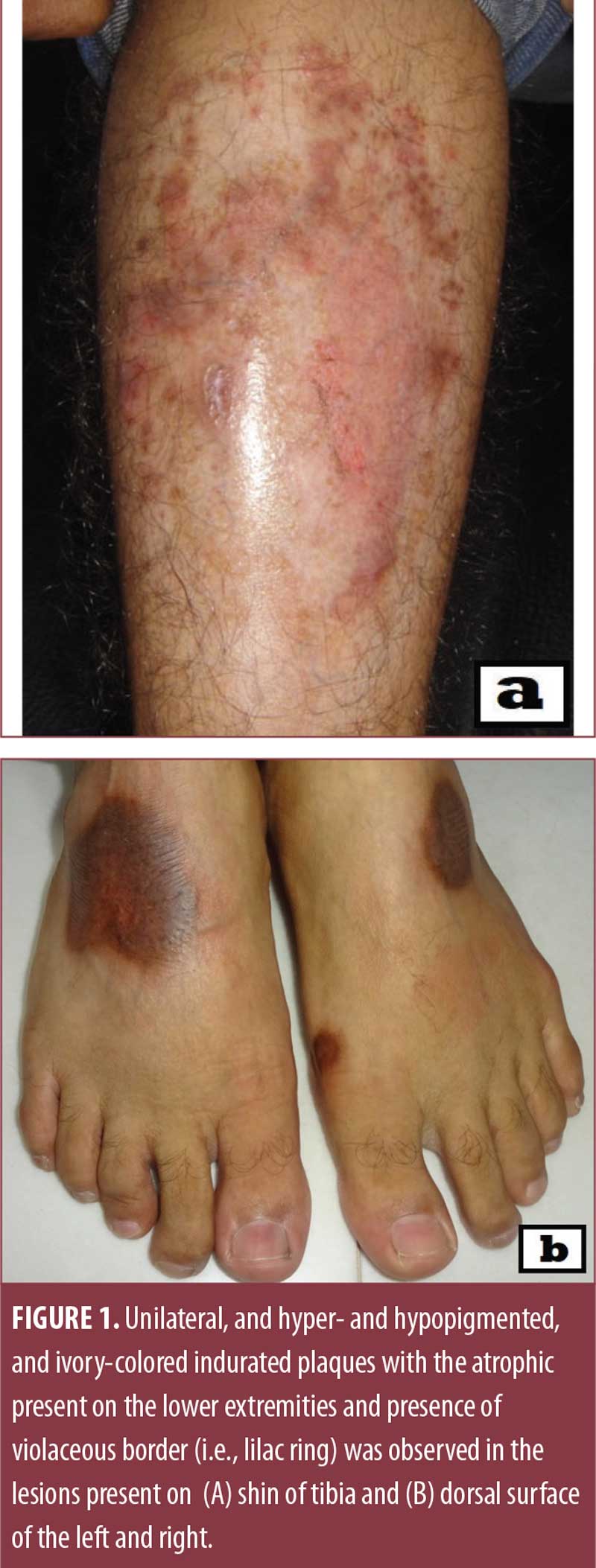
A 28-year-old man presented with a six-year history of dark-coloured, oval-shaped lesions on his lower extremities. There was no known family history of skin disorders and no history of infection, trauma, drug use, or toxic exposure. All vitals were normal and laboratory data were unremarkable. Physical examination revealed four, extensive, unilateral, and hyper- and hypopigmented ivory-colored indurate plaques with the atrophy present on the lower extremities. One lesion, which was oval-shaped and ranged from 15cm to 20cm in diameter, was present on the shin of the tibia (Figure 1A). The other two lesions, also round-shaped and ranging from 2cm to 6cm in diameter, were present on the dorsal surface of the left foot (Figure 1A). The fourth lesion was present on the dorsal surface of the right foot (Figure 1B). All lesions were surrounded by a violaceous border (i.e., lilac ring). There was no burning sensation, no itching, no numbness, no swelling, and no warmth over the lesions.

A punch biopsy was performed (Figure 2) and, based on clinical and histopathological findings, the definite diagnosis of morphea was made. After the diagnosis, the patient was started on methotrexate (15mg weekly, oral), topical steroids (once a day), topical tacrolimus ointment (0.1%) once a day, and clobetasol propionate (0.05%) once a day. After one year of therapy, the side effects of skin atrophy and surrounding hypopigmentation were observed, with no improvement in the lesions. To overcome this, the patient was prescribed topical tacrolimus (0.1%) for one year. After six months, no significant improvement was observed. Meanwhile, the patient was treated with fractional CO? laser after six months of topical tacrolimus ointment (0.1%) therapy. The laser parameter for fractional CO? laser was the density of 12×12, and energy ranging from 10–50 mJ. In total, six sessions were given. Concurrently, three intramuscular injections of triamcinolone acetonide (10mg intramuscularly, once a month) were administered.


 Two weeks after the introduced therapy, the lesion initially turned into brownish patches (Figure 3A, 3B). After six months of combined treatment of fractional CO? with topical tacrolimus ointment (0.1%) and intramuscular injections of triamcinolone acetonide, there was significant improvement in the observed skin lesions (Figure 4A, 4B). All lesions were completely resolved. After discontinuing treatment, the lesion present on the dorsal surface of the right foot gradually reappeared (Figure 5A, 5B, 5C) within a few weeks. At this time, the PDO threads technique was introduced. PDO mono threads of 29mm thickness and 10mm lengths were inserted in 5 to 6 entry points (Figure 6). PDO threads were absorbed into the body over six months by hydrolysis. The patient was prescribed topical tacrolimus ointment (0.1%) once a day for six months after thread therapy. Clinically, the lesions were softer to palpation and lighter in color after six months (Figure 7), and no recurrence was observed at one-year follow up.
Discussion
The term scleroderma is referred to a spectrum of disorders characterized by thickening of the skin and subcutaneous tissue in which two clinical categories are identified: systemic sclerosis, in which visceral lesions are present, and morphea, in which lesions are limited to the skin.12 As per epidemiological data, the incidence of morphea is accounting for 0.4 to 2.7 per 100,000 individuals and is highly predominant in women compared to men, with an estimated ratio of 2 to 3:1.13
Peterson et al6 divided the morphea into five categories: plaque, generalized, bullous, linear, and deep. Plaque morphea is a superficial type of morphea that is confined frequently to the dermis and occasionally to the superficial panniculus. The various subtypes of plaque have been mentioned earlier. Among them, morphea en plaque is the most prevalent of plaque group. It involves only one or two anatomic sites that can be found on the back, upper and lower extremities, the buttocks, and the face, neck, or scalp. It is characterized by an insidious onset of one or more oval or round circumscribed areas of indurations, which are larger than 1cm in diameter, and varying degrees of pigmentary changes. A violaceous or erythematous halo (i.e., lilac ring), which is often evident early during disease, corresponds with inflammatory activity. With disease progression, the skin becomes sclerotic and the center of the lesion becomes ivory-colored as the inflammation subsides. After several months or years, the skin softens and becomes atrophic. A residual area of hypo- or hyperpigmentation ensues. Morphea en plaque is more common on the trunk than on the extremities and occasionally involves the face. Notably, distinctive borders are usually separate the plaques from the surrounding normal skin.6 Our patient fits most of the criteria for plaque morphea.
The pathogenesis of morphea is unknown. It is postulated to be multifactorial, including genetic, autoimmune, and environmental factors, which leads to microvascular injury. This stimulates an imbalance between collagen production and degradation. Triggers such as mechanical trauma at lesional sites, suggesting koebnerizing phenomena or infection with Borrelia spp., particularly European strains, have been notified in the literature.13,14 Patients with morphea commonly have positive autoantibodies with a high prevalence of positive ANA titers, homogenous pattern; single-stranded antibody; antihistone antibodies; anti-topoisomerase II alpha antibody; and rheumatoid factor.13
Excess collagen deposition is thought to be activated by vascular injury via factors reported earlier.13,15 The vascular theory stated endothelial injury causes a release of inflammatory cytokines and subsequent up-regulation of the expression of adhesion molecules and E-selectins. This upregulation recruits T-cells that produce pro-fibrotic cytokines, mainly interleukin 4 (IL-4), interleukin 6 (IL-6), and transforming growth factor-beta (TGF-?), leading to increased collagen production and extracellular matrix deposition favouring a type 2 helper T-cell response. TGF-? also decreases protease production, primarily through inhibition of matrix metalloproteinases, and increases protease inhibitors. This causes an imbalance of collagen production and breakdown, thus, favoring fibrosis and a subsequent hardening of the skin.
Based on clinical findings, a provisional diagnosis of morphea can often be made. However, many times a biopsy is used for conclusive confirmation and defining the depth of involvement. Differential diagnosis of morphea includes other fibrosing disorders, such as drug reaction, mycosis fungoides, scleromyxedema, myxedema, scar, radiation dermatitis, amyloidosis, chronic graft-versus-host disease, lipodystrophy, sarcoidosis, and nephrogenic systemic fibrosis related to this context histopathologic finding are the cornerstone. Additionally, laboratory abnormalities can provide hints in the diagnosis of morphea. Eosinophilia, circulating antibodies such as antinuclear antibodies, anti-single-stranded DNA antibodies, and antihistone antibodies have been reported in morphea (not done in our case). For plaque morphea, a punch biopsy is usually sufficient.16
Although no standard therapeutic strategy for morphea exists, different topical and systemic treatment options give beneficial results. Topical corticosteroids and calcipotriene therapy should be considered in the active phase of the disease. This therapy was effective in the first described morphea patient. UVA 1 irradiation and PUVA can be used as monotherapy or as a part of combined treatment. Recent studies have shown satisfactory efficacy of systemic corticosteroids, methotrexate, cyclosporine A, mycophenolate mofetil, azathioprine, TNF-? inhibitors, and other medications used to treat morphea. The existing pharmacological regimens combined with physical and surgical therapy should be an integrated part of the treatment, to avoid the development of indivertible damage.16 Based on empirical experience in the dermatology field; herein the case of reoccurrence of morphea was managed with thread technique.
Currently, three main types of threads are available: PDO, polylactic acid (PLA), and polycaprolactone (PCA). PDA threads are made of a synthetic, biodegradable polymer, and the longest thread has been used in surgery for many years. Mono, cog, and screw threads are the most commonly used PDO threads in the medical field. As per the literature review, PDO threads can be used for lifting, rejuvenation (improving skin texture and wrinkles), volumizing, and even reducing fat. Use of PDO threads in treatment of morphea was not documented. PDO threads are absorbed into the body over six months by hydrolysis and better suited to younger patients.17
While the lesions in this present case were treated with combined therapy of methotrexate, topical steroids, topical tacrolimus ointment and clobetasol propionate, no significant improvement was observed. Nevertheless, no recovery was observed with only tacrolimus ointment therapy. When the lesions were treated with combined therapy of tacrolimus, fractional CO? laser, and triamcinolone acetonide, significant improvement was observed.
The lesions present on the dorsal surface of the right foot reappeared after discontinuation of introduced therapy. At this point, the patient was treated with blend of PDO mono thread injection and topical tacrolimus ointment, which resulted in significant improvement in the recurrent lesion. The probable mechanism behind this may be tacrolimus acts specifically on inflammatory cells. However, morphea lesions are not inflammatory, and thus may not resolve completely with tacrolimus alone.2,11 Introduction of PDO mono thread in the lesion can trigger the natural self-healing capabilities to induce an inflammatory reaction. Therefore, tacrolimus works well with this lesion.
Conclusion
The PDO threads technique described here is a minimally invasive technique that was well tolerated in our patient. The procedure is quick, mostly pain free, and provides beneficial outcomes. Use of this technique with tacrolimus ointment may be beneficial for clinicians treating morphea in the future.
References
- Laxer RM, Zulian F. Localized scleroderma. Curr Opin Rheumatol. 2006;18:606–613.
- Omair MA, Johnson SR. Inflammatory arthritis, sacroiliitis, and morphea: evidence of a systemic inflammatory disease. Case Rep Rheumatol. 2013;2013:347694.
- Zulian F. Systemic manifestations in localized scleroderma. Curr Rheumatol Rep. 2004;6:417–424.
- Chung L, Lin J, Furst DE, et al. Systemic and localized scleroderma. Clin Dermatol. 2006;24:374–392.
- Leitenberger JJ, Cayce RL, Haley RW, et al. Distinct autoimmune syndromes in morphea: a review of 245 adult and pediatric cases. Arch Dermatol. 2009;145:545–550.
- Peterson LS, Nelson AM, Su WPD. Classification of morphea (localized scleroderma). Mayo Clin Proc. 1995;70:1068–1076.
- Dytoc M, Ting PT, Man J, et al. First case series on the use of imiquimod for morphoea. Br J Dermatol. 2005;153:815–820.
- Dytoc MT, Kossintseva I, Ting PT. First case series on the use of calcipotriol-betamethasone dipropionate for morphoea. Br J Dermatol. 2007;157:615–618.
- Cunningham BB, Landells ID, Langman C, et al. Topical calcipotriene for morphea/linear scleroderma. J Am Acad Dermatol. 1998;39:211–215.
- Batchelor R, Lamb S, Goulden V, et al. Photodynamic therapy for the treatment of morphoea. Clin Exp Dermatol. 2008;33:661–663.
- Kroft EB, Groeneveld TJ, Seyger MM, et al. Efficacy of topical tacrolimus 0.1% in active plaque morphea: randomized, double-blind, emollient-controlled pilot study. Am J Clin Dermatol. 2009;10:181–187.
- Savoia P, Zaccagna A, Bernengo MG. Guess what? Inflammatory disseminated morphea profunda. Eur J Dermatol. 1999;9:654–656.
- Fett N, Werth VP. Update on morphea: Part I. Epidemiology, clinical presentation, and pathogenesis. J Am Acad Dermatol. 2011;64:217-28; quiz 229.
- Grabell D, Hsieh C, Andrew R, et al. The role of skin trauma in the distribution of morphea lesions: A cross-sectional survey of the morphea in adults and children cohort IV. J Am Acad Dermatol. 2014;71:493–498.
- Badea I, Taylor M, Rosenberg A, et l. Pathogenesis and therapeutic approaches for improved topical treatment in localized scleroderma and systemic sclerosis. Rheumatology. 2009;48:213–221.
- Nguyen JV, Werth VP, Werth VP, et al. Morphea workup updated. Medscape. May 21, 2018. Available from: https://emedicine.medscape.com/article/1065782-workup#c6.
- Wong V, Rafiq N, Kalyan R. Hanging by a thread: choosing the right thread for the right patient. J Dermatol Cosmetol. 2017;1:86–88.
OX40/OX40L Costimulatory Pathway: A Potential Therapeutic Target for Allergic Contact Dermatitis?