Dear Editor:
We read the recent article, “Recurrence of Localized Pemphigus Foliaceus Induced by Knee Replacement,” with great interest.1 The authors attribute the presentation of unilateral pemphigus foliaceus (PF) in a previously uninvolved site to an immunocompromised district (ICD), where edema and nerve injury paved the way for a buildup of immunoglobulins and immune cells. We noted a similar case presentation in a 70-year-old man with a history of PF who presented with an acute eruption of erosions at the healed surgical site following a right total-knee arthroplasty and highlight a different perspective behind the pathogenesis of this recurrence. We discuss alternative theories that might induce localized PF distinct from the concept of ICD.

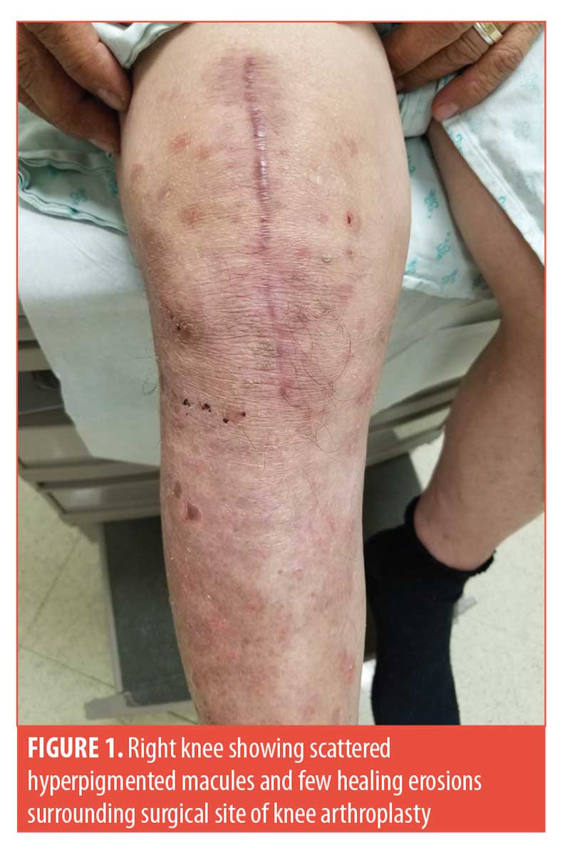
A 70-year-old man, with a history of PF, presented with an acute eruption of erosions at the healed surgical site and subsequent generalized PF 98 days after a right total-knee arthroplasty (Figure 1). He had previously been in remission on adjuvant therapy comprising 1,000mg of mycophenolate mofetil (MMF) twice daily and 100mg of doxycycline once daily for 11 months. Before surgery, the patient had tapered and stopped MMF due to his surgeon’s concern of an increased risk of postoperative complications while on immunosuppressants. Repeat biopsy and direct immunofluorescence (DIF) were deferred. Repeat indirect immunofluorescence (IIF) revealed anti-intercellular antibodies and ELISA for desmoglein-1 was positive at 290U. The patient was started on 60mg of prednisone with subsequent taper and restarted on MMF 1,000mg twice daily. At two months post-eruption, the patient remained clear on MMF 1,000mg twice daily and was tapered off of prednisone.
There are several theories proposed to explain the development of pemphigus at postoperative sites, including epitope spreading, exposure of “hidden” antigens, disruption of normal keratinocyte differentiation, and local changes in vascular and connective tissue function.2–5 Trauma to the dermoepidermal junction might result in increased exposure of epidermal antigens resulting in epitope spreading and increased antigen presentation.2 Disruption of normal keratinocyte differentiation in regions of prior trauma (i.e., scars) might result in increased susceptibility of keratinocytes to circulating pemphigus autoantibodies via defective or deficient levels of cadherins. Lastly, direct Nikolsky’s sign must also be considered, whereby trauma to clinically normal skin results in blistering due to subclinical autoantibody deposition.4 Intraoperative shearing forces, postoperative edema, or rehabilitation devices might provoke clinical unmasking of previously normal skin. Therefore, pemphigus arising rapidly in the area of trauma would support Nikolsky’s sign over other theories.
Recently, ICD has been posited when a previously injured site exhibits an immune reaction, but it does not explain the pathogenesis behind the recurrence in the patients described in our studies. Daneshpazhooh et al3 analyzed case reports of pemphigus vulgaris occurring secondary to trauma, including surgery, dental procedures, and blunt physical trauma. Two cases of localized PF exacerbations following Mohs micrographic surgery have also been reported.4,5 The latency period from traumatic injury to the onset of PV ranged from less than one week to three years, and the extent of the lesions varied from restricted to the site of trauma to generalized skin and/or mucosal lesions.3 The variable latency period of pemphigus development following surgery likely points to different underlying mechanisms. Given the time course in our patient, we speculate that antigen unmasking in the setting of immune dysregulation from surgical stress led to a relapse of pemphigus with local inflammation inducing the Koebner phenomenon.
Unfortunately, no evidence-based practice currently exists for preventing post-operative relapse.6 In our experience, administering 20mg of prednisone for two weeks following a surgical procedure can prevent relapse in most cases. However, the risks of prednisone on suboptimal wound healing and infection must be weighed with the prevention of relapse.
With regard,
Payal M. Patel, MD; Frederick T. Gibson, BA; and Kyle T. Amber, MD
Affiliations. All authors are with the Department of Dermatology at the University of Illinois at Chicago in Chicago, Ilinois.
Funding. No funding was provided for this study.
Disclosures. The authors have no conflicts of interest relevant to the content of this article.
REFERENCES
- Milani-Nejad N, Chung C, Kaffenberger J. Recurrence of localized pemphigus foliaceus induced by knee replacement. J Clin Aesthet Dermatol. 2019;12(5):12.
- Balighi K, Daneshpazhooh M, Azizpour A, et al. Koebner phenomenon in pemphigus vulgaris patients. JAAD Case Rep. 2016;2(5):419–421.
- Daneshpazhooh M, Fatehnejad M, Rahbar Z, et al. Trauma-induced pemphigus: a case series of 36 patients. J Dtsch Dermatol Ges. 2016;14(2):166–171.
- Rotunda AM, Bhupathy AR, Dye R, Soriano TT. Pemphigus foliaceus masquerading as postoperative wound infection: report of a case and review of the Koebner and related phenomenon following surgical procedures. Dermatol Surg. 2005;31(2):226–231
- Tolkachjov SN, Frith M, Cooper LD, Harmon CB. Pemphigus Foliaceus demonstrating pathergy after Mohs micrographic surgery. Dermatol Surg. 2018;44(10):1352–1353.
- Lavie CJ, Thomas MA, Fondak AA. The perioperative management of the patient with pemphigus vulgaris and villous adenoma. Cutis. 1984;34(2):180–182

