 J Clin Aesthet Dermatol. 2020;13(8):49–50
by Brian Wanner, DO; Stephanie Saridakis, DO; and Dawn Sammons, DO
Dr. Wanner is a dermatology resident, PGY-3, at OhioHealth Riverside Methodist Hospital in Columbus, Ohio. Dr. Saridakis is an intern, PGY-1, at OhioHealth Riverside Methodist Hospital in Columbus, Ohio. Dr. Sammons is Dermatology Residency Program Director at OhioHealth Riverside Methodist Hospital in Columbus, Ohio.
FUNDING: No funding was provided for this study.
DISCLOSURES: The authors have no conflicts of interest relevant to the content of this article.
ABSTRACT: Pyoderma gangrenosum (PG) is a rare ulcerative skin disease commonly associated with pathergy and systemic comorbidities. We present the case of a patient who experienced two episodes of PG following consecutive dermatologic surgeries to the left hand. The initial PG ulcerations occurred simultaneously following Mohs surgery and a standard elliptical excision. Five months later, her PG recurred after Mohs surgery. Our patient denied a history of PG, however, further questioning elicited a medical history significant for Crohn’s disease. Dermatologists and Mohs surgeons should consider the diagnosis when evaluating patients with poor postoperative wound healing. Unfortunately, a delay in diagnosis often occurs, as the presentation of postsurgical PG can mimic other common skin conditions. Awareness of PG prior to dermatologic surgery is critical to prevent further postoperative complications and unnecessary debridement.
Keywords: Pyoderma gangrenosum, Mohs surgery, postoperative pyoderma gangrenosum
Pyoderma gangrenosum (PG) is a rare, ulcerative, neutrophilic dermatosis commonly associated with pathergy (i.e. an exacerbation or induction of skin lesions after minor trauma) and systemic comorbidities, such as inflammatory bowel disease, rheumatoid arthritis, and hematologic dyscrasias.1–4 Compared to idiopathic PG, postoperative PG, which develops at prior surgical sites, occurs at uncommon locations and is less likely associated with systemic disease.
The pathogenesis of PG involves increased and aberrant neutrophilic function and commonly occurs following trauma or surgical procedures. Postoperatively, PG can mimic other dermatologic disorders and must be considered in the differential diagnosis of non-healing postoperative wounds.1 Here, we report a rare case of two consecutive episodes of PG in the left hand of a patient following dermatologic surgery.
Case Report
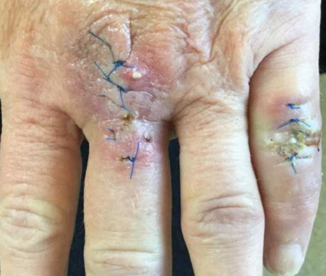
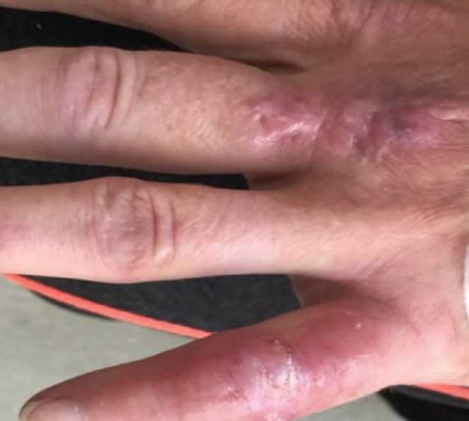
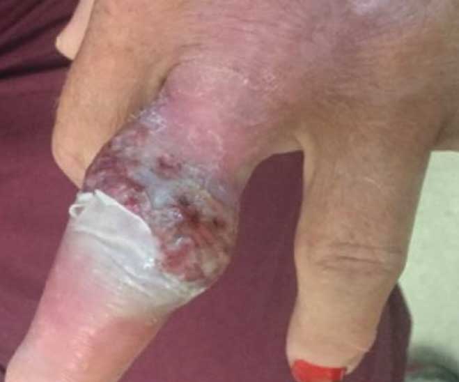
A 56-year-old woman presented to the dermatology clinic for examination of lesions on the dorsum of her left metacarpophalangeal joint and left fifth digit. The patient’s medical history included hypertension, anxiety, depression, cutaneous squamous cell carcinoma, and Crohn’s disease. Biopsies of each lesion were performed, which revealed squamous cell carcinoma KA type (SCC-KA) over the MCP joint and an atypical cystic squamous proliferation over the fifth digit. The patient elected for Mohs surgery for the SCC-KA and elliptical excision for the atypical squamous proliferation. Two weeks after surgery, the wounds were erythematous to violaceous, sore to touch, and swollen (Figure 1). Postoperative infection was suspected, and the patient was started on Bactrim DS and Mupirocin ointment. The sutures were removed one week later after improvement in erythema and pain. After another week, the lesions were healing appropriately with minimal swelling and erythema (Figure 2). The patient was lost to follow-up, but presented a month later with non-healing, purulent ulcerations with undermined, violaceous borders in the previous surgical sites (Figure 3). A diagnosis of PG was made based on the clinical presentation. The patient was started on clobetasol cream twice daily and a 30-day prednisone taper with complete resolution of the lesions.
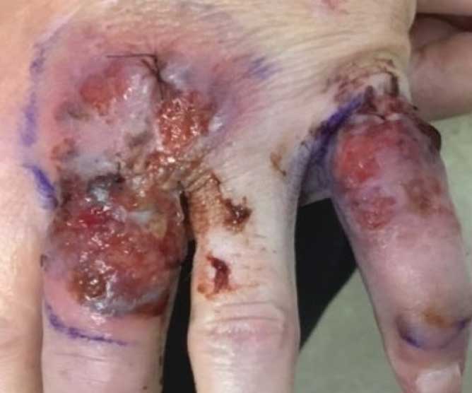
Five months later, the patient elected for Mohs surgery to a keratoacanthoma of the left third digit (located distal to the previous SCC-KA). She again developed a purulent ulcer with undermined, violaceous borders at the surgical site consistent with PG (Figure 4). A 30-day steroid taper, clobetasol cream, and dapsone were prescribed with complete resolution of her ulcerations.
Discussion
The pathogenesis of PG incorporates multiple factors: neutrophilic dysfunction, inflammatory cytokines, and genetic predisposition (e.g. pyogenic arthritis, pyoderma gangrenosum and acne [PAPA] syndrome).2,3 In addition, PG is associated with disorders that either lead to neutrophilic dysfunction or are driven by neutrophilic dysfunction (i.e. inflammatory bowel disease, rheumatoid arthritis).2 Treatment of PG is often aimed at suppressing the immune system with topical or systemic glucocorticoids.
On top of a complex pathogenesis, PG is a difficult postsurgical diagnosis. The diagnosis might be delayed since it is a rare entity and sometimes mimics more prevalent dermatoses, such as cutaneous infections, skin cancer, arthropod bites, vasculitis, and ulcerative inflammatory disorders.3 As our case demonstrates, it is important for clinicians to consider PG in the setting of non-healing postoperative wounds refractory to antibiotics. Recognizing PG is also important to prevent further exacerbation through wound debridement.3,4
An interesting aspect of our case was recurrence of postoperative PG on the left hand. Unfortunately, few studies have looked at the risk of developing postoperative PG or the risk of developing recurrent postoperative PG. A study by Xia et al6 showed 15.1 percent of patients experienced postoperative recurrence or exacerbation of PG, and the risk of postoperative recurrence or exacerbation of PG was significantly associated (P<0.05) with procedure type and having chronic PG. Our patient had no history of PG; however, she did receive procedures associated with a higher risk of recurrent PG: Mohs surgery and skin excision.
Our patient also had a history of Crohn’s disease, which is a well-known risk factor for PG.1–6 While postsurgical PG was not at the top of our differential, this case demonstrates the necessity to always consider it. In 1 to 2 weeks following surgery, both surgical sites demonstrated common complications: pain and delayed wound healing. These findings, along with the rarity of postsurgical PG, led to a delay in diagnosis. It is important for dermatologists and general practitioners to recognize PG, as delaying the diagnosis can lead to additional morbidity.4,5
Conclusion
We presented the case of a 56-year-old woman with a history of Crohn’s disease who experienced subsequent episodes of PG of the left hand following dermatologic surgeries. The diagnosis should be considered during the evaluation of postsurgical patients with a history of inflammatory bowel disease and/or delayed postoperative wound healing. Recognition of this delicate patient population in the dermatology clinic is essential to prevent recurrent episodes and morbidity.
References
- Almukhtar R, Armenta AM, Martin J, et al. Delayed diagnosis of post-surgical pyoderma gangrenosum: A multicenter case series and review of literature. Int J Surg Case Rep. 2018;44:152–156.
- Braswell SF, Kostopoulos TC, & Ortega-Loayza AG. Pathophysiology of pyoderma gangrenosum (PG): an updated review. J Am Acad Dermatol. 2015;73(4):691-698.
- Ahronowitz I, Harp J, & Shinkai K. Etiology and management of pyoderma gangrenosum. Am J Clin Dermatol. 2012;13(3):191–211.
- Tolkachjov SN, Fahy AS, Wetter DA, et al. Postoperative pyoderma gangrenosum (PG): The Mayo Clinic experience of 20 years from 1994 through 2014. J Am Acad Dermatol. 2015;73(4), 615–622.
- Arivarasan, K, Bhardwaj, V, Sud, S, Sachdeva, S, & Puri, AS. Biologics for the treatment of pyoderma gangrenosum in ulcerative colitis. Intest Res. 2016;14(4):365–368.
- Xia FD, Liu K, Lockwood S, et al. Risk of developing pyoderma gangrenosum after procedures in patients with a known history of pyoderma gangrenosum—A retrospective analysis. J Am Acad Dermatol. 2018;78(2):310–314.

