 J Clin Aesthet Dermatol. 2023;16(10):40–42.
by Sara Holt, DO; Lisa Fronek, DO, FAAD; and Kristin Witfill, DO, FAAD
All authors are with the HCA Healthcare and USF Morsani College of Medicine GME Programs at HCA Florida Largo Hospital in Largo, Florida. Additionally, Dr. Fronek is with Bighorn Mohs Surgery and Dermatology Center at Scripps Clinic in La Jolla, California.
FUNDING: No funding was provided for this article.
DISCLOSURES: Dr. Witfill received an honorarium from Galderma as a speaker and instructor for this study. This research was supported (in whole or in part) by HCA Healthcare and/or an HCA Healthcare affiliated entity. The views expressed in this publication represent those of the author(s) and do not necessarily represent the official views of HCA Healthcare or any of its affiliated entities.
ABSTRACT: The chin is recognized as an essential element in overall facial balance and contributes to the perception of youthfulness. Age-related chin retrusion and resorption can alter the shape and projection of the chin, disrupting facial harmony. Hyaluronic acid-based dermal fillers offer a temporary, non-surgical option to correct mild to moderate chin retrusion and resorption. In this case series of three patients, we sought to describe a modified injection technique of the chin with hyaluronic acid-based filler using a tapered lateral to medial approach aimed at correcting mild to moderate chin retrusion and resorption. The aesthetic improvement in appearance, compared to pretreatment, was measured by the investigators using the Global Aesthetic Improvement Scale. Each patient received either a score of 4 or 5 indicating improvement from baseline or achievement of optimal corrective results, respectively. An advantage of using this approach is that starting from a lateral to medial approach with a needle allows the injector to rebuild the chin compartment using precise volumes of filler, requiring minimal total filler volume to achieve optimal corrective results. While this report demonstrates promising results in aesthetic outcomes, further research is required to demonstrate reproducible results and long-term efficacy.
Keywords. Chin, lateral to medial, dermal filler, hyaluronic acid, nonsurgical augmentation
The chin is recognized as an essential element in overall facial balance and contributes to the perception of youthfulness.1 Age-related changes of the shape and projection of the chin can disrupt facial harmony. Reduction in chin size is due to age-related mandibular bone resorption, resulting in loss of mandibular height and length.2 This, along with volume loss and descent of soft tissue, contribute to the disruption of the shape of the chin, the loss of its projection, and exaggeration of the prejowl sulcus.3,4
There are numerous options for chin augmentation including surgical techniques like genioplasty, alloplastic implants, and autologous fat transfer. However, surgical augmentation of the chin has declined in recent years, which is likely due to increasing popularity of minimally invasive treatment options such as dermal fillers.5 Hyaluronic acid (HA)-based dermal fillers offer a temporary, non-surgical alternative to correct mild to moderate chin retrusion and resorption. We sought to describe a modified injection technique of the chin with HA-based filler using a tapered lateral to medial approach. The technique focuses on re-volumizing mild to moderate chin retrusion and resorption by restoring age-related losses from bone and the deep fat compartments.
Methods
Injection technique. This injection technique is indicated for patients who present with mild to moderate chin retrusion and resorption. Each treatment was performed in an outpatient setting, lasting approximately 15 to 30 minutes. Written consent was obtained from each patient.
Prior to injection, the overlying skin was cleansed of makeup, and topical anesthetic cream composed of 7% benzocaine, 7% lidocaine and 7% tetracaine was applied for approximately 30 minutes. A topical antiseptic ethanol solution was then used to sterilize injection areas, followed by chlorhexidine.
Using a sterile marker, the boundaries of the treatment zone were outlined (Figure 1). The superior boundary of the chin compartment was identified by outlining the mentolabial crease, and the lateral boundary was identified by outlining the labiomandibular fold. To partition the medial chin from the lateral chin, a straight line was drawn vertically from the nasal ala to the mandible. Within the lateral chin compartment, the superior and inferior border of the mandible was demarcated. To reduce the risk of intravascular injection, care was taken to mark the submental arteries, located either at or paramedian to the mandibular symphysis.

Injection sites were outlined as shown in Figure 2. Cumulative filler volume ranged from 1.1 to 1.5mL. Each site was injected with HA 20mg/mL with lidocaine 3mg/mL using a 27-gauge, 0.5-inch needle in a supraperiosteal plane using a tapered technique, as follows.

Beginning from lateral to medial, three 0.025 to 0.05mL injections were evenly placed along the superior base of the mandible within the area outlined by the labiomandibular fold laterally and the medial aspect of the chin. This was repeated on the contralateral side.
Next, within the chin compartment, four 0.1 to 0.15mL injections were placed in a paramedian fashion, creating two rows as outlined in Figure 2. The inferior row was located bilaterally at the superior base of the mandible and the superior row was located directly above.
Finally, a 0.2mL injection was placed at the gnathion, followed by a 0.2 to 0.3mL injection at the pogonion.
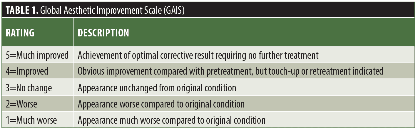
The aesthetic improvement in appearance, compared to pretreatment, was measured by the investigators using the Global Aesthetic Improvement Scale (GAIS). The GAIS is a five-point scale that is rated as follows: 1=much worse, 2=worse, 3=no change, 4=improved, 5=much improved (Table 1).
Case Series
Case 1. A 46-year-old woman presented seeking improvement in chin shape and what she described as chin dimpling. Upon examination, there was mild chin retrusion with blunted anterior projection as well as the appearance of peau d’orange texture changes (Figure 3A, 3B). The patient was determined to be a candidate for the lateral to medial injection technique, which resulted in significant improvement in chin shape and projection post treatment (Figure 3C, 3D). This patient received a score of 5 on the GAIS at her two-week follow-up appointment.

Case 2. A 64-year-old woman presented with complaints of chin regression. Physical examination was notable for moderate chin retrusion and loss of chin contour. After one treatment with the lateral to medial technique, the patient’s anterior chin projection and shape significantly improved, receiving a score of 4 on the GAIS at her two-week follow-up appointment.
Case 3. A 63-year-old woman presented with the chief complaint of a flat chin with dimpling at rest and with movement. Physical exam was notable for moderate chin retrusion with blunting of the chin’s shape and hallowing in the prejowl sulcus. After a single treatment with the lateral to medial technique, anterior chin projection significantly improved, along with a more contoured and evenly textured chin, especially in the lateral compartments. This patient received a GAIS score of 5 at her two-week follow-up appointment.
Discussion
The chin is recognized as an essential element in the aesthetics of the lower third of the face and is commonly targeted to combat age-related changes.1 These age-related changes include reduction in chin size due to mandibular bone resorption resulting in loss of mandibular height and length.2 This chin retrusion, along with volume loss and descent of soft tissue, contribute to the disruption of the shape of the chin, the loss of its projection, and exaggeration of the prejowl sulcus.3,4
A thorough understanding of chin anatomy is crucial when evaluating and treating patients with dermal HA replacement. The shape of the chin is described using anatomical landmarks: the pogonion marks the most anteriorly projected point on the chin, the menton marks the most inferiorly projected point on the chin, and the gnathion lies in between the two.5 The boundaries of the chin compartment include the mentolabial crease superiorly and the labiomandibular folds laterally.5 The prejowl sulcus is a concave space that separates the chin compartment from the jowl.5 The primary blood supply to the chin consists of the mental arteries (terminal branches of the maxillary artery) which exit the mental foramina approximately at the mid-pupillary line, accompanied by the mental nerves.5 The submental artery, a branch of the facial artery, also supplies the chin as it crosses over the mandible near the mandibular symphysis.5
Techniques using either cannulas or needles are well described; however, data on using a tapered lateral to medial approach is lacking. In the present case series, the authors found that filler volume ranged from 1.1 to 1.5mL in total. The advantage of using this approach is that starting from a lateral to medial approach with a needle allows the injector to rebuild the chin using precise volumes of filler, requiring minimal total filler volume to achieve optimal corrective results. Conversely, using a medial to lateral approach, the injector is at risk of injecting excess product laterally. This results in the injector needing to correct this through injection of additional product medially to maintain the chin’s natural shape and projection at the pogonion. With the technique outlined in this study, this game of “catch-up” can be avoided.
Sahan et al6 used a lateral to medial approach; however, they used a cannula instead of a needle. The authors assessed chin augmentation in 50 patients using a single midline entry point with HA filler in a retrograde lateral to medial tunneling approach. Median injected filler volume was 2.25mL. A disadvantage of using a cannula over a needle is that many authors think a cannula does not allow for as much precision that a needle offers.7,8 However, needles are often more painful and have a higher risk of complications like bruising.6 It is the authors’ preference to use a needle because it allows for greater injection accuracy, and with basic understanding of chin anatomy and location of underlying vessels, risk of intravascular injection and/or trauma can be minimized.
Conclusion
Using a lateral to medial approach in a tapered design with supraperiosteal HA filler, the injector is able to re-mold and restore age-related losses in projection and shape while maintaining facial symmetry. In doing so, minimal total filler volume is needed to achieve optimal corrective results. While this report demonstrates promising results in aesthetic outcomes, further research is required to demonstrate reproducible results and long-term efficacy.
References
- Lee EI. Aesthetic alteration of the chin. Semin Plast Surg. 2013;27(3):155–160.
- Pessa JE, Slice DE, Hanz KR, et al. Aging and the shape of the mandible. Plast Reconstr Surg. 2008;121(1):196–200.
- Shaw RB Jr, Katzel EB, Koltz PF, et al. Aging of the mandible and its aesthetic implications. Plast Reconstr Surg. 2010;125(1):332–342.
- Mendelson B, Wong CH. Changes in the facial skeleton with aging: implications and clinical applications in facial rejuvenation. Aesthetic Plast Surg. 2012;36(4):753–760.
- Vanaman Wilson MJ, Jones IT, Butterwick K, et al. Role of Nonsurgical Chin Augmentation in Full Face Rejuvenation: A Review and Our Experience. Dermatol Surg. 2018;44(7): 985–993.
- Sahan A, Karaosmanoglu N, Ozdemir Cetinkaya P. Chin augmentation with the use of cannula from a single, midline entry point: Evaluation of 50 patients. J Cosmet Dermatol. 2020;19(6):1301–1306.
- Moradi A, Shirazi A, David R. Nonsurgical chin and jawline augmentation using calcium hydroxylapatite and hyaluronic acid fillers. Facial Plast Surg. 2019;35(2):140–148.
- Kontis TC, Lacombe VG, eds. Cosmetic injection techniques: a text and video guide to neurotoxins and fillers. New York, NY: Thieme; 2019:164–166.
OX40/OX40L Costimulatory Pathway: A Potential Therapeutic Target for Allergic Contact Dermatitis?