J Clin Aesthet Dermatol. 2020;13(1):38–40
 by Penelope Hirt, MD; Suchismita Paul, MD; Weena Phuthongkam, MD; and Lawrence Schachner, MD
All authors are with the Department of Dermatology and Cutaneous Surgery at the University of Miami, Miller School of Medicine in Miami, Florida. Dr. Phuthongkam is also with the Department of Pediatrics at the Faculty of Medicine Vajira Hospital at Navamindradhiraj University in Bangkok, Thailand.
FUNDING: No funding was provided for this study.
DISCLOSURES: The authors have no conflicts of interest relevant to the content of this article.
ABSTRACT: Oral mucosal involvement in patients with morphea is quite rare. Unilateral nevoid telangiectasia is a rare vascular dermatosis characterized by unilateral telangiectasia distributed in a dermatomal pattern, especially on the upper trunk and extremities. We describe a case of a 10-year-old female patient that presents with morphea on her left chin and upper lip extending to the inner oral mucosa as well as a unilateral nevoid telangiectasia on her left cheek as an early presentation of localized scleroderma. This case is significant as it demonstrates a striking clinical presentation of morphea and an interesting early presentation of morphea as a unilateral nevoid telangiectasia that improved after treatment with oral methotrexate and prednisone.
KEYWORDS: Morphea, localized scleroderma, morphea with oral involvement, nevoid telangiectasia, nevus, telangiectasia
Morphea is a localized inflammatory skin disease that presents with sclerosis of the skin and underlying tissues due to excessive collagen deposition.1 Morphea is the preferred designation for localized scleroderma (LSc) and has been differentiated from systemic sclerosis (SSc) based on the absence of internal organ involvement with almost exclusively cutaneous findings, though there might be some underlying involvement of muscle and bone.1 Although scleroderma is a spectrum of disorders that can occur at any age, the clinical manifestations in the pediatric population differ from that in adults.1 Morphea is the most frequent form of scleroderma in childhood.2 A recent epidemiological study in the United Kingdom reported an incidence rate of 3.4 cases per million children per year and the majority presented with the linear subtype of morphea.2 Morphea represents one percent of all office visits in our pediatric dermatology clinic.3 We present a case of a 10-year-old female patient that presents with morphea on her left chin and upper lip extending to the inner oral mucosa as well as a unilateral nevoid telangiectasia on her left cheek as an early presentation of localized scleroderma.
Photoconsent was obtained for use of photos in this article.
Case presentation
A 10-year-old female patient presented to our pediatric dermatology clinic with a one-year history of hypopigmented patches on her upper lip and left chin (Figure 1) as well as telangiectasia on her left cheek (Figure 2). She was previously diagnosed with vitiligo and treated with low potency topical corticosteroids for one year with no improvement. The patient also had small telangiectatic patches on her left cheek for almost two years, suggestive of unilateral nevoid telangienctasia (UNT) (Figure 2A). Clinical examination showed well-demarcated, atrophic, hypopigmented patches with mild surrounding erythema on the left chin and the upper lip (Figure 1) extending to the inner mucosa, including the gingivae with underlying tooth shifted superiorly (Figure 2E). There was evidence of malocclusion, but no tongue atrophy or facial asymmetry were present. A diagnosis of linear morphea with oral mucosa involvement was made. On both lower extremities, reticulated patches were observed and no leg length discrepancy was appreciated, consistent with largely resolved cutis marmorata telangiectatica congenita. There was no family history of autoimmune diseases, thyroid diseases, or Type 1 diabetes mellitus. Laboratory studies revealed normal complete blood count and estrogen levels, slight hypertriglyceridemia, and low vitamin D levels.
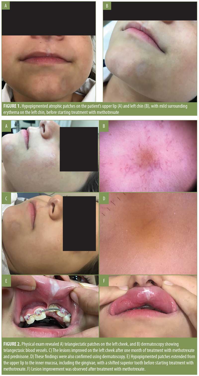
The patient was started on calcipotriene 0.005% cream, desonide 0.05% cream, and triamcinolone 0.1% paste for the oral mucosa and ointment for the face, as well as vitamin D 2000 units daily. After six weeks, the topical therapies yielded no improvement, and the patient was started on methotrexate 10mg once a week, with folic acid daily, and prednisone, starting with 30mg daily and tapering down over the next five months. The topical treatments were not discontinued. Both the UNT on the left cheek and skin lesions on the upper lip and left chin began to improve after treatment with methotrexate and prednisone.
Discussion
Morphea has been associated with various complications due to skin, muscle, and bone atrophy, such as growth defects and deformities that are mostly seen in patients with linear and deep morphea.1 Linear morphea can extend over joint lines and cause flexion contractures, limb growth retardation, muscle atrophy, and extracutaneous complications—especially articular and neurologic.2 Linear morphea involving the frontoparietal area—called en coup de sabre—can extend down to the cheek, nose, and upper lip (Romberg-Parry), including the mouth and gingival tissues.4 Based on our literature review, few cases of morphea with oral mucosal involvement have been reported.
A cross-sectional, multicenter study performed in Denmark showed a higher incidence of odontostomatologic abnormalities in patients with linear morphea of the face.5 In that study, 16 patients were investigated (9 female, 7 male) aged between 6.5 and 21.9 years. All patients reported at least one of the following odontostomatologic complications: malocclusion (94%), overgrowth tendency of the anterior lower third of the face (82%), gnatologic alterations (69%), dental anomalies (63%), skeletal asymmetry (56%), bone involvement (50%), and temporomandibular joint involvement (19%).5
Another case reported a mobile upper right central incisor and marked gingival recession in a 36-year-old woman with en coup de sabre.4 In 2015, Van der Veken et al6 described a 19-year-old woman with progressive recession of Teeth 11 and 12, who presented with en coup de sabre involving the nose and upper lip on the right side of her face. A six-year-old Turkish girl with left-sided en coup de sabre had malformed left maxillary incisors with short roots and lack of eruption.7 In 2016, Dixit et al8 reported the case of a 20-year-old man with morphea on the left side of his face and chin, who also had tongue atrophy, relative microdontia, thinning of the ramus/body of the mandible, and sclerotic lesions on trunk. The patient was diagnosed with mixed morphea with Parry-Romberg syndrome. A second case presented by Dixit and colleagues was that of a 53-year-old woman who was diagnosed with systemic sclerosis but also had oral manifestations characterized by edentulous arches, xerostomia, and candidiasis. Oral mucosal involvement was also described in a 13-year-old girl with progressive recession on the attached gingiva of her upper left incisors and morphea on the left upper lip, vermilion, and lip mucosa.9
There are a variety of treatment options for morphea, such as moderate-to-high potency topical corticosteroid, intralesional corticosteroid, topical calcipotriol, psoralen and ultraviolet A (PUVA), and topical tacrolimus.10 Systemic therapy is usually required in severe cases or when there is musculoskeletal involvement.10 A randomized, controlled clinical trial used oral methotrexate (15mg/m2/week) and prednisone (1mg/kg/day, maximum 50mg) for the first three months with promising results.10 Our patient was initially treated with calcipotriene 0.005% cream for the lesion on the chin, triamcinolone 0.1% paste for the oral mucosa and ointment for the face, and desonide 0.05% cream, without great benefit; oral methotrexate and prednisone were then used with good results.
UNT is a vascular dermatosis characterized by unilateral telangiectasias distributed in a dermatomal pattern, especially on the upper trunk and extremities.11,12 There are two types: congenital and acquired.13 The congenital type is transmitted in an autosomal dominant pattern and is believed to be influenced by maternal estrogens.13 The acquired type occurs in association with hyperestrogenism, such as puberty, pregnancy, oral contraceptives, and liver diseases.13 Our patient had normal estrogen levels and the UNT improved after methotrexate and prednisone treatment. To our knowledge, only a few cases have been reported that showed “telangiectasia-like” lesions as an early presentation of morphea.14,15 In 2006, Zulian et al14 reported one case of congenital localized scleroderma that started as a salmon patch, and in 2011, Nijhawan et al15 reported four patients with morphea whose lesions started as acquired Port-wine stains. We believe acquired UNT and Port-wine stains are precursor lesions to morphea.
Conclusion
The presented case is unique in that it demonstrates a striking clinical presentation of morphea with oral mucosa involvement and also highlights UNT as an early presentation of morphea.
References
- Zulian F, Cuffaro G, Sperotto F. Scleroderma in children: an update. Curr Opin Rheumatol. 2013;25(5):643–650.
- Herrick AL, Ennis H, Bhushan M, et al. Incidence of childhood linear scleroderma and systemic sclerosis in the UK and Ireland. Arthritis Care Res. 2010;62(2):213–218.
- Schachner L, Ling NS, Press S. A statistical analysis of a pediatric dermatology clinic. Pediatr Dermatol. 1983;1(2):157–164.
- Pace C, Ward SE, Pace A. A rare case of frontal linear scleroderma (en coup de sabre) with intra-oral and dental involvement. Br Dent J. 2010;208(6):249–250.
- Trainito S, Favero L, Martini G, et al. Odontostomatologic involvement in juvenile localised scleroderma of the face. J Paediatr Child Health. 2012;48(7): 572–576.
- Van der Veken D, De Haes P, Hauben E, et al A rare cause of gingival recession: morphea with intra-oral involvement. Oral Surg Oral Med Oral Pathol Oral Radiol. 2015;119(5):e257–264.
- Hørberg M, Lauesen SR, Daugaard-Jensen J, Kjær I. Linear scleroderma en coup de sabre including abnormal dental development. Eur Arch Paediatr Dent. 2015;16(2):227–231.
- Dixit S, Kalkur C, Sattur AP, et al. Scleroderma and dentistry: Two case reports. J Med Case Rep. 2016;10(1):297.
- Niklander S, Marín C, Martínez R, Esguep A. Morphea “en coup de sabre”: an unusual oral presentation. J Clin Exp Dent. 2017;9(2):
e315–e318. - Zulian F, Vallongo C, Patrizi A, et al. A long-term follow-up study of methotrexate in juvenile localized scleroderma (morphea). J Am Acad Dermatol. 2012;67(6):1151-1156.
- Afsar FS, Ortac R, Diniz G. Unilateral nevoid telangiectasia with no estrogen and progesterone receptors in a pediatric patient. Indian J Dermatol Venereol Leprol. 2008;74(2):163–164.
- Oliveira A, Velho G, Sanches M, Selores M. Unilateral nevoid telangiectasia–report of two cases. Int J Dermatol. 2014;53(1):e32–33.
- Wilkin JK, Smith JGJ, Cullison DA, et al. Unilateral dermatomal superficial telangiectasia. Nine new cases and a review of unilateral dermatomal superficial telangiectasia. J Am Acad Dermatol. 1983;8(4):468–477.
- Zulian F, Vallongo C, de Oliveira SK, et al. Congenital localized scleroderma. J Pediatr. 2006;149(2):248–251.
- Nijhawan RI, Bard S, Blyumin M, et al. Early localized morphea mimicking an acquired port-wine stain. J Am Acad Dermatol. 2011;64(4):
779–782.
OX40/OX40L Costimulatory Pathway: A Potential Therapeutic Target for Allergic Contact Dermatitis?