J Clin Aesthet Dermatol. 2021;14(2):44–45.
by Cory Pettit, MD, and Joy Mosser-Goldfarb, MD
Drs. Pettit and Mosser-Goldfarb are with the Division of Dermatology, Department of Internal Medicine, The Ohio State University Wexner Medical Center in Columbus, Ohio.
FUNDING: No funding was provided for this article.
DISCLOSURES: The authors report no conflicts of interest relevant to the content of this article.
ABSTRACT: Morphea is a localized form of scleroderma that presents with dermal thickening and fibrotic plaques in the absence of internal organ involvement. Like many autoimmune conditions, these plaques have many different phases, starting out as active, red plaques before later burning out, leaving white, fibrotic plaques behind. Many drugs have been shown to induce morphea, including bleomycin and bromocriptine. We present a case of minocycline-induced reactivation of previously burned out morphea plaques. Minocycline is an important drug in dermatology and the reporting of new adverse events is important so as to help clinicians better weigh the risks and benefits of the drug for specific populations.
KEYWORDS: Fibrotic plaques, scleroderma, minocycline, morphea, autoimmune
Morphea is a specific type of scleroderma that presents with dermal thickening and fibrotic plaques in the absence of internal organ involvement.1 The lesions evolve through different phases, beginning with an inflammatory phase, which consists of erythematous, fibrotic plaques. Soon after, the center becomes white, while the edges remain red and active.2 As the active phase subsides and the lesion enters a latent phase, the edges of the plaque lose their red hue and the indurated lesion becomes uniformly white or hyperpigmented. Lesions entering this phase are said to have “burnt out.” The pathogenesis of morphea is multifactorial; however, drug-induced cases have been reported.3 We present a case of morphea reactivation due to minocycline in a patient with previously latent lesions.
Case Presentation
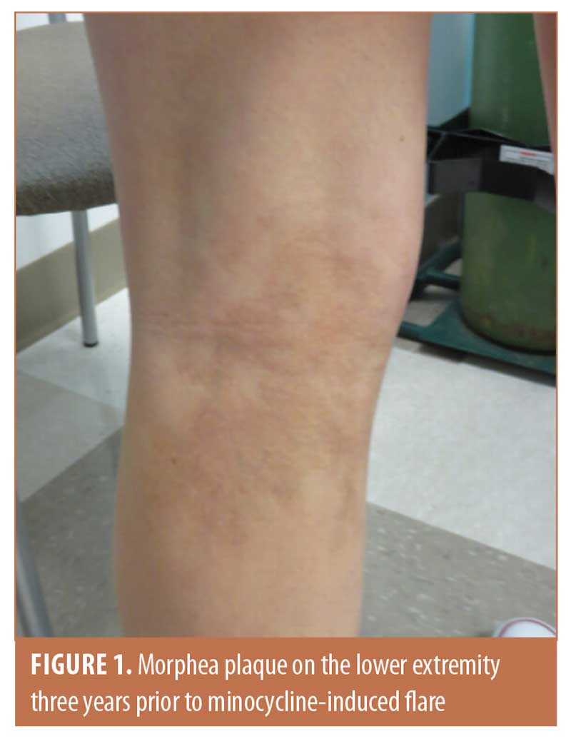
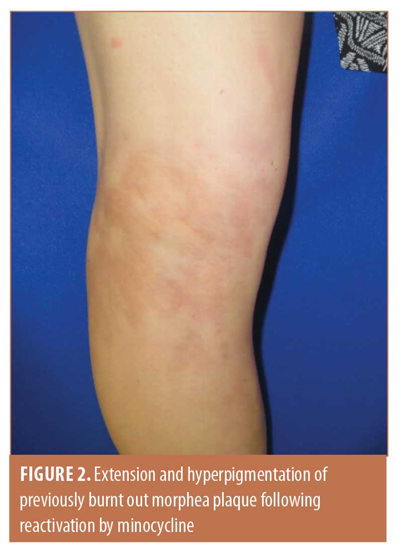
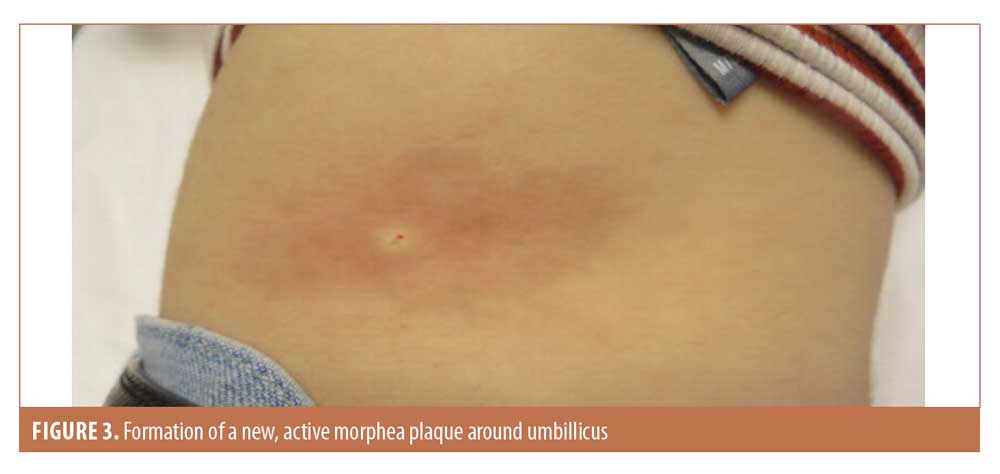
Our patient was a 16-year-old female with a history of linear morphea on the posterior left leg (Figure 1). Her disease had been stable for years and appeared to be burned out. Minocycline was initiated for inflammatory acne refractory to Epiduo. Two weeks following minocycline initiation, the morphea lesions of her lower extremity reactivated and began growing and became more hyperpigmented (Figure 2). In addition, she developed new active morphea plaques under her umbilicus and on the left side of her chest under the rib cage (Figure 3). She did not report any systemic symptoms. Punch biopsy was performed and showed some perivascular lymphoplasmacytic cells and unremarkable dermal collagen. The patient’s antinuclear antibody test was positive, with a 1:640 titer and homogenous/speckled pattern. Additionally, her anti-histone antibody test was negative, erythrocyte sedimentation rate was elevated to 18, anti-Sjögren’s syndrome A and anti-Sjögren’s syndrome B tests were negative, and complete blood count was normal.


Minocycline was discontinued and mometasone 0.1% cream and tacrolimus 0.1% ointment were prescribed. Following discontinuation of the minocycline, the patient’s lesions again became stable over the course of a few months and no additional new lesions were reported.
Discussion
There are many drugs that have been reported to induce morphea, including bleomycin, balicatib, D-penicillamine, bromocriptine, and even vitamin K injections.1,3–5 While there are no reported cases of minocycline-induced morphea in the literature, minocycline has been implicated as a cause of other types of autoimmune diseases, including lupus, autoimmune hepatitis, serum sickness-like reactions, and vasculitis.6 The exact mechanism of this phenomenon has not been identified but could be related to minocycline’s action as an immunomodulator. Minocycline has various effects on the immune and inflammatory pathways and, in general, is thought of as anti-inflammatory. It does, however, increase the expression of various cytokines, including interleukin-6, a cytokine thought to be involved in the pathogenesis of scleroderma.6,7
Besides acting as an antibiotic and immunomodulator, minocycline has many other effects on the body, including inhibiting metalloproteinases (MMPs).6 MMPs form a crucial part of connective tissue homeostasis, breaking down excess extracellular collagen.8 Recent studies have suggested that a reduction in metalloproteinase activity—and, therefore, a reduction in the breakdown of extracellular collagen—may play an important role in the pathogenesis of scleroderma.8,9 In addition, another important step in the pathogenesis of scleroderma and morphea is vasospasm and the decrease of angiogenesis, leading to ischemic injury and free-radical generation, which damages cellular proteins and provides epitopes for the autoantibodies present in the disease.10 Recent studies have also demonstrated that tetracyclines, including minocycline, may decrease angiogenesis,11 making this and MMP inhibition other interesting possible mechanisms by which minocycline could induce or exacerbate morphea.
Conclusion
Minocycline is an important drug in dermatology, often forming an integral part of inflammatory acne-treatment regimens. As such, thorough monitoring and reporting of new types of side effects is paramount to avoid initiating therapy with the drug in patients who are most at risk for serious side effects. This case is the first to add morphea to the ever-growing list of autoimmune conditions that may be caused or exacerbated by minocycline therapy.
References
- Browning JC. Pediatric morphea. Dermatol Clin. 2013;31(2):229–237.
- Fett N , Werth VP. Update on morphea: part I. Epidemiology, clinical presentation, and pathogenesis. J Am Acad Dermatol. 2011;64:217–228; quiz 29–30.
- Peroni A, Zini A, Braga V, et al. Drug-induced morphea: report of a case induced by balicatib and review of the literature. J Am Acad Dermatol. 2008;59(1):125–129.
- Liddle BJ. Development of morphoea in rheumatoid arthritis treated with penicillamine. Ann Rheum Dis. 1989;48(11):963–964.
- Guidetti MS, Vincenzi C, Papi M , Tosti A. Sclerodermatous skin reaction after vitamin K1 injections. Contact Dermatitis. 1994;31(1):45–46.
- Hess EV. Minocycline and autoimmunity. Clin Exp Rheumatol. 1998;16(5):519–521.
- Desallais L, Avouac J, Frechet M, et al. Targeting IL-6 by both passive or active immunization strategies prevents bleomycin-induced skin fibrosis. Arthritis Res Ther. 2014;16(4):R157.
- Mattila L, Airola K, Ahonen M, et al. Activation of tissue inhibitor of metalloproteinases-3 (TIMP-3) mRNA expression in scleroderma skin fibroblasts. J Invest Dermatol. 1998;110(4):416–421.
- Tomimura S, Ogawa F, Iwata Y, et al. Autoantibodies against matrix metalloproteinase-1 in patients with localized scleroderma. J Dermatol Sci. 2008;52(1):47–54.
- Chen K, See A , Shumack S. Epidemiology and pathogenesis of scleroderma. Australas J Dermatol. 2003;44(1):1–7; quiz 8–9.
- Sapadin AN , Fleischmajer R. Tetracyclines: nonantibiotic properties and their clinical implications. J Am Acad Dermatol. 2006;54(2):258–265.

