 by Diane Irvine Duncan, MD, FACS
Dr. Duncan is with Plastic Surgical Associates in Fort Collins, Colorado.
J Clin Aesthet Dermatol. 2022;15(8):28–32.
FUNDING: No funding was provided for this article.
DISCLOSURES: The author reports no conflicts of interest relevant to the content of this article.
ABSTRACT: Background. High-intensity focused electromagnetic field (HIFEM) and radiofrequency (RF) are established stand-alone techniques used in body contouring. However, data on the simultaneous combination and effects of these modalities for inner thigh treatment is lacking.
Objective. To investigate the efficacy and safety of the HIFEM+RF procedure for non-invasive fat reduction in the inner thigh, as well as toning and strengthening of the inner thigh muscles.
Methods. Sixteen women with an average age of 47.31±12.51 years were recruited. Each patient received four 30-minute bilateral treatments on the inner thighs once a week. Magnetic resonance imaging (MRI) scans of the treated area were evaluated at baseline, one-, and three-month follow-up visits for subcutaneous fat and muscle thickness changes. Anthropometric data and digital photographs were collected. Subject satisfaction and therapy comfort were evaluated.
Results. The results peaked at three months, showing significant changes in both treated tissues. On average, the fat thickness was reduced by 27.4 percent (p<0.001), while muscle tissue showed an average increase of 23.2 percent. Thigh circumference was reduced on average by 1.52cm, with a maximum change of –1.8cm, observed at 5cm below the gluteal crease. The treatment was considered comfortable with high patient satisfaction.
Conclusion. Analysis of magnetic resonance images and thigh circumference showed that therapy combining HIFEM and RF is highly effective for subcutaneous fat reduction and increased muscle thickness in inner thighs.
The inner thigh in the female body is one area that, for various reasons (mainly genetic predisposition), frequently presents fat deposits that are most common in women.1–3 Besides cumbersome lifestyle changes (e.g., diet and exercise), invasive fat reduction methods are undesirable due to the need for anesthesia, substantial downtime, and the possibility of surgery-related risks. Modern-day beauty standards and aesthetic appearance have propelled interest in new and safer techniques for non-invasive body contouring. As the public’s demand for greater efficacy and safety has grown steadily over the past decades, so too has the search for and development of new measures for fat reduction. The main fat-reduction devices approved for use in aesthetic medicine employ cryolipolysis, radiofrequency (RF), high-intensity focused ultrasound (HIFU), lasers, or the recently introduced high-intensity focused electromagnetic field (HIFEM).4–8
Simultaneous application of HIFEM and synchronized RF might be one possibility to treat both fat and muscles in the same session. The HIFEM generates an electromagnetic field that activates neuromuscular tissue by induced electric current, leading to supramaximal muscle contractions, creating a significant energy demand that forces muscles to utilize the stored energy in adipocytes in the form of free fatty acids. Consequently, this reduces size and, under the extreme muscle load, the number of adipocytes.6 The synchronized RF generates electromagnetic energy, which can be adjusted so that the energy predominantly accumulates in the fat tissue, leading to selective heating of adipocytes within a temperature range of 42°C to 45°C, inducing apoptosis.9,10
Recent studies investigating the effect of the novel combination of HIFEM and RF technologies have shown that such therapy is effective for non-invasive fat reduction and muscle toning of the abdomen.11
Since the two fields (HIFEM and RF) do not interfere with each other due to the unique technical solution, the technology remains entirely safe.12 This procedure, therefore, may present a new shift in the field of aesthetic medicine, showing considerable potential to provide greater satisfaction regarding the outcome of aesthetic appearance. However, the efficacy of synchronized RF and HIFEM needs to be ascertained in other body areas. This study aims to investigate the efficacy and safety of the HIFEM+RF procedure for non-invasive fat reduction in the inner thigh and toning and strengthening of the inner thigh muscles.
Methods
Study population and design. Sixteen women interested in non-invasive treatment for aesthetic improvement of inner thigh fat and muscle participated in this prospective, open-label, single-arm, nonrandomized study based at one site. Selection criteria included women with a body mass index (BMI) less than or equal to 35kg/m2. Exclusion criteria were pregnancy, postpartum period, breastfeeding, injury in the treatment area, or any other medical conditions that contraindicated the application of electromagnetic fields and RF, such as cardiovascular disease, malignant tumor, metal, or electronic implants. After receiving detailed instructions about the study and signing informed consent, qualifying subjects were recruited as participants in the study. The study design and treatment protocol were approved by the Advarra Institutional Review Board and followed the ethical guidelines of the 1975 Declaration of Helsinki.13
The group had an average age of 47.31±12.51 years (24–69 years) and a BMI averaging 27.38±4.79kg/m2 (21.30–35.00kg/m2) before treatment. The group comprised all Fitzpatrick Skin phototypes I to VI; one subject had Type I, five subjects had Type II, two subjects had Type III, four subjects had Type IV, one subject had Type V, and three subjects had Type VI. Each subject received four 30-minute bilateral treatments on the inner thighs with the Emsculpt Neo (BTL Industries Inc.; Boston, Massachusetts) device simultaneously delivering HIFEM and RF energies through a single applicator. During the treatment, patients were comfortably seated with their legs apart. The small applicators, designed to treat more localized body areas, were positioned on the inner thighs and remained fixed in the same position throughout the treatment session. The power of the magnetic field (0–100%) was adjusted according to the subject’s tolerability. The intensity of the RF energy was set to 100 percent (i.e., maximum power) from the start. Patients were regularly asked about the therapy comfort during the whole treatment time, and the energy settings were adjusted accordingly. The treatments were administered once a week, and no anesthesia was required. All of the patients were instructed to maintain their daily routine and not to change their lifestyles.
Data collection and evaluation. Digital photographs, thigh circumference, weight, BMI measurement, and magnetic resonance imaging (MRI) of the treatment area were done at baseline, one-, and three-month follow-up visits.
MRI was the primary evaluation method for assessing the treatment effects. The medical-grade system with an intensity of 1.5T (Siemens Magnetom Aera, Siemens Healthineers AG) was used to scan the lower body area from the iliac crest to the upper half of the femur (T2 weighted, slice thickness of 5mm, spacing 7mm, 400×512 matrix). Patients were scanned in the supine position, while keeping their thighs apart. Hip adductors located at the measured level beneath the treated fat tissue (namely m. gracilis, m. adductor longus, and m. adductor magnus) were assessed. The latero-medial diameter of m. gracilis and anterior-posterior diameter of m. adductor magnus and m. adductor longus were measured.
Furthermore, thigh circumference was measured on both limbs with a stretch-resistant tape at the level of the pelvic crease and 5cm and 10cm below this level. The average inner thigh circumference was calculated per visit, and posttreatment values were compared to baseline.
The secondary aim was to determine patient satisfaction with the aesthetic outcome and comfort with HIFEM+RF treatment. After the last treatment and at each follow-up visit, the patient’s satisfaction with the treatment results was assessed with a five-point Likert scale Subject Satisfaction Questionnaire. The comfortability of treatment was evaluated by a five-point Likert scale Therapy Comfort Questionnaire and a 10-point visual analog scale (VAS) assessing the pain level after each treatment session. Throughout the study, the occurrence of potential adverse events and side effects were closely monitored. All data were statistically analyzed using repeated-measures analysis of variance (ANOVA) with Tukey post-hoc test and paired and two-sample t-tests, with significance level α=0.05.
Results
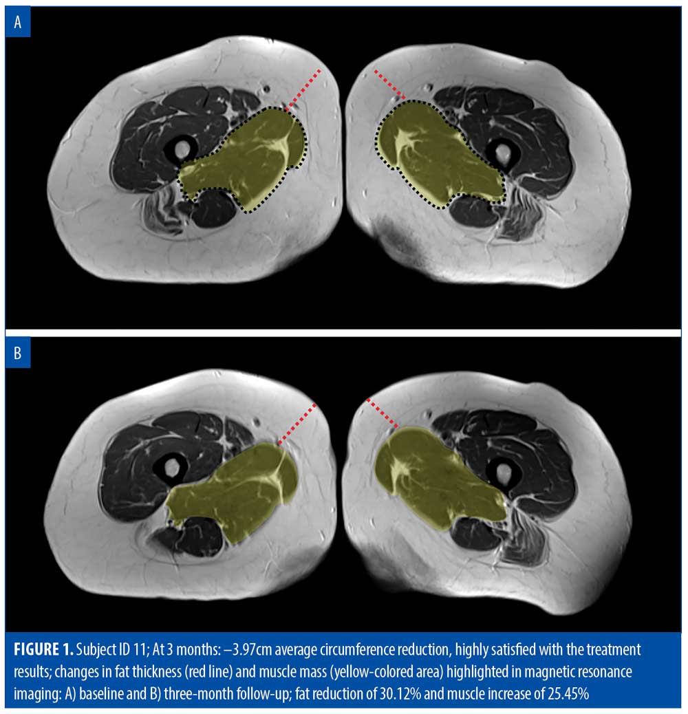
MRI evaluation. Fat. Evaluation of scans at one month showed an average fat reduction of 22.59 percent (–8.39±1.50mm) on the inner thigh (p<0.001). The results were fairly consistent, with only one patient showing a reduction lower than 20 percent, thirteen patients showing a reduction higher than 20 percent, and fifteen patients showing a reduction higher than 25 percent. All patients had responded to treatment at this stage.
At three months, the results further improved significantly (n=16, p<0.001) in comparison to baseline, as the patients lost, on average, 10.12±1.66mm of fat, corresponding to 27.38 percent of fat, since the baseline visit. All 16 patients had surpassed 20 percent fat reduction. Thirteen out of the 16 patients showed a reduction higher than 25 percent, of which three patients had fat reduction over 30 percent. Table 1 shows a detailed summary of MRI measurements. An example of a patient MRI scan can be seen in Figure 1.


Muscle tissue. The overall muscle thickness was increased, on average, by 21.1 percent after one month. Similar to the fat tissue, the results in the muscles continued to improve at the three-month follow-up. The average increase in muscle thickness was 23.2 percent at three-month follow-up. Each muscle that was measured showed a similar increase in thickness. Analyzing the results revealed statistically significant differences (p<0.001) in each muscle thickness, comparing baseline to three-month follow-up. A detailed summary of MRI measurements is shown in Table 1.
Satisfaction and comfort. The subject satisfaction questionnaire analysis revealed a very high patient satisfaction of 94 percent (15 out of 16) with the treatment results at three-months posttreatment. Furthermore, the patients found the treatments comfortable, with a score of 1.5 on a 10-point VAS (0=no discomfort, 10=unbearable pain). By the end of the first treatment, all patients reached 100 percent intensity of the RF and HIFEM fields. There were no severe adverse events associated with the therapy.
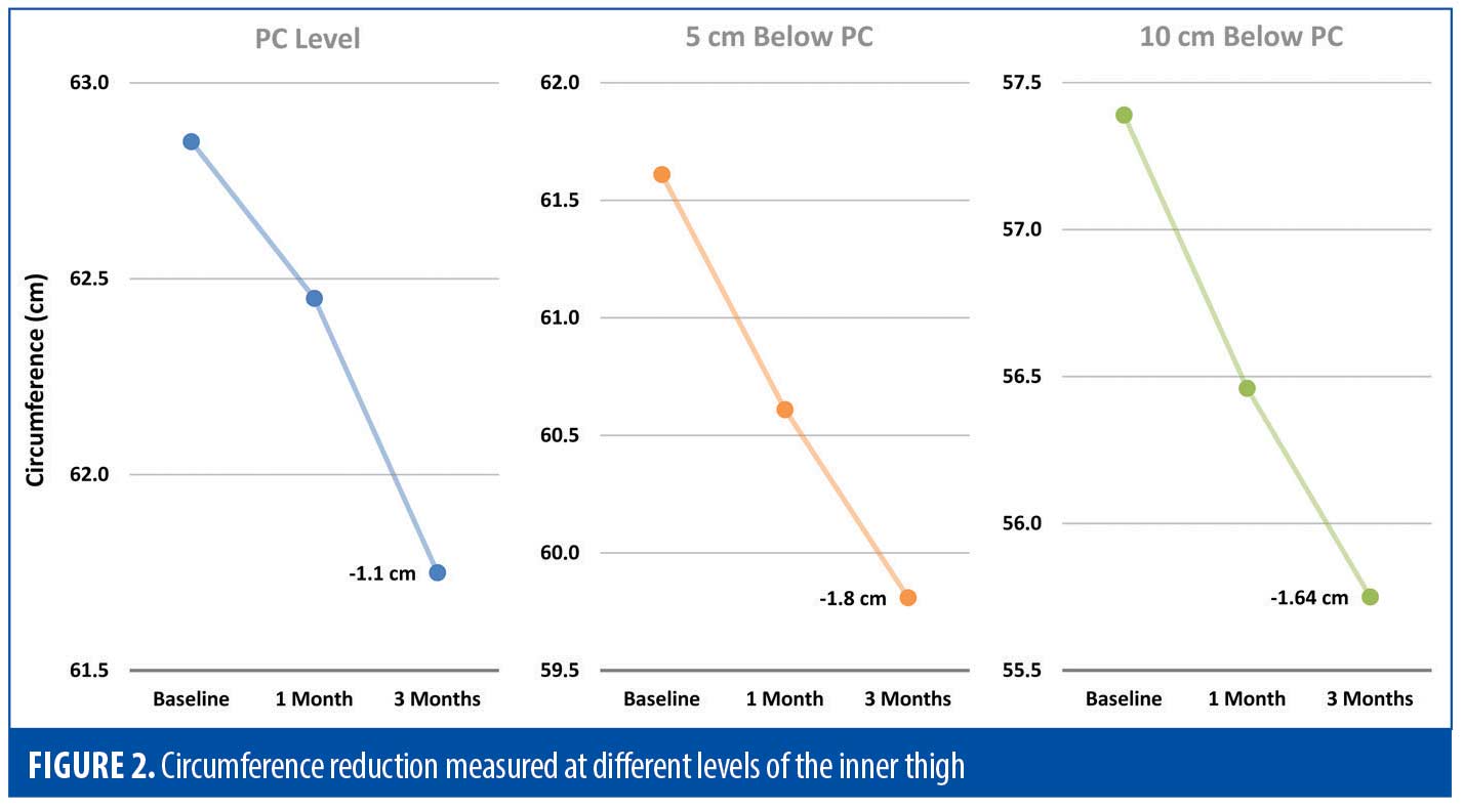
Thigh circumference and weight. The thigh circumference was reduced by 0.78±0.32cm and 1.52±0.36cm at one-month and three-month follow-up visits, respectively. The greatest magnitude of the difference in circumference (–1.80 cm at 3 months) was 5cm below the pelvic crease (Figure 2). Changes between baseline and three-month follow-up were statistically significant (p<0.01).

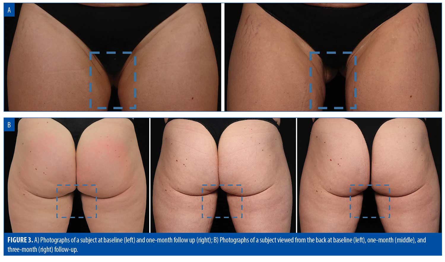
Finally, there was no significant fluctuation in the BMI or weight between any of the time points. Digital photographs showed notable improvement, as seen in the example of patient outcomes (Figures 3a and 3b).

Discussion
The treatments with a novel device that simultaneously delivers HIFEM and synchronized RF energies resulted in a statistically significant reduction in fat layer thickness and thigh circumference and an increase in inner thigh muscle mass/thickness. These three measured parameters showed continuous improvement up to three months, at which point there was a peak in the observed results.
The main benefits of treatment using combined HIFEM+RF are fat reduction in the inner thigh (achieved through subcutaneous fat lipolysis/apoptosis) and improved muscle tone, due to increased muscle thickness.14 Compared to other modalities employing non-invasive fat reduction techniques in the inner thigh, the HIFEM+RF device used in this study produced noticeably more significant outcomes. The mean fat reduction in a study by Boey et al15 was 20 percent (3.3mm), and in another trial by Zelickson et al,16 it was just 2.8mm. In contrast, our study device resulted in an overall fat reduction of 8 to 10mm, representing 22.6 to 27.4 percent relative improvement. Statistical analysis verified the significance of achieved results.
The muscles in this area, which are necessary to stabilize one’s lower back, core, hips, and knees, are also difficult to train, requiring intense effort to strengthen when exercising.17–20 Hence, the study targeted postpubescent women. Predecessors of the HIFEM+RF device used in this study showed muscle strengthening in other parts of the body in previously published literature. In the abdomen, investigations revealed a muscle mass increase of 20.56 percent based on histological analysis.21 An assessment of this same treatment by various imaging techniques (i.e., computer tomography, MRI, and ultrasound) revealed an average muscle thickness increase peaking at three months posttreatment in the range of 14.8 to 21.3 percent, depending on the treated patient group and body part.22,23 Documented results suggest even higher efficacy of the novel HIFEM+RF device when used for toning and shaping the inner thigh region, showing statistically significant differences (p<0.05) in muscle thickness at three-month follow-up. This coincides with the peak period of muscle growth seen in preceding studies. Hence, it can be inferred that treatment with the HIFEM+RF device resulted in increased muscle thickness, which was most prominent three months after the final treatment.
The study also reported a decrease in inner thigh circumference. The group’s total average decrease in thigh circumference was 0.78cm after one month and 1.52cm after three months (p<0.05). This can be attributed to the simultaneous use of HIFEM+RF energies, targeting both the fat and muscle tissues in the same therapy, thus leading to desirable effects of fat reduction and muscle toning. The studied device showed noticeable results in a shorter time frame, out-performing findings from previous studies, especially for inner thigh fat treatment. In a study conducted by Zelickson et al,16 the thigh showed a circumferential decrease of 0.90cm after 16 weeks. Similarly, Sadick et al24 found a 0.5cm decrease in the upper thigh after eight weeks. In this study, at each follow-up, there was a notable reduction of the thigh circumference, implying that the placement of the applicator over the inner thighs during the treatment was suitable for the fat tissue reduction and produced a desirable outcome when considering the shape and appearance of the inner thigh.
The majority of patients were satisfied with the fat reduction and aesthetic results of the treatment after three months. The 94 percent satisfaction rating is slightly higher than the patient satisfaction outcomes in recent HIFEM+RF studies. Moreover, most group members found the experience very comfortable, emphasizing the procedure’s safety, since no serious adverse events were observed. Most non-invasive fat reduction treatments are associated with a range of much more undesirable, long-lasting side effects, such as numbness and tenderness in the treatment area, taking as long as 8 to 18 weeks to resolve in certain cases.15,16 This clearly shows the benefit of the HIFEM+RF device over other methods intended for inner thigh fat reduction.
The study population was adequate for administering the treatment in the inner thigh area since female patients experience cumbersome fat deposits in this body part.1 Even though the peak of muscle mass increase and fat reduction prominence was noted at three months after the final treatment, as in the recent HIFEM studies+RF, it is advisable that follow-up beyond the three months might be scheduled to monitor the longevity and sustainability of the treatment effect.11 Nonetheless, this study employed several methods of analysis and evaluation outcomes to assure the validity of the results. The most marked limitation faced was recruiting more patients. Due to the study taking place during the COVID-19 pandemic, many patients were wary of health concerns, restrictions on movement, and social distancing measures.
Conclusion
Treatment with simultaneous application of HIFEM and synchronized RF is effective and safe for non-invasive fat reduction and muscle enhancement in the inner thigh. The changes induced at the level of muscle and fat tissue peaked at three months and led to the overall improvement in the aesthetic appearance. Results imply that this application is more effective than using these energies alone.
References
- Rask-Andersen M, Karlsson T, Ek WE, et al. Genome-wide association study of body fat distribution identifies adiposity loci and sex-specific genetic effects. Nat Commun. 2019;10(1):339.
- Karastergiou K, Smith SR, Greenberg AS, et al. Sex differences in human adipose tissues – the biology of pear shape. Biol Sex Differ. 2012;3(1):13.
- El-Khatib HA. Unusual distribution of the lower body fatty tissue: classification, treatment, and differential diagnosis. Ann Plast Surg. 2008;61(1):2–8.
- Rzepecki AK, Farberg AS, Hashim PW, et al. Update on noninvasive body contouring techniques. Cutis. 2018;101(4):285–288.
- Mordon S, Plot E. Laser lipolysis versus traditional liposuction for fat removal. Expert Review of Medical Devices. 2009;6(6):677–688.
- Kennedy J, Verne S, Griffith R, et al. Non-invasive subcutaneous fat reduction: a review. J Eur Acad Dermatol Venereol. 2015;29(9):1679–1688.
- Kim KH, Geronemus RG. Laser lipolysis using a novel 1,064 nm Nd:YAG Laser. Dermatol Surg. 2006;32(2):241–248; discussion 247.
- Fakhouri TM, El Tal AK, Abrou AE, et al. Laser-assisted lipolysis: a review. Dermatol Surg. 2012;38(2):155–169.
- Zarkovic D, Kazalakova K. Repetitive peripheral magnetic stimulation as pain management solution in musculoskeletal and neurological disorders a pilot study. Int J Physiotherapy. 2016;3(6):721–725.
- Mulholland RS, Paul MD, Chalfoun C. Noninvasive body contouring with radiofrequency, ultrasound, cryolipolysis, and low-level laser therapy. Clinics in Plastic Surgery. 2011;38(3):503–520.
- Jacob C, Kent D, Ibrahim O. Efficacy and safety of simultaneous application of HIFEM and synchronized radiofrequency for abdominal fat reduction and muscle toning: a multicenter magnetic resonance imaging evaluation study. Dermatol Surg. 2021;47(7):969–973.
- Duncan D. A novel technology combining RF and magnetic fields: technical elaboration on novel RF electrode design. AJBSR. 2020;11(2):147–149.
- World Medical Association. World Medical Association Declaration of Helsinki: ethical principles for medical research involving human subjects. JAMA. 2013;310(20):2191.
- Weiss RA, Bernardy J, Tichy F. Simultaneous application of high-intensity focused electromagnetic and synchronized radiofrequency for fat disruption: histological and electron microscopy porcine model study. Dermatol Surg. 2021;47(8):1059–1064.
- Boey GE, Wasilenchuk JL. Fat reduction in the inner thigh using a prototype cryolipolysis applicator. Dermatol Surg. 2014;40(9):1004–1009.
- Zelickson BD, Burns AJ, Kilmer SL. Cryolipolysis for safe and effective inner thigh fat reduction. Lasers Surg Med. 2015;47(2):120–127.
- Kiel J, Kaiser K. Adductor Strain. In: StatPearls. StatPearls Publishing; 2021.
- Khan IA, Bordoni B, Varacallo M. Anatomy, bony pelvis and lower limb, thigh gracilis muscle. In:StatPearls. StatPearls Publishing; 2021.
- Hudelmaier M, Wirth W, Himmer M, et al. Effect of exercise intervention on thigh muscle volume and anatomical cross-sectional areas-auantitative assessment using MRI: exercise intervention and thigh muscles. Magn Reson Med. 2010;64(6):1713–1720.
- Rønnestad BR, Hansen EA, Raastad T. Effect of heavy strength training on thigh muscle cross-sectional area, performance determinants, and performance in well-trained cyclists. Eur J Appl Physiol. 2010;108(5):965–975.
- Duncan D, Dinev I. Noninvasive induction of muscle fiber hypertrophy and hyperplasia: effects of high-intensity focused electromagnetic field evaluated in an in-vivo porcine model: a pilot study. Aesthet Surg J. 2020;40(5):568–574.
- Jacob CI, Rank B. Abdominal remodeling in postpartum women by using a high-intensity focused electromagnetic (HIFEM) procedure: an investigational magnetic resonance imaging (MRI) pilot study. J Clin Aesthet Dermatol. 2020;13(9 Suppl 1):S16–S20.
- Katz B, Duncan D. Lifting and toning of arms and calves using high-intensity focused electromagnetic field (HIFEM) procedure documented by ultrasound assessment. J Drugs Dermatol. 2021;20(7):755–759.
- Sadick N, Magro C. A study evaluating the safety and efficacy of the VelasmoothTM system in the treatment of cellulite. J Cosmet Laser Ther. 2007;9(1):15–20.

