 J Clin Aesthet Dermatol. 2024;17(1):28–32.
by Hemali B. Gunt, PhD and Stanley B. Levy, MD
Dr. Gunt is with Burt’s Bees in Durham, North Carolina. Dr. Levy is with the Department of Dermatology at Duke University’s Medical Center in Durham, North Carolina.
FUNDING: No funding was provided for this study.
DISCLOSURES: Dr. Gunt is an employee of Burts’ Bees. Dr. Levy serves as a scientific advisor to Burt’s Bees.
ABSTRACT: Objective. To examine racial differences in lip hydration values– a retrospective analysis.
Methods. Baseline lip hydration data collected as Corneometer® CM 825 measurements were culled from sixteen clinical studies conducted under a standard protocol. Data for the three largest subject groups were compared by ANOVA. Possible weather and age effects were also examined.
Results. The groups ranked, in order of increasing lip hydration: Black < Caucasian < Hispanic. Two smaller groups not included in the ANOVA, Asian-American and Native-American, had baseline lip hydration values numerically comparable to the Hispanic group. The observed hydration trend was consistent with some literature reports of skin hydration differences due to race measured in other body areas.
Limitations. This work had two primary limitations: (1) the studies were conducted by four different clinical research laboratories at different times of the year; (2) the studies relied on the self-classification of race.
Conclusion. Given that there is lack of information in the literature on lip biophysical properties, the results of this analysis shed new light on and suggest that racial differences in lip hydration exist, as have been reported for other body areas.
Keywords: Lip hydration, race, vermilion, biophysical properties.
Stratum corneum water content is essential for skin to remain healthy and function properly. Water acts as a plasticizer; the stratum corneum becomes brittle if its water content drops below about 10 percent.1 It also influences epidermal morphology, lamellar body secretion, and barrier homeostasis.2–5 Water plays an important role in the desquamation of corneocytes from the skin surface via proteases that degrade desmosomal linkages between adjacent corneocytes.4,6,7 Water is also critical to the enzymatic degradation of filaggrin to form natural moisturizing factor (NMF)6,8,9 and might influence stratum corneum lipid phase transformations.10
Despite having important functional and aesthetic roles,11–13 the biophysical properties of the lip (vermilion) are not well-studied compared to skin on the rest of the body. Histologically, the vermilion is covered by stratified squamous epithelium, which is less keratinized. This outermost layer is thinner on the lips than on the rest of the body, and lips lack sebaceous glands, hair follicles or melanin.14 However, both lips and body skin have Cathespin-D- and chymotrypsin-like proteinase enzymes that are involved in desquamation.15 Thus, water’s role in biological processes in the lip epithelium is likely similar to its role in the stratum corneum. Water is also critical to lip comfort and health, maintaining pliability and prevent chapping.16
Numerous studies have examined the possible relationship between race and body skin hydration; however, corresponding studies on the lips are lacking. Apart from scientific interest this information might have practical value in lip moisturizer selection, as opposed to moisturizers used to treat dry skin on the rest of the skin.17 This paper presents a retrospective analysis that was conducted using data derived from sixteen clinical studies to probe the possible relationship between self-identified race and lip hydration.
Methods
Data source. Baseline lip hydration data was collected from sixteen clinical studies conducted at four testing facilities located in different parts of the United States. The studies were conducted under a standard protocol that included a three-day washout period during which subjects were prohibited from using any products on their lips and were instructed to avoid excessive sun exposure.
Subjects. All subjects provided written informed consent as required by the International Council for Harmonization/Good Clinical Practice (ICH/GCP) Guidelines. Healthy adult females who normally used a lip product at least three times weekly were recruited for each study. A screening questionnaire was utilized to confirm eligibility and subjects’ self-identified race. A specific level of lip dryness and damage
was not required for enrollment; however, individuals with moderate to severe dryness, fissuring, or photodamage on the lips were excluded from participation.
Lip measurement procedure. Subjects acclimated for at least 30 minutes in a controlled environment upon arriving at the facility for their baseline evaluation. During this time, and until measurements were completed, they were instructed to keep their lips gently closed and to breathe exclusively through their nose to minimize potential measurement confounding due to humidity from the breath. A Corneometer®CM 825 was used to measure lip hydration (as capacitance) at each facility. This instrument is an accepted method to assess skin surface hydration and reported to have good intra- and inter-instrument measurement agreement.18,19 Three measurements were taken across the lower lip surface (left side, middle, right side) and then averaged to obtain a single value that reflected the degree of lip hydration.
Weather information. Weather can influence the water content and condition of skin and lips;20–23 however, local climatological data were not collected during the conduct of these studies. Daily data recorded at National Oceanic and Atmospheric Administration (NOAA) reporting stations located near each test facility were used to estimate the temperature, relative humidity, and dew point present over the 3-day period before and after each study’s stated baseline visit date providing a basis to evaluate the possible effect of weather on the measured baseline hydration values.
Dataset analysis. The aggregated baseline data from the sixteen studies comprised 794 lip hydration values. Measurements made on subjects who self-identified as Middle Eastern or Pacific Islander were removed from consideration as there was only a single individual in each group. Measurements made on individuals who self-identified as being of mixed racial background were also removed due to lack of specificity, leaving a total of 769 lip hydration values measured on individuals who self-identified with one of five racial groups (Table 1). The numbers of subjects who self-identified as Asian- or Native American were considerably less than those identifying with the other three groups. For this reason, the data for these two groups were used to calculate summary statistics but were excluded from further analysis. The numbers of subjects in the remaining three groups are still disparate; however, each of these groups was represented across the range of studies.
Seventeen percent of the subjects had participated in multiple studies. In most cases these subjects participated in two studies; however, the multiple participations were typically spaced by several months and in some instances by a year or more. Given the exploratory nature of this work we decided to proceed with two datasets. Since subjects’ multiple participations were well-spaced, the first approach was to use the full set of hydration values to maximize the amount of available data. In the second approach, which was more conservative and sacrificed some data to better satisfy the assumption of independence that underlies some statistical analyses, only data from the first study subjects with multiple participations were enrolled in were retained. This approach was chosen to avoid selection bias. This left a reduced dataset comprising 641 lip hydration values (Table 1).
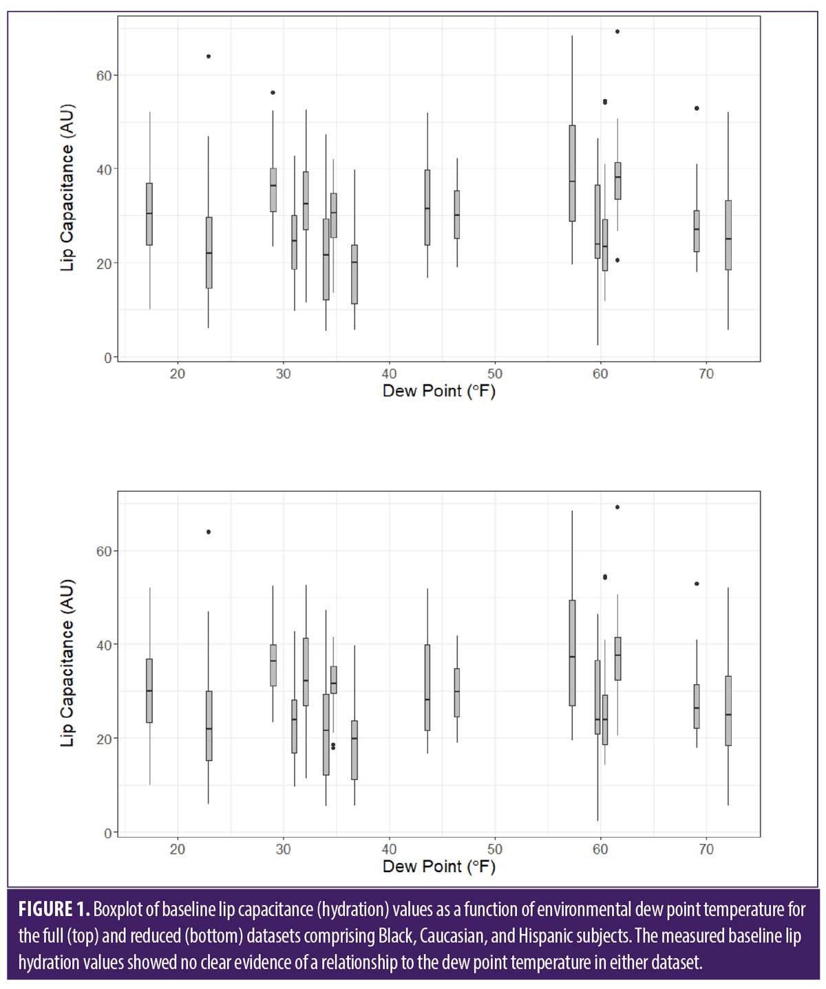
Data analysis. Summary statistics by self-identified race were calculated for the five groups in both datasets (Table 2). As already noted, further analysis focused only on the Black, Caucasian, and Hispanic subject groups. Weather data for the period around each study baseline was summarized and boxplots were used to visualize the relationship between environmental weather dew point and baseline lip hydration for these groups (Figure 1). A similar procedure was used to assess the potential effect of subject age. After evaluating underlying assumptions, a Type II ANOVA using ‘race’ as the independent variable and ‘study’ as a covariate was used to assess possible lip hydration differences due to race.24,25 In cases of overall significance the Tukey method was used to generate pairwise comparisons while controlling for Type I error. A two-sided α-level of 0.05 was used throughout.
Results
Influence of weather conditions. The sixteen studies that served as the data source for this exercise were conducted in different geographic locations and at different times throughout the year. Not surprisingly, environmental weather conditions around the time of the studies’ baseline visits showed considerable variation: the temperature ranged from 33.9–99.6°F; the relative humidity ranged from 17.1–86.6%; and the dew point ranged from 17.3–72.1°F. No significant positive or negative relationship between lip hydration and environmental dew point temperature was apparent in boxplots of baseline lip hydration values for the three analysis groups as a function of dew point temperature (Figure 1); this was confirmed by regression analysis (results not shown). Baseline lip hydration values similarly showed no clear positive or negative relationship to the environmental temperature or relative humidity (results not shown).
Influence of subjects’ age. Although the protocol specified no upper age limit for study eligibility, more than 98 percent of subjects enrolled were between the ages of 20 and 50 years. Age data summary statistics for the three analysis groups are shown in Table 3. The mean ages are not statistically different for the full dataset, but the difference between the mean ages for Black and Hispanic subjects reached statistical significance (P=0.04) for the reduced data set.
No relationship between baseline lip hydration and age was apparent when lip hydration values for subjects in the full or reduced data sets were plotted versus age at the time of enrollment (results not shown). This finding is consistent with work by Caisey et al. and by Kobayashi and Tagami, which also found no significant relationship between lip hydration and subject age.26,27
Influence of subjects’ race. Lip hydration summary statistics by self-identified race for the full and reduced datasets are shown in Table 2. The mean values fall into two groups, one group comprising Black and Caucasian subjects, the other group comprising Hispanic subjects (along with the Asian-American and Native American subjects). Mean values measured on the former group are lower than those measured on the latter group by about 4 to 5 units, but there is relatively little distinction between values within each group.
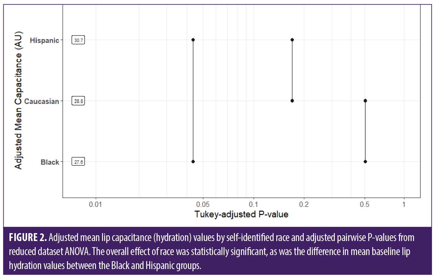
Analysis of the full dataset for the three largest groups showed no significant overall effect of race, although the result approached statistical significance (P=0.07). These groups were ranked, in order of increasing mean baseline lip hydration: Black < Caucasian < Hispanic. A similar pattern was observed when the reduced dataset was considered. Analysis of data for the three largest groups showed that they again ranked, in order of increasing mean baseline lip hydration: Black < Caucasian < Hispanic. However, in this case the overall effect of race on baseline lip hydration reached statistical significance (P=0.05). Pairwise comparisons showed that the mean baseline lip hydration in Black and Hispanic subjects was statistically different (Figure 2).
Discussion
The structural properties and exposed location of the lips cause their condition to be quite susceptible to environmental influences. Estimates of the environmental weather conditions present around the time of baseline visits in the sixteen studies considered here showed considerable variation; yet lip hydration values measured at baseline showed no apparent weather dependence. This might reflect the fact that the weather condition values used in this work were estimated from historical data, that subjects were not located near the NOAA reporting stations, or that the conditions present were not as severe as in some previous reports. Acclimation in a controlled environment might also have a role. Skin responds rapidly to environmental changes; e.g. Tsukahara et al and Cravello et al observed significant changes in hydration and other body skin properties in as little as 30 minutes following environmental changes.23,28 The lip surface, having a less efficient barrier than body skin, is expected to respond to environmental changes at least as quickly.27 Guidelines for skin impedance measurements recommend that subjects acclimate under controlled environmental conditions for at least 20 minutes before measurements are made;29 subjects in the studies considered here acclimated for at least 30 minutes. Weather can induce visible changes in skin or lips (e.g., cracking) that will not resolve rapidly, but subjects with such extreme damage were excluded from these studies. Thus, the absence of a marked weather effect on baseline measurements across this study set could reflect subjects’ forced exposure to standard environmental conditions before measurements were made.
ANOVA performed on the full dataset for the three largest subject groups showed no significant overall effect of race on baseline lip hydration. However, the ‘race’ term in the model was statistically significant when the reduced dataset was analyzed, and pairwise comparisons showed that the difference in baseline lip hydration for the African American and Hispanic groups reached statistical significance (P=0.04, Figure 2). Analysis of both datasets yielded the same ranking of the three groups in terms of mean baseline lip hydration: Black < Caucasian < Hispanic. Mean baseline lip hydration values for the Asian-American and Native-American groups, while not included in the ANOVA due to their relatively small sample sizes, were comparable to the Hispanic group (Table 2). Previous studies that examined racial differences in stratum corneum hydration on other areas of the body reported results that are generally consistent with the ranking we found. For example, Johnson and Corah reported higher skin resistance, and thus lower water content, in Black skin compared to White skin.30 Berardesca et al reported that three racial groups ranked: Black < White < Hispanic in terms of conductance measured on the volar forearm, Hispanic subjects having significantly higher hydration.31 Diridollou et al reported a higher dryness index measured by hydration imaging in aged (>51 years) African American compared to Chinese and Mexican skin.32 Sugino et al reported that stratum corneum water content was highest in Asians and lower in Caucasian, African American, and Hispano American groups.33 Wan et al, in a review of literature related to moisturizing different racial skin types, reported that the moisture content of Black skin was less than that of White skin, which was in turn less than that of Asian skin.17 And Young et al reported that skin hydration measured on the faces of African nursing students was significantly lower than that measured on Caucasian students.34
However, there are also stratum corneum hydration results reported on different racial groups that are inconsistent with our ranking. The study by Berardesca et al also measured skin conductance on the dorsal forearm, and found that on that site the ranking was White < Hispanic < Black, with White subjects significantly less hydrated.31 And the study by Diridollou et al, which found differences in aged subjects, found no significant difference between the dryness index measured by capacitance imaging on young (18–50 years) African American, Chinese, Caucasian, or Mexican skin. Warrier et al. reported significantly greater hydration on the cheek for Black skin compared to White skin, but found no statistical difference on the forearm or leg.35 Grimes et al also found no significant hydration difference between African American and White skin on the forearm.36 Manuskiatti et al found no difference in stratum corneum hydration measured on Black or White skin across ten anatomical sites.37 Voegeli et al reported that overall skin hydration values measured on the faces of four racial groups ranked: Indians < Chinese < Caucasians < Black Africans.38 And Hillebrand et al found significantly higher skin hydration measured on the cheeks of African Americans, Latinos, and East Asians than on Caucasians.39 These researchers also noted that differences between the groups were body site and age dependent. The lack of consensus in these studies shows that establishing whether real differences in stratum corneum hydration due to race exist will require more study, regardless of body site.
We believe the work presented herein provides a useful initial assessment that supports the existence of differences in lip hydration based on race. It incorporated a relatively large dataset aggregated from lip hydration measurements collected in a series of sixteen clinical studies, which provided a reasonable basis for the retrospective analysis. However, this approach has several potential limitations. First, the studies were conducted at four different clinical research facilities and at different times of the year, although the studies followed a standard protocol and used the same model instrument for measurements. External weather conditions present were estimated and had no apparent effect on the baseline measurements, but other factors unique to a study or site might have had an impact. A ‘study’ term was incorporated into the analysis model to help account for such occult influences. Finally, the studies required subjects to self-identify their race and some of the classification options were broad. How well the resultant selections reflected subjects’ true ancestry is uncertain.
Conclusion
Aggregated data from a series of sixteen clinical studies was used to investigate possible differences in lip hydration between several racial groups. The data analysis showed that the three largest groups ranked, in order of increasing lip hydration: Black < Caucasian < Hispanic. This ranking is consistent with some, but not all, literature related to racial differences in stratum corneum hydration measured on other areas of the body. The retrospective analysis presented is a first step in understanding possible lip hydration differences related to race; however, the lack of literature consensus on racial differences in stratum corneum hydration on other body areas shows that further, more structured study of this topic is required, regardless of body site.
References
- Blank IH. Factors which influence the water content of the stratum corneum. J Invest Dermatol. 1952;18(6):433–440.
- Denda M, Sato J, Masuda Y, et al. Exposure to a dry environment enhances epidermal permeability barrier function. J Invest Dermatol. 1998;111(5):858–863.
- Sato J, Denda M, Chang S, et al. Abrupt decreases in environmental humidity induce abnormalities in permeability barrier homeostasis. J Invest Dermatol. 2002;119(4):900–904.
- Sato J, Denda M, Nakanishi J, et al. Dry condition affects desquamation of stratum corneum in vivo. J Dermatol Sci. 1998;18(3):163–169.
- Sato J, Yanai M, Hirao T, et al. Water content and thickness of the stratum corneum contribute to skin surface morphology. Arch Dermatol Res. 2000;292:412–417.
- Harding CR, Watkinson A, Rawlings AV, et al. Dry skin, moisturization and corneodesmolysis. Int J Cosmet Sci. 2000:22(1):21–52.
- Rawlings A, Sabin R, Harding C, et al. The effect of glycerol and humidity on desmosome degradation in stratum corneum. Arch Dermatol Res. 1995;287(5):457–464.
- Scott IR, Harding CR. Filaggrin breakdown to water binding compounds during development of the rat stratum corneum is controlled by the water activity of the environment. Dev Biol. 1986;115(1):84–92.
- Katagiri C, Sato J, Nomura J, et al. Changes in environmental humidity affect the water holding property of the stratum corneum and its free amino acid content, and the expression of filaggrin in the epidermis of hairless mice. J Dermatol Sci. 2003;31(1):29–35.
- Silva CL, Topgaard D, Kocherbitov V, et al. Stratum corneum hydration: Phase transformations and mobility in stratum corneum, extracted lipids and isolated corneocytes. Biochim Biophys Acta BBA – Biomembr. 2007;1768(11):2647–2659.
- Merinville E, Grennan GZ, Gillbro JM, et al. Influence of facial skin ageing characteristics on the perceived age in a Russian female population. Int J Cosmet Sci. 37(S1):3–8.
- Stephen ID, McKeegan AM. Lip colour affects perceived sex typicality and attractiveness of human faces. Perception. 2010;39(8):1104–1110.
- Gunn DA, Rexbye H, Griffiths CEM, et al. Why some women look young for their age. PLOS ONE. 2009;4(12):e8021.
- Zugerman C. The lips: anatomy and differential diagnosis. Cutis. 1986;38(2):116–120.
- Hikima R, Igarashi S, Ikeda N, et al. Development of lip treatment on the basis of desquamation mechanism. Int J Cosmet Sci. 2004;26(3):165.
- Gaul LE, Underwood GB. Relation of dew point and barometric pressure to chapping of normal skin. J Invest Dermatol. 1952;19(1):9–19.
- Wan DC, Wong VW, Longaker MT, et al. Moisturizing different racial skin types. J Clin Aesthetic Dermatol. 2014;7(6):25–32.
- Heinrich U, Koop U, Leneveu‐Duchemin MC, et al. Multicentre comparison of skin hydration in terms of physical-, physiological- and product-dependent parameters by the capacitive method (Corneometer CM 825). Int J Cosmet Sci. 2003;25(1-2):45–53.
- O’goshi K, Serup J. Inter-instrumental variation of skin capacitance measured with the Corneometer®. Skin Res Technol. 2005;11(2):107–109.
- Gaul LE, Underwood GB. Relation of dew point and barometric pressure to chapping of normal skin. J Invest Dermatol. 1952;19(1):9–19.
- Spencer TS, Linamen CE, Akers WA, et al. Temperature dependence of water content of stratum corneum. Br J Dermatol. 1975;93(2):159–164.
- Devillers C, Piérard GE, Quatresooz P, et al. Environmental dew point and skin and lip weathering. J Eur Acad Dermatol Venereol JEADV. 2010;24(5):513–517.
- Cravello B, Ferri A. Relationships between skin properties and environmental parameters. Skin Res Technol. 2008;14(2):180–186.
- Shaw RG, Mitchell-Olds T. ANOVA for unbalanced data: an overview. Ecology. 1993;74(6):1638–1645.
- Langsrud Ø. ANOVA for unbalanced data: Use Type II instead of Type III sums of squares. Stat Comput. 2003;13(2):163–167.
- Caisey L, Gubanova E, Camus C, et al. Influence of age and hormone replacement therapy on the functional properties of the lips. Skin Res Technol. 2008;14(2):220–225.
- Kobayashi H, Tagami H. Functional properties of the surface of the vermilion border of the lips are distinct from those of the facial skin. Br J Dermatol. 2004;150(3):563–567.
- Tsukahara K, Hotta M, Fujimura T, et al. Effect of room humidity on the formation of fine wrinkles in the facial skin of Japanese. Skin Res Technol. 2007;13(2):184–188.
- Berardesca E. European Group for Efficacy Measurements on Cosmetics and Other Topical Products (EEMCO). EEMCO guidance for the assessment of stratum corneum hydration: electrical methods. Skin Res Technol Off J Int Soc Bioeng Skin ISBS Int Soc Digit Imaging Skin ISDIS Int Soc Skin Imaging ISSI. 1997;3(2):126–132.
- Johnson LC, Corah NL. Racial differences in skin resistance. Science.1963;139(3556):766–767.
- Berardesca E, de Rigal J, Leveque JL, et al. In vivo biophysical characterization of skin physiological differences in races. Dermatologica.1991;182(2):89–93.
- Diridollou S, Rigal JD, Querleux B, et al. Comparative study of the hydration of the stratum corneum between four ethnic groups: influence of age. Int J Dermatol. 2007;46(s1):11–14.
- Sugino K, Imokawa G, Maibach HI. Ethnic difference of stratum corneum lipid in relation to stratum corneum function. J Invest Dermatol. 1993;100(4):587.
- Young MM, Franken A, du Plessis JL. Transepidermal water loss, stratum corneum hydration, and skin surface pH of female African and Caucasian nursing students. Skin Res Technol. 282 2019;25(1):88–95.
- Warrier AG, Kligman AM, Harper RA, et al. A comparison of black and white skin using noninvasive methods. J Soc Cosmet Chem. 1996;47.
- Grimes P, Edison BL, Green BA, et al. Evaluation of inherent differences between African American and white skin surface properties using subjective and objective measures. Cutis. 2004;73(6):392–396.
- Manuskiatti W, Schwindt DA, Maibach HI. Influence of age, anatomic site and race on skin roughness and scaliness. Dermatology. 1998;196(4):401–407.
- Voegeli R, Rawlings AV, Seroul P, et al. A novel continuous colour mapping approach for visualization of facial skin hydration and transepidermal water loss for four ethnic groups. Int J Cosmet Sci. 2015;37(6):595–605.
- Hillebrand GG, Levine MJ, Miyamoto K. The age-dependent changes in skin condition in ethnic populations from around the world. Ethnic Skin and Hair. Dermatology: Clinical & Basic Science Series. Informa Healthcare; 2007:105–122.
OX40/OX40L Costimulatory Pathway: A Potential Therapeutic Target for Allergic Contact Dermatitis?