 J Clin Aesthet Dermatol. 2020;13(5):29–30
by Justin W. Marson, MD and Hilary E. Baldwin, MD
Dr. Marson is with the Department of Medicine at University of California Irvine in Orange, California. Dr. Baldwin is with the Acne Treatment and Research Center in Morristown, New Jersey and Rutgers Robert Wood Johnson Medical Center in Piscataway, New Jersey.
FUNDING: No funding was provided for this study.
DISCLOSURES: Dr. Baldwin is on the speakers bureau for Galderma, Valeant, Sun, Mayne, and Bayer and is an investigator for Galderma and Valeant. The other author has no conflicts of interest relevant to the content of this article.
ABSTRACT: Background. Keloids are dense, fibrous tumors that arise from the dysregulation of normal wound healing, ultimately outgrowing the initial traumatic lesion.
Objective. We present a modified technique for the excision of dumbbell-shaped keloids on the earlobe using tools common to every dermatologist’s office.
Methods. This was an observational report on the outcomes of dumbbell keloid excision using a #15 blade and punch biopsy. Eligible individuals were those with dumbbell-shaped keloids located on the earlobe. All procedures were conducted at an urban dermatology clinic.
Results. When combining the technique with continual compression earrings and intralesional corticosteroids, excellent cosmetic outcomes and minimal recurrence were achieved.
Conclusion. The pairing of a #15 blade and punch biopsy has been demonstrated to produce a more user-friendly method for dumbbell keloid excision by dermatologists and clinicians without advanced surgical training.
Keywords: Keloid, dumbbell-shape, auricular, earlobe, core excision, surgical excision
Keloids are dense, fibrous tumors that result from the dysregulation of normal wound healing and classically “outgrow” the original traumatic lesion.1 Typically, keloids occur around the ears and high tension sites (e.g., chest, upper back, shoulders, neck).1 Studies have indicated that there is a familial predisposition for developing keloids, which are more commonly found in individuals of African and Asian descent.2 Ear piercing is a common inciting factor in forming dumbbell-shaped keloids of the earlobe, especially in menarcheal or postmenarcheal women.3
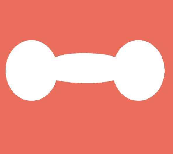
Dumbbell-shaped keloids visually take after their namesake: there are two spheroidal ends (i.e., buttons) that are connected by a narrower, elongated cylindrical core positioned anteroposteriorly through the earlobe (Figure 1). A surgical technique for the “core excision” of dumbbell-shaped keloids of the earlobe described by Salasche et al4 requires the use of a curved blade (#67 Beaver blade) and considerable surgical finesse. Here, we present a modified technique using tools common to every dermatologist’s office that, when paired with compression earrings and intralesional corticosteroids, has yielded excellent cosmetic outcomes and minimal recurrence in our clinical experience.

Description of Technique and Follow-up
After initial cleansing with an alcohol swab, the anterior and posterior buttons are anesthetized with 1% lidocaine with epinephrine. The area is then recleansed with chlorhexidine gluconate. After allowing for adequate time for anesthesia, a #15 blade is used to shave both buttons from the earlobe, leaving the central core in the earlobe flush with surrounding nonlesional skin (Figure 1). At this point, a disposable wooden tongue depressor is placed behind the earlobe for stability and a punch excision is made through the entire thickness of the earlobe to the tongue depressor (Figure 2A). This punch excises the remaining core with at least a 1-mm margin around the core to obtain negative margins (Figure 2B).

Prior to closure, 0.5mL of triamcinolone acetonide 40mg/mL is injected circumferentially into the walls of the defect. The anterior and posterior aspects of the remaining circular defect are then closed separately using a 4-0 Prolene (Johnson & Johnson, New Brunswick, New Jersey) or nylon suture. To avoid creating an angulated defect on the inferior aspect of the earlobe, the anterior portion is closed with two simple sutures parallel to the long axis of the ear, while the posterior defect is closed with two simple sutures parallel to the short axis of the ear, perpendicular to the closure on the anterior defect (Figure 3). Hemostasis is obtained intraoperatively using manual pressure with woven 4×4 gauze.

Immediately following the excision, patients are instructed to wear compression earrings continuously for up to 12 hours a day for upwards of six months until the surgical site lies flat without evidence of elevation or extension past the surgical site.5 Two weeks postprocedure, patients are instructed to return for suture removal and administration of intralesional triamcinolone acetonide 40mg/mL. Four weeks postprocedure, patients again are asked to return for intralesional triamcinolone acetonide 40mg/mL. At this point, patients can return monthly for up to six months for repeat intralesional injections as needed until the lesional site lies flat with adjacent normal skin and is without protuberance or other signs of recurrence.
Discussion
Recent studies suggest that complete surgical excision of the keloid is thought to remove the inflammatory nidus, thereby decreasing chance of recurrence. Chong et al6 found that complete removal of a keloid with negative margins confirmed with dermatopathology results in a smaller defect and no recurrence. The authors suggest that complete removal of the proliferative, inflammatory core with clear margins reduces recurrence rates and improves cosmesis.6
A previous technique published by Salasche et al4 achieved the same result, albeit using more specialized instrumentation and surgical finesse. The use of a #15 blade (e.g., DermaBlade from AccuTec Blades, Verona, Virginia, or other preferred flat excisional tool) and punch biopsy allow for the procedure to be conducted in the average dermatologist’s office using common tools. Combined with early and repeated intralesional corticosteroids and at least 12 hours of continuous daily use of pressure earrings, we have observed low incidence of recurrence.5
Conclusion
Core excision of dumbbell-shaped keloids with negative margins, continuous use of compression earrings, and repeated intralesional corticosteroids appears to provide an efficacious therapy with low incidence of recurrence. The pairing of a #15 blade and punch biopsy represents a more user-friendly treatment method that could be performed by dermatologists and clinicians without advanced surgical training.
Acknowledgements
The authors would like to thank Bo Tang, MD for providing her artistic talents to depict our procedural methodology.
References
- Ogawa R. Keloid and hypertrophic scars are the result of chronic inflammation in the reticular dermis. Int J Mol Sci. 2017;18(3). pii: E606.
- Baldwin HE. Prevention of Keloids. In: Norman R. Preventative Dermatology, vol. 1. Berlin, Germany: Springer; 2010.
- Lane JE, Waller JL, Davis LS. Relationship between age of ear piercing and keloid formation. Pediatrics. 2005;115(5):1312–1314.
- Salasche SJ, Grabski WJ. Keloids of the earlobes: a surgical technique. J Dermatol Surg Oncol. 1983;9(7):552–556.
- Park TH, Seo SW, Kim JK, Chang CH. Outcomes of surgical excision with pressure therapy using magnets and identification of risk factors for recurrent keloids. Plast Reconstr Surg. 2011;128(2):431–439.
- Chong Y, Kim CW, Kim YS, et al. Complete excision of proliferating core in auricular keloids significantly reduces local recurrence: a prospective study. J Dermatol. 2018;45(2):139–144.
OX40/OX40L Costimulatory Pathway: A Potential Therapeutic Target for Allergic Contact Dermatitis?