 by Rashmi Sarkar, MD, MNAMS; Pallavi Ailawadi, MD, DNB; and Shilpa Garg, DNB
Dr. Sarkar is a professor at the Department of Dermatology, Maulana Azad Medical College in New Delhi, India. Dr. Ailawadi is Senior Resident at the Maulana Azad Medical College and LokNayak Hospital in New Delhi, India. Dr Garg is Consultant Dermatologist, Sir Gangaram Hospital in New Delhi, India.
Funding: No funding was provided for this article.
Disclosures: The authors have no conflicts of interest to relevant to the content of this article.
Abstract: Melasma is a common skin condition that affects both men and women. However, it is more commonly seen in women and dark-skinned individuals, such as in Hispanics, Asians, and African Americans who live in areas with intense ultraviolet radiation. Melasma is less common in men, but it negatively affects the quality of life in men as much as it does in women. While melasma has been studied in detail in women, however, there is a paucity of studies on the clinico- etiopathology and therapeutics of melasma in men. This article reviews and discusses important clinical, etiological, and treatment aspects of melasma in men. The authors recommend that clinicians educate their patients on the causes, prevention and treatment methods, and recurrence rates of melasma. The authors also recommend that clinicians take into careful consideration each patient’s preferences and expectations when creating treatment regimens, as these might differ greatly among men and their female counterparts.
J Clin Aesthet Dermatol. 2018;11(2):53–59
Introduction
Melasma is a common skin condition characterized by the presence of symmetrical, irregular, light to dark brown hyperpigmentation involving sun-exposed areas, especially on the face. Although melasma can affect all races and both sexes, it is more commonly seen in women of child-bearing age and in dark-skinned individuals living in areas with intense ultraviolet (UV) radiation.1,2 Hyperpigmentation on exposed areas such as the face can be a source of cosmetic concern for patients, that can negatively impact quality of life (QOL). Melasma in women has been studied in detail, but despite several similarities, there are certain differences in clinical, etiological, and treatment aspects of melasma in men that still need to be studied. Understanding characteristics of melasma specific to men will allow for better management of the disorder among male patients. This article aims to highlight the important clinical, etiological and treatment aspects of melasma in men.
Methods
The information was collected through an extensive literature search from the databases Pubmed and Cochrane Library. The keywords used for the search were melasma, etiology and men. Articles published within the last 20 years were included. However, some older publications were included in order to describe the evolution of melasma in men. Poorly designed studies and those with conflicting results were excluded. Signed photoconsent was obtained from patients whose photographs are included herein.
Epidemiology
The exact prevalence of melasma is not known among the general population, including men and women. This could be due to underreporting by the affected patients due to its asymptomatic nature and because many patients choose to treat it with over-the-counter products rather than consult with a dermatologist.3 The global prevalence of melasma varies according to ethnicity, skin type, and intensity of sun exposure. It has been found to be more common in Hispanics, Asians, and African Americans than in Caucasian populations.1,4 Further, it is more common in individuals with dark skin and Fitzpatrick skin types IV, V and VI.5,6
The majority of the studies reporting the prevalence of melasma are based on clinical samples rather than population samples. The prevalence of melasma among the general population is reported to be 1.8 percent in Ethiopia7, 2.88 percent in Saudi Arabia8, 3.4 percent in Lebanon9 and 8.2 percent in United States.10 South Asian countries have a relatively higher prevalence of melasma than in other countries, as seen in Nepal (6.8%) and China (13.61% ).11,12
Research has shown that melasma is more common in women than in men. Studies in Brazil, Singapore, and India have shown a difference as high as 39:114, 21:115, and 6:1, respectively.13
In Puerto Rico, a study by Vazquez et al16 found that men made up only 10 percent of the cases of melasma. In contrast to this low incidence of melasma in Caucasian men, the researchers found that melasma was more commonly reported in men of Hispanic, Asian, and Indian origin.1,2,4 In the study by Pichardo et al6, which included Hispanic men with melasma, data were pooled from three studies: one examining 25 poultry workers, one examining 54 farm workers, and one examining 300 Hispanic farm workers. The prevalence of melasma was 36 percent, 7.4 percent, and 14 percent in the three study groups, respectively. There was a mean prevalence of 14.5 percent, larger than the prevalence of melasma reported in a random sampling of Hispanic women (8.2%).10 The difference could be partially due to the difference in study design between the two studies; in the study on Hispanic women by Werlinger et al,10 melasma was self-diagnosed by patients over telephone interviews, which could result in inaccurate reporting.10 Therefore, further studies are required to better quantify the sex-specific prevalence of melasma among the Hispanic population.
In Indian patients, two studies show a higher prevalence of melasma in men (25.8% [n=120] and 20.5% [n=200]).5,17 This increased prevalence among Indian men compared to Caucasian men could be attributed to their darker complexion and the tropical Indian climate.
Etiopathogenesis
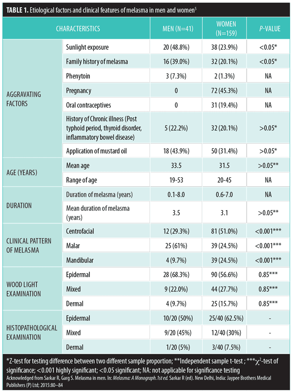
The exact etiology of melasma is not known. However, some of the most common factors include sun exposure, hormonal influences, and genetic susceptibility. Other less common causes include cosmetics, photosensitizing drugs, food items, thyroid diseases, hepatopathies, ovarian tumors, parasitic infestations, and stressful events.13,14,18,19 Melasma is caused by a complex interplay of environmental factors in a genetically predisposed individual. The etiological factors causing melasma in men are likely the same as those implicated in women, except for certain differences (Table 1).
Genetic etiology of melasma in terms of familial predisposition is a notable risk factor. The occurrence of melasma has been described in a pair of identical twins,20 suggesting a common genetic component. This has been confirmed in various studies, with positive family history being present in 10 to 70 percent of patients in studies on melasma among Iranian, Singaporean, Hispanic, and African-American populations.18,21,22
In a multicenter study (9 centers across the world) by Ortenne et al,18 48 percent of 324 patients with melasma reported family history in at least one relative, with 97 percent being first-degree relatives. In other studies, the prevalence varied from 56 percent in Brazil (n=3012) to 33 percent in India (n=312) and 10 percent in Singapore (n=205).23,24 A majority of these studies focused on women and there are only a few studies that document the presence of family history in men with melasma. However, a family history was reported in among a large percentage of male patients in the study by Vazquez et al (70.4%). In this study, family history of melasma was reported among close family members (e.g., mother, siblings, aunts, and uncles) however, none of the patients reported melasma in the father.16
In an Indian study, 39 percent of men had a family history of melasma, compared to 20.1 percent in women (Table 2).5 Further, a study by Keeling et al, documented the presence of family history of melasma in a first- or second-degree female relative in all five male patients, while two patients also had an affected male relative.25

However, to date, there have been no gene association studies on melasma. The variation in positive genetic predisposition across various studies and populations could be due to the multifactorial causation of melasma, suggesting that the development of the disease might be related to epigenetic hormonal control and other environmental stimuli, such as UV radiation.
Sun exposure is known to be an important etiological factor in causing melasma, irrespective of sex.18 UV radiation (UVA and UVB) increases proliferation and melanocyte activity, causing epidermal pigmentation, that occurs more intensely in melasmic areas with melasma than in unaffected skin.26,27 This is further substantiated by the findings that melasma usually improves during the winter and worsens during the summer months(or during other periods of intense sun exposure). Moreover, prevalence is high in tropical regions and high elevation areas.17 Recently, infrared radiation and visible light have been found to cause melasma, although not as severely as UV radiation.28
In a majority of studies on men with melasma, sun exposure was documented as the main cause. In a study by Sarkar et al,5 48.8 percent of the male patients reported sun exposure compared to 23.9 percent of female patients. Among these 41 men with melasma, 24 (58.5%) were outdoor workers and 12 (29.3%) lived in high elevation regions of north India. Similar findings were reported in two other studies where 45.16 percent (n=31) and 81.4 percent (n=) of the subjects had histories of chronic sun exposure.16,17
In women, hormonal factors, such as pregnancy, oral contraceptive pills, hormonal therapy, and mild ovarian dysfunction, are considered to be some of the most common etiological factors in the development of melasma.29 Hormonal imbalances between estrogen and testosterone might play a role in the development of melasma in men. Estrogen is known to lower blood testosterone and suppress the secretion of leuteinizing hormone (LH) and follicle stimulating hormone (FSH), which further increases estrogen levels by reversing the LH- and FSH- induced suppression. The effects of estrogen on melanocytes and induction of pigmentation in melasma are well-documented.31 Several aspects of melanocyte function respond directly to estrogenic stimulation, which takes place through the estrogen receptors present on the melanocytes in the cytoplasm and nucleus. Estrogen increases melanin synthesis by stimulating the activity of tyrosinase enzyme. Further, it increases the extrusion of melanin from the cells. Several studies have found increased estrogen levels in women with melasma. Tadokoro et al32 indicated that testosterone affects human melanocytes by reducing the level of intracellular cyclic adenosine monophosphate and tyrosinase activity, thereby decreasing melanogenesis.
The two studies noted low testosterone levels in Indian men with melasma (Table 2). In one of these studies, hormonal evaluation in men with melasma revealed increased LH and low testosterone in four men (9.7%).5 The second study evaluated 15 men with melasma (aged 20–40 years, 2-month to 1.5-year melasma duration, 11 age-matched controls), and found a significantly high level of circulating LH coupled with low levels of testosterone and normal LH/FSH ratio compared to controls. These findings suggests the presence of subtle testicular resistance in men with melasma.30 Presence of mild subclinical ovarian dysfunction was reported in a study of nine women with idiopathic melasma; the women showed increased levels of LH along with lower estradiol levels compared to normal age- and sex-matched controls.31 This is in contrast to a majority of studies that have reported increased levels of estrogen in female patients, which implies that melasma patients have some degree of mild endocrinopathy.
Besides the endogenous hormonal factors, exogenous or iatrogenic administration of hormonal medications have shown to induce melasma. Several cases of melasma following hormone therapy (e.g., estrogen therapy,33 fosfestrol tetrasodium,34 an androgenic agent, Andro-6,35 and finasteride36) have been reported. Finasteride is hypothesized to increase testosterone, which is available for aromatization to estradiol, resulting in the subsequent induction of melasma pigmentation.36
The use of cosmetics and consumption of certain drugs and other photosensitizing substances have also been implicated in melasma induction. An Indian study observed that the use of mustard oil for body and hair massage was more common in men with melasma (43.9%) than women (31.4%), though the difference was statistically insignificant (Table 1).5 Mustard oil acts as a photosensitizer, which can lead to facial pigmentation, predominantly on the forehead and the temporal region of the face. Labeled as toxic melanoderma in India, this form of pigmentation is difficult to differentiate from melasma. However, since the use of mustard oil was found in a significant number of patients in this study, one might postulate that mustard oil plays a role in the development of melasma among patients who use it. 5 Further studies are needed to substantiate the relationship between mustard oil and melasma.
The possibility of exogenous substances causing melasma in men is lower than that of women due to comparatively lesser use of cosmetics among men. Studies have not been able to establish any association between the melasma and the use of any chemical. In the study by Vazquez et al,16 use of cosmetics like soaps, shaving creams, aftershave, and perfumes were documented in 25 (92.6%) men with melasma. Use of a single cosmetic could not be identified among the patients, and none of the patients attributed the development of melasma to the use of cosmetics.16
The drug phenytoin has shown to be a causative agent in 6.45 percent and 7.3 percent of men with melasma in two Indian studies.5,17 Recently, imatinib has also been shown to induce melasma.37
Certain chronic illnesses have been identified in men with melasma (3 men had a thyroid disorder, 1 had inflammatory bowel disease, and 1 had recently recovered from typhoid). In women with melasma, chronic illness was observed in 20.1 percent (n=32) (Table 2). However,the association needs to be substantiated by further studies.5 Further, the association of melasma with thyroid diseases has also been suggested, with higher incidence of thyroid autoimmunity occurring in patients with melasma, but the results have been conflicting.38,39
Clinical Features
Melasma typically presents as well-defined, symmetrical, brown-black hyperpigmented patches on sun-exposed areas of the skin. The clinical presentation of melasma in men is similar to the presentation in women, except for a few subtle differences. The age of onset of melasma in male patients is variable, ranging from 18 to 72 years, with the average age of onset being 30.7 years.6,16,17 In the study by Sarkar et al,5 there was no difference in the mean age of melasma between men (33.5 years) and women (31.5 years) (Table 1).
In a study of Hispanic men, melasma was present across all age groups, but was most prevalent in men over 31 years old (70%).6 Two other Indian studies reported the mean age of men with melasma as 33.5 years and 34.5 years. Of the 41 men with melasma, 51.2 percent (21/41) were between 31 and 40 years of age, 26.8 percent (11/41) were between 19 and 30 years, and 22 percent (9/41) were aged 41 years or older.5 In contrast to these findings, Vazquez et al16 observed a higher average age (38.8 years) of melasma in men (Table 3).
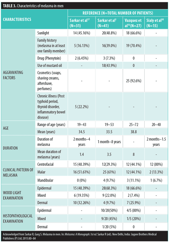
The mean duration of melasma reported in the two Indian studies were 1.4 years17 and 3.55 years in contrast to a longer duration of eight years reported Pichardo and colleagues.6 A shorter duration of melasma ranging between 2 months to 1.5 years was reported in another study (Table 3).30
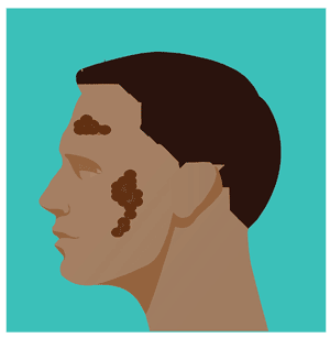
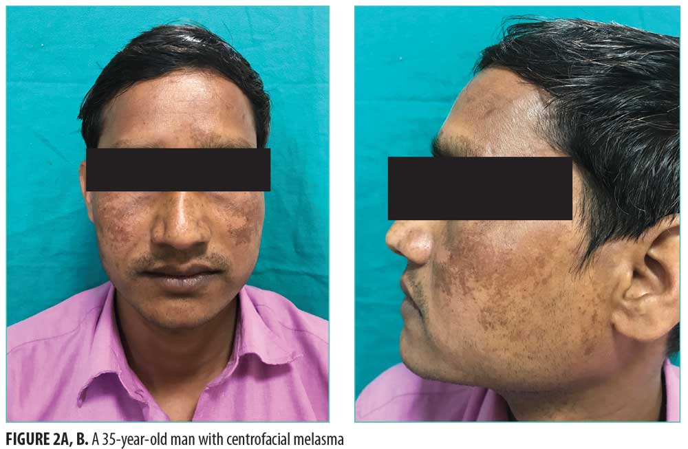
According to the predominant localization of the lesions on the face, three patterns of facial melasma are recognized clinically: malar (Figure 1), centrofacial (Figures 2A and 2B), and mandibular. In the centrofacial pattern, the forehead, cheeks, nose, upper lip, and chin are affected, whereas the malar pattern distributes melasma on the cheeks and nose, and the mandibular pattern covers the mandibular ramus. Among women, the centrofacial pattern is most common. Among men, the malar pattern is most common, representing 44.1 to 61 percent of male patients. The second most common pattern among men is the centrofacial variant.5,16,17 Clinically, melasma is most commonly seen on the face, but melasma on other sun-exposed areas such as the arms, neck, sternal regions, and back, have been seen in women. Extra-facial melasma affecting the upper body occurs mainly among elderly, menopausal women, and might be associated with hormone replacement therapy.34,40,41

A few cases of melasma on the neck and forearms have also been reported in men.34,42 However, no other similarities were documented in these male patients.
Similar to women, woods lamp examination of melasma revealed the epidermal pattern (accentuation of pigmentation) to be the most common in men across three studies (48.4–68.3%).5,16,17
Histopathology
The diagnosis of melasma is generally made clinically. However, skin biopsy with histopathological examination (HPE) can be done. The histopathology of melasma in men is similar to that in women.5 In epidermal melasma, increased melanin seen in the basal and suprabasal layers, whereas dendritic melanocytes and melanophages are present in the dermal type. In an Indian study, HPE was done in 48.8 percent (20/41) of the male patients, and the findings revealed that epidermal melasma was the most common pattern (50%), followed by mixed melasma (45%). Dermal melasma was the least common (5%) (Table 3). Other features included solar elastosis in 17 subjects (85%), flattening of rete ridges in nine (45%) and chronic inflammatory infiltrate in six (30%) male patients with no evidence of basal layer degeneration.5 In concordance with this study, two other studies also reported epidermal melasma as the predominant histopathological type seen in men (Table 3).16,17 The study by Jang et al43 also found epidermal melasma and increased elastotic material in the lesional dermis compared to the nonlesional dermis was more common among male patients, however, the difference was not statistically significant.43
Further, it has been demonstrated that stem cell factor (SCF) and its receptor c-kit have an important role in the pathogenesis of melasma. Recently, in a study of 60 Korean women with melasma, researchers observed increased expression of SCF around the dermal fibroblast and c-kit in the basal layer of the lesional skin compared to normal skin.44 In order to study these factors in men, Jang et al43 compared the HPE characteristics of eight men to 10 women with melasma and found a significant increase in SCF and c-kit expression in the lesional skin of the men. Further, the lesional to nonlesional ratio of SCF in the epidermis was increased in the men compared to the women. This suggests that chronic UV radiation might be associated with signaling of paracrine cytokines, which could play an important role in the mechanism of melasma in male patients. Additionally, the investigators found that vascularity in male patients with melasma was higher than that in female patients, suggesting that chronic UV exposure could play a large role in the development melasma in men.43
Melasma and Quality of life
Melasma is a can have a significant impact on the psychosocial well-being of the patient. Patients with melasma commonly report feelings of shame, low self-esteem, sadness, dissatisfaction, and decreased motivation to go out.45 In the study by Pichardo et al,6 there was a statistically significant difference in the Dermatology Life Quality Index (DLQI) in men with melasma compared to those without melasma (7.5 vs. 2.8) in a group of poultry workers, indicating a moderately poor QoL (Table 4). However, the men the other two groups (men in cross-sectional farm worker study and men in the longitudinal farm worker study) did not report such a difference.6 Further studies need to be done to better quantify the impact of the melasma on the QOL of male patients.
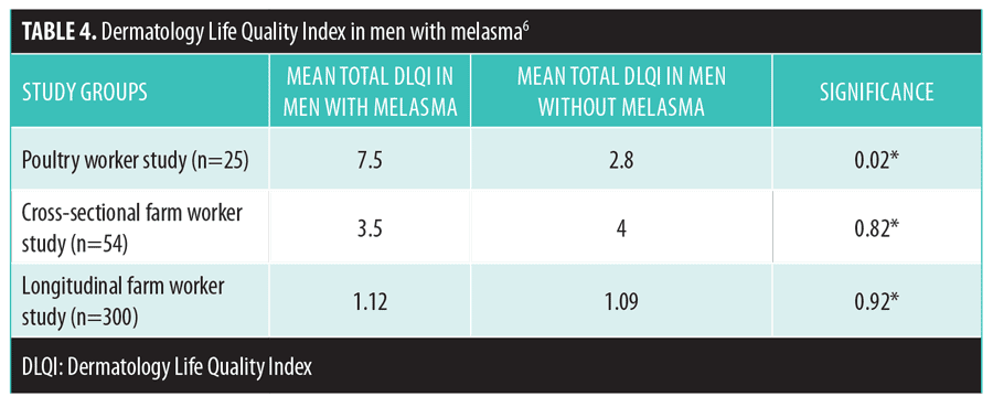
Studies on women with melasma have shown that the QOL score has a poor correlation with the clinical severity of the disease, suggesting that a patient’s subjective perception of the disfiguration goes beyond the clinical dimension of the dyschromia.45,46
Diagnosis
While the diagnosis of melasma is usually a straightforward process, certain facial melanosis can closely mimic melasma and must be ruled out before a definitive diagnosis of melasma can be made. The differential diagnosis of melasma in men includes post-inflammatory hyperpigmentation, pigmented contact dermatitis, toxic melanoderma, lichen planus pigmentosus, frictional melanosis, acanthosis nigricans, and maturational pigmentation. Certain other conditions such as nevus of Ota, nevus of Hori, freckles, solar lentigo, and Becker’s melanosis might also create confusion, especially when these conditions are bilateral. Further, these conditions can coexist with melasma and should be identified before prescribing treatment. Pigmentary demarcation lines (PDL) are an important differential and might be confused with melasma.1 A detailed medical history, thorough clinical examination of the skin, dermoscopy, and histopathology are helpful in arriving at the correct diagnosis.
Investigations
Woods lamp examination, dermoscopy, mexametry, confocal microscopy, and HPE of a skin biopsy specimen are helpful tools for distinguishing melasma from other dermatological conditions. Wood’s lamp examination highlights the difference in pigmentation of the affected skin and type of accentuation helps to classify the type of melasma. Another emerging diagnostic modality is dermoscopy, a simple noninvasive modality. The standard dermoscopic findings of melasma include a fine brown reticular pattern superimposed on a background of faint light brown structureless areas. Further, a vascular component can also be seen in a large number of patients.47 Melanin in the superficial epidermis presents as a dark brown, well-defined pigment network, with shades of light brown and irregularity within the network. Sparing of the appendegeal openings is seen when melanin is located in the lower layers of the epidermis. A blue or bluish-gray color is seen in dermal melasma when pigment is located in the dermis.48 The objective melanin content can be measured with a narrowband reflectance spectrophotometer (Mexameter®, CK Electronics, Cologne, Germany) or in vivo reflectance confocal microscopy.49
Management
Many clinicians might assume that their male patients are not as concerned about the appearance of their skin as women are and that they will be reluctant to follow a stringent skin care plan. However, clinicians should bear in mind that if a male patient is seeking treatment for a dermatological condition, such as melasma, then cosmesis might at least be of partial concern to him, and thus the patient might be very motivated to adhere to a prescribed treatment regimen. To encourage the greatest degree of treatment adherence, clinicians should take into careful consideration each patient’s individual needs when creating treatment regimens, as preferences and expectations might differ greatly among men and their female counterparts. For example, use of products with strong fragrances or an oily base or use of camouflaging agents might not be acceptable to some patients. Additionally, some patients might find the idea of applying skin care products several times a day or even once daily unappealing, and clinicians should be aware of this before developing their treatment plans. Additionally, patient counseling is an integral component of melasma management, and clinicians should educate their patients on its causes, prevention and treatment methods, and recurrence rates.
Sun avoidance is the most important part of melasma treatment, both for current improvement and future prevention of recurrence. The use of broad spectrum sunscreens (UVA and UVB) along with an inorganic sunscreen (physical block) like zinc oxide or titanium dioxide with a minimum sun protection factor of 15 should be encouraged.50 Regardless of sex, physicians should counsel all patients regarding protection from sun exposure, with emphasis on optimal and regular application of sunscreen and use of hats and clothing that block the sun. Physicians must be extra attentive to male patients, who have shown to be less successful in adhering to sunscreen application guidelines.51
There is a paucity of literature on treatment of melasma in men. Most of the guidelines for melasma have been based on studies done predominantly in women. The management recommendations for men are similar to those for women, and there are no separate recommendations for the men.
Various treatment options for melasma include the use of topical and oral depigmenting agents, chemical peels, and surgical modalities, such as dermabrasion and lasers. Although hydroquinone (HQ) and triple combination creams (HQ+retinoic acid [RA]+corticosteroid [CS]) remain the gold standard of treatment, alternate treatment options for topical use include dual combinations (HQ+RA, CS+RA; HQ +CS), kojic acid, azelaic acid, arbutin, mequinol, ascorbic acid, tranexamic acid, rucinol, lignin peroxidase, orchid extracts, and licorice extract. Other therapeutic modalities include chemical peels along with laser light therapies. In a study by Keeling et al,25 five men with melasma were treated with a combination of 2% mequinol and 0.01% retinoic acid solution, which resulted in complete clearance in four patients and moderate improvement in one patient at 12 weeks.
Medical treatment is the mainstay of management of melasma in men, and combinations of modalities can be used to optimize results. According to a literature review by Krupashankar et al,50 topical therapy with triple combination cream should be the first line of treatment for patients with melasma. Monotherapies and dual therapies have lower efficacy, slower onset of action, and are recommended to patients unable to access triple therapy or who have sensitivity to the ingredients.
Another important component in the armamentarium against melasma are chemical peels, such as glycolic acid, mandelic acid, lactic acid, jessners peels, trichloroacetic acid, and retinoid peels. However, caution should be exercised in using peels for melasma in darker skin types, as there are higher rates of adverse effects like irritation and post-inflammatory hyperpigmentation.52
Laser and light treatment used for treating melasma include Q-switched neodymium: yttrium-aluminum-garnet (QS Nd: YAG) laser (1064,532nm), Q switched ruby (694nm), Q switched alexandrite (755nm), copper bromide laser, erbium: YAG laser, 1550nm erbium-doped fractional laser, and intense pulsed light.53 Among these, the nonablative1550nm fractional laser is approved by the United States Food and Drug Administration (FDA) for the treatment of melasma and is associated with decreased downtime and lower risk of complications.54 Various studies have reported conflicting results with lasers in the treatment of melasma. Although lasers can produce rapid and significant results in some patients, adverse effects such as irritation, post-inflammatory hyperpigmentation, mottled hypopigmentation, and rebound hyperpigmentation can occur, particularly in dark-skinned individuals. There is no consensus in the literature regarding the safety, efficacy, or durability of laser-based treatments and, thus, they are not considered as first-line treatments for melasma in either sex. Their use should be restricted to cases unresponsive to topical therapy or chemical peels.
Conclusion
Melasma has traditionally been considered to be a pigmentation disorder of the female sex, but the occurrence in men is not uncommon. It appears to affect dark-skinned men of Asian and African- American origin more frequently than previously thought. Melasma has a multifactorial origin that is exacerbated by environmental factors such as sunlight, especially in those genetically predisposed to the condition. The etiopathogenesis of melasma in men is similar to that of women, except for hormonal factors, which are more prevalent in women. The role of mustard oil needs to be better substantiated. Malar melasma is most common in men.
The treatment of melasma is challenging, often unsatisfactory, and needs to be continued indefinitely to avoid recurrence. To ensure optimal adherence, clinicians should consider individual patient needs, preferences, and expectations when devising a treatment plan. Strict sun protection, including sunscreens, do offer some protection against relapse, but it is not guaranteed. Further studies on melasma in men belonging to different population groups would go a long way in a better understanding of the differences from their female counterparts.
References
- Pandya AG, Guevara IL. Disorders of hyperpigmentation. Dermatol Clin. 2000;18:91–8.
- Grimes PE. Melasma: etiological and therapeutic considerations. Arch Dermatol. 1995;131:1453–1457.
- Al-Hamdi KI, Hasony HJ, Jareh HL. Melasma in Basrah: A clinical and epidemiological study. MJBU. 2008;26:1–5.
- Taylor SC. Epidemiology of skin diseases in people of color. 2003; 71:271–275.
- Sarkar R, Puri P, Jain RK, et al. Melasma in men: a clinical, aetiological and histological study. J Eur Acad Dermatol Venereol. 2010;24: 768–772.
- Pichardo R, Vallejos Q, Feldman SR, et al. The prevalence of melasma and its association with quality of life in adult male Latino migrant workers. Int J Dermatol. 2009;48: 22–26.
- Hiletework M. Skin diseases seen in Kazanchis health center. Ethiop Med J. 1998;36:245–254.
- Parthasaradhi A, Al Gufai AF. The pattern of skin disease in Hail region, Saudi Arabia. Ann Saudi Med. 1998;18:558–561.
- Tomb RR, Nassar JS. Profile of skin diseases observed in a department of dermatology (1995–2000). J Med Liban. 2000;48:302–309.
- Werlinger KD, Guevara IL, Gonzalez CM, et al. Prevalence of self-diagnosed melasma among pre-menopausal Latino women in Dallas and Forth Worth, Tex. Arch Dermatol. 2007;143:424–425.
- Walker SL, Shah M, Hubbard VG, et al. Skin disease is common in rural Nepal: results of a point prevalence study. Br J Dermatol. 2008;158:334–338.
- Wang R, Wang T, Cao L et al. Prevalence of melasma in Chinese Han and Chinese Yi: a survey in Liangshan district. Chin J Dermatovenereol. 2010;24:546–548.
- Achar A, Rathi SK. Melasma: a clinico-epidemiological study of 312 cases. Indian J Dermatol. 2011;56:380–382.
- Hexsel D, Lacerda DA, Cavalcante AS, et al. Epidemiology of melasma in Brazilian patients: a multicenter study. Int J Dermatol. 2013;53:440–444.
- Goh CL, Dlova CN. A retrospective study on the clinical presentation and treatment outcome of melasma in a tertiary dermatological referral centre in Singapore. Singapore Med J. 1999;40:455–458.
- Vazquez M, Maldonado H, Benmaman C, et al. Melasma in men. a clinical and histologic study. Int J Dermatol. 1988;27:25–27.
- Sarkar R, Jain RK, Puri P. Melasma in Indian males. Dermatol Surg. 2003;29: 204.
- Ortonne JP, Arellano I, Berneburg M, et al. A global survey of the role of ultraviolet radiation and hormonal influences in the development of melasma. J Eur Acad Dermatol Venereol. 2009;23:1254–1262.
- Tamega AA, Miot LD, Bonfietti C, et al. Clinical patterns and epidemiological characteristics of facial melasma in Brazilian women. J Eur Acad Dermatol Venereol. 2013;27:151–156.
- Hughes BR. Melasma occurring in twin sisters. J Am Acad Dermatol. 1987;17:841.
- Sheth VM, Pandya AG. Melasma: a comprehensive update: part I. J Am Acad Dermatol. 2011;65:689–697.
- Moin A, Jabery Z, Fallah N. Prevalence and awareness of melasma during pregnancy. Int J Dermatol. 2006;45: 285–288.
- Tamega Ade A, Miot LD, Bonfietti C, Gige TC, Marques ME, Miot HA. Clinical patterns and epidemiological characteristics of facial melasma in Brazilian women. J Eur Acad Dermatol Venereol. 2013;27:151–156.
- Goh CL, Dlova CN. A retrospective study on the clinical presentation and treatment outcome of melasma in a tertiary dermatological referral centre in Singapore. Singapore Med J. 1999;40:455–458.
- Keeling J, Cardona L, Benitez A et al. Mequinol 2%? tretinoin 0.01% topical solution for the treatment of melasma in men: a case series and review of the literature. 2008;81:179–183.
- Guinot C, Cheffai S, Latreille J, et al. Aggravating factors for melasma: a prospective study in 197 Tunisian patients. J Eur Acad Dermatol Venereol. 2010;24: 1060–1069.
- Videira IF, Moura DF, Magina S. Mechanisms regulating melanogenesis. An Bras Dermatol. 2013;88:76–83.
- Mahmoud BH, Ruvolo E, Hexsel CL, et al. Impact of long-wavelength UVA and visible light on melanocompetent skin. J Invest Dermatol. 2010;130:2092–2097.
- Perez M, Sanchez JL, Aquilo F. Endocrinologic profile of patients with idiopathic melasma. J Invest Dermatol. 1983; 81: 543–545.
- Sialy R, Massan I, Kaur I, et al. Melasma in men: a hormonal profile. J Dermatol. 2000; 27: 64–65
- Mahmood K, Nadeem M, Aman S, et al. Role of estrogen, progesterone and prolactin in the etiopathogenesis of melasma in females. J Pak Assoc Dermatol. 2011;21: 241–47.
- Tadokoro T, Rouzaud F, Itami S, et al. The inhibitory effect of androgen and sex-hormone-binding globulin on the intracellular cAMP level and tyrosinase activity of normal human melanocytes. Pigment Cell Res. 2003;16:190–219.
- Ogita A, Funasaka Y, Ansai S, Kawana S, Saeki H. Melasma in a male patient due to estrogen therapy for prostate cancer. Ann Dermatol. 2015;27:763–764.
- O’Brien TJ, Dyall-Smith D, Hall AP. Melasma of the forearms. Australas J Dermatol. 1997;38:35–37.
- Burkhart CG. Chloasma in a man due to oral hormone replacement. 2006;5:46–47.
- Famenini S, Gharavi NM, Beynet DP. Finasteride associated melasma in a Caucasian male. J Drugs Dermatol. 2014;13:484–486.
- Ghunawat S, Sarkar R, Garg VK. Imatinib induced melasma-like pigmentation: Report of five cases and review of literature. Indian J Dermatol Venereol Leprol. 2016;82:409–412.
- Rostami M, Iranparvar M, Maleki N, et al. Evaluation of autoimmune thyroid disease in melasma. J Cosmet Dermatol. 2015;14:167–171.
- Muller I, Rees DA. Melasma and endocrine disorders. Pigmentary Disorders. 2014;S1:001.
- Ritter CG, Fiss DV, Borges da Costa JA, de Carvalho RR, Bauermann G, Cestari TF. Extra-facial melasma: clinical, histopathological, and immunohistochemical casecontrol study. J Eur Acad Dermatol Venereol. 2013;27:
1088–271094. - Madke B, Kar S, Yadav N et al. Extrafacial melasma over forearms. Indian Dermatol Online J. 2016;7:344–345.
- Lonsdale-Eccles AA, Langtry JA. Melasma on the nape of the neck in a man. Acta Derm Venereol. 2005;85: 181–182.
- Jang YH, Sim JH, Kang HY, et al. The histopathologic characteristics in male melasma: comparison with female melasma and lentigo. J Am Acad Dermatol. 2012; 66: 642–649.
- Kang HY, Hwang JS, Lee JY et al. The dermal stem cell factor and c-kit are overexpressed in melasma. Br J Dermatol. 2006; 154: 1094–1099.
- Ikino JK, Nunes DH, Silva VPMD, et al. Melasma and assessment of the quality of life in Brazilian women. An Bras Dermatol. 2015;90:196–200.
- Yalamanchili R, Shastry V, Betkur J. Clinico-epidemiological study and quality of life assessment in melasma. Indian J Dermatol. 2015;60:519.
- Rendon MI, Benitez AL, Gaviria JI. Telangiectatic melasma: a new entity? J Cosmet Dermatol. 2007;20(1):17–21
- Manjunath KG, Kiran C, Sonakshi S et al. Melasma: through the eye of a dermoscope. Int J Res Dermatol. 2016;2:113–117.
- Sarkar R, Arora P, Garg VK, Sonthalia S, Gokhale N. Melasma update. Indian Dermatol Online J. 2014;5:
426–435 - Krupashankar DS, Godse K, Aurangabadkar S, et al. Evidence-based treatment for melasma: expert opinion and a review. Dermatol Ther (Heidelb). 2014; 4(2):165–186.
- Buller DB, Anderson PA, Walkosz BJ, et al. Compliance with sunscreen advice in a survey of adults engaged in outdoor winter recreation at high-elevation ski areas. J Am Acad Dermatol. 2012; 66: 63–70.
- Salam A, Dadzie OE,Galadari H. Chemical peeling in ethnic skin: an update. Br J Dermatol 2013;169(Suppl. 3):82–90.
- Arora P, Sarkar R, Garg VK, et al. Lasers for treatment of melasma and post-inflammatory hyperpigmentation. J Cutan Aesthet Surg. 2012;5: 93–103.
- Sheth VM, Pandya AG. Melasma: a comprehensive update part II. J Am Acad Dermatol. 2011; 65: 699–714.

