 Dermatology by Dermatologists

Dear Editor:
The cosmetic/aesthetic industry is a rapidly evolving, multibillion dollar entity valued at $10.4 billion in 2011, quadrupling from 1995 to 2010, and it is projected to exponentially increase. With the advent of energy-based devices and injectable products has come an expansion in the number of providers performing these services, many of whom are non-physicians as well as physicians practicing outside of the scope of their training in residency.1 Patients are often unaware of the differences in training pertaining to the individual performing an ambulatory cosmetic procedure, and board certification or specialization like dermatology or plastic surgery (PS) is currently not necessary in aesthetics. Gibson et al. analyzed the variation of medical directorship and oversight present in medical spas (MS); of the 247 MS that they researched, only 47 (20.2%) were associated with a dermatology or PS practice, raising concern for unforeseen ramifications in patient safety.2 In addition to aesthetic training, dermatologists are experts at identifying high risk lesions through rigorous training in cutaneous malignancies.
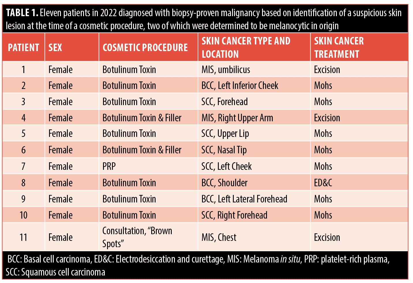
Although a non-dermatological provider may perform an adequate job in cosmetic procedures, other significant cutaneous findings may be missed by one who is not trained in dermatology. There have been many encounters in our dermatology clinic where skin malignancies were inadvertently discovered during a cosmetic encounter (Table 1). Identification of the cancerous lesion is just the beginning of the management; a biopsy, appropriate histopathological examination, and treatment are all routine in the practice of a dermatologist. Continuity is established with the patient, and the care can be monitored appropriately as their risk for new skin cancer is increased. Additionally, melanomas and other skin cancers may be inappropriately treated with cosmetic lasers without histological assessment.3
Most dermatology programs have assigned reading (70.7%) and lectures (81.4%) on procedures such as fillers, botulinum toxin, sclerotherapy, and lasers, with 79.7 percent of dermatology residents reporting they participate in formal cosmetic training sessions, and 73 percent getting hands-on technical skills training from their attending.4 In contrast, PS residents report breast augmentation, breast reduction and abdominoplasty as their main procedures for training. Skin resurfacing, fillers, and botulinum toxin injections, however, were reported by senior residents as areas of concern due to lack of overall training and subsequent discomfort integrating into practice upon graduation.5 While plastic surgeons, surgical oncologists, and other surgeons often treat skin malignancies, dermatologists get in-depth training and experience in identification and diagnosis of these tumors on physical examination.
While the field of aesthetics is increasing in utilization, and multiple providers outside of dermatology often deliver these services, dermatologists have a unique perspective on the diagnosis of skin disease, including skin malignancies. Although many cosmetic visits to a dermatologist’s office may not include a full skin examination, skin diseases may be incidentally identified on these visits, benefiting our patients. An evaluation by a dermatologist is therefore encouraged prior to receiving aesthetic treatments.
With regard,
by David Crasto, DO; Drew Taylor, MD; Eduardo Weiss, MD; and Stanislav Tolkachjov, MD
Affiliations. Dr. Crasto is a PGY-4 Dermatology resident at Larkin Community Hospital in South Miami, Florida. Dr. Taylor is with Epiphany Dermatology in Aspen, Colorado. Dr. Weiss is with Miami Miller School of Medicine in Miami, Florida. Dr. Tolkachjov is with Epiphany Dermatology in Dallas, Texas.
Keywords. Cosmetics, fillers, botox, dermatologist, non-melanoma skin cancer, med spas
Funding. No funding was provided for this article.
Disclosures. Dr. Weiss is a speaker for Allergan and Galderma. Dr. Tolkachjov is an investigator and a speaker for Bioventus and Castle Biosciences and on the medical advisory board for Illumisonics.
References
- Ahn CS, Davis SA, Dabade TS, et al. Cosmetic procedures performed in the United States: a 16-year analysis. Dermatol Surg. 2013 Sep;39(9):1351–1359.
- Gibson JF, Srivastava D, Nijhawan RI. Medical Oversight and Scope of Practice of Medical Spas (Med-Spas). Dermatol Surg. 2019 Apr; 45(4): 581–587.
- Delker S, Livingstone E, Schimming T, et al. Melanoma diagnosed in lesions previously treated by laser therapy. J Dermatol. 2017 Jan; 44(1): 23–28.
- Group A, Philips R, Kelly E. Cosmetic dermatology training in residency: results of a survey from the residents’ perspective. Dermatol Surg. 2012 Dec;38(12):1975–1980.
- Morrison CM, Rotemberg SC, Moreira-Gonzalez A, et al. A survey of cosmetic surgery training in plastic surgery programs in the United States. Plast Reconstr Surg. 2008 Nov;122(5):1570–1578.
Gnathophyma: Two Cases and Review of the Literature
Dear Editor:
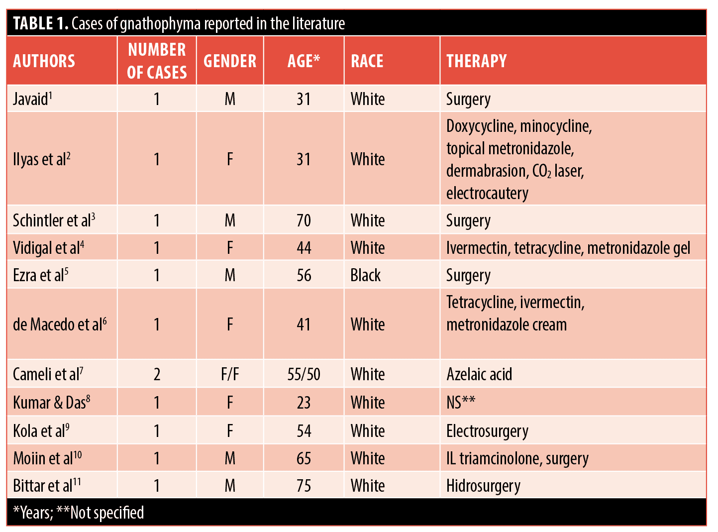
Phyma is considered an uncommon presentation of the last stage of rosacea, but it can also appear independent from rosacea. The most common phyma is rhinophyma; however, phyma of the forehead (e.g., metophyma), the eyelids, uni- or bilaterally (e.g., blepharophyma), the ears, uni- or bilaterally (e.g., otophyma) and the chin (e.g., gnathophyma) can occur. Phyma is characterized clinically by telangiectasias and disfiguring, erythematous papules, nodules and plaques. The consistency is parenchymatous and hard. Gnathophyma, in particular, has been very rarely reported in the literature; to our knowledge, only 11 articles including a total of 12 patients have been published (Table 1).1–11 We describe two cases of gnathophyma with an accompanying review of the literature.
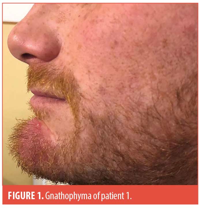
Patient 1. A 33-year-old White male patient was admitted to our clinic with a diagnosis of gnathophyma. The patient stated that he was in good general health and that he was not in therapy with systemic drugs. He also declared that the dermatitis appeared at the age of 28 years and that it was unsuccessfully treated with topical metronidazole and ivermectin for four months, as well as oral doxycycline, 40mg/day, and limecycline, 600mg/day, for three months. The patient did not have a history of acne or rosacea. Dermatological examination revealed an enlargement of the chin with slight erythema (Figure 1), and a parenchymatous, hard consistency. No lesions in other sites were found.
General physical examination did not reveal anything pathological. Laboratory tests were within normal ranges, except for high levels of total cholesterol (320mg/dl). Tests for Helicobacter pylori were negative. Microscopic examinations of scrapings from the chin revealed only one Demodex folliculorum/cm2. Histopathologic examination of a 4mm punch biopsy showed numerous hypertrophic sebaceous glands, a perivascular and perifollicular lymphoplasmocytic infiltrate in upper and mid dermis, and fibrosis. It was decided not to prescribe oral isotretinoin because of high total cholesterol levels. The patient did not want to move forward with a surgical approach.
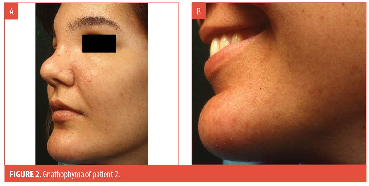
Patient 2. An 18-year-old White female patient was admitted with a diagnosis of gnathophyma. The patient reported being well and that she was not in therapy with systemic drugs. She claimed that the dermatitis appeared one year earlier and that it had not yet been treated. The patient had no history of acne or rosacea. Dermatological examination showed an enlargement of the chin without the presence of abnormal coloring, but with parenchymatous-hard consistency (Figure 2a and 2b). No lesions in other sites were found. General physical examination did not reveal anything pathological. Laboratory tests were within normal ranges. Tests for Helicobacter pylori were not performed. Microscopic examinations of scrapings from the chin were negative for Demodex folliculorum. A biopsy was not performed. The patient’s parents did not want to move forward with any laser or surgical treatment options. The patient was treated with doxycycline 40mg/day for three months; however, clinical results were very poor. The patient was lost to follow up.
We identified 12 cases of gnathophyma reported in the literature. All patients were White, except one Black patient.5 Seven patients were female, and five were male. All patients were adults, with an age ranging from 23 to 75 years (mean age: 49.6 years). Suprisingly, gnathophyma appeared to be very rarely associated with rosacea.1,2,4–10 As previously mentioned, both of our patients discussed here had negative medical history and clinical picture for rosacea. In two patients, gnathophyma was associated with severe tooth decay.2,7 In one patient, gnathophyma significantly improved following treatment of bacterial plaque and gingivitis.7 Demodex folliculorum was detected in only 2 out of 12 patients.1,4 Often, pharmacological treatments were found to be inadequate as treatment.2,4 Various laser and surgical approaches appeared to be more effective.1–3,5,9–11 However, no controlled clinical studies have been published so far.
With regard,
by Stefano Veraldi, MD, PhD; Rossana Schianchi, MD; and Gianluca Nazzaro, MD
Affiliations. Dr. Veraldi is with Dermatological Centre in Milan, Italy. Dr. Schianchi is with the European Institute of Dermatology in Milan, Italy. Dr. Nazzaro is with Dermatology Unit, Foundation IRCCS Ca’ Granda Ospedale Maggiore Policlinico in Milan, Italy.
Keywords. Phyma, rosacea, gnathophyma
Funding. No funding was provided for this article.
Disclosures. The authors report no conflicts of interest relevant to the content of this article.
References:
- Javaid M. Rosacea of the chin. J R Soc Med. 1997;90:528.
- Ilyas EN, Hanson MR, Lawrence N, et al. Gnatophyma: a rare rosacea phyma variant. J Am Acad Dermatol. 2006;55:165–166.
- Schintler MV, Arbab E, Aberer W, et al. Surgical management of extensive gnathophyma. J Eur Acad Dermatol Venereol. 2006;20:1325–1327.
- Vidigal MR, Kakihara CT, Gatti TR, et al. Gnatophyma: a rare variant of phyma. Clin Exp Dermatol. 2008;33:743–744.
- Ezra N, Greco JF, Haley JC, et al. Gnathophyma and otophyma. J Cutan Med Surg. 2009;13:266–272.
- de Macedo ACL, Dias Pacheco Sakai F, et al. Gnatophyma – a rare form of rosacea. An Bras Dermatol. 2012;87:903–905.
- Cameli N, Cavallotti C, Muscardin L, et al. Two cases of gnatophyma, an unusual form of rosacea. Skin Appendage Disord. 2017; 2:180–182.
- Kumar P, Das A. Gnathophyma. Skinmed. 2018;16:45.
- Kola E, Alimehmeti M, Belba G. Rhinophyma and gnathophyma concomitantly in a 54-year-old female – A clinical-histopathology correlation and review of the literature. J Clin Rev Case Rep. 2018;3:1–3.
- Moiin A, Mahmood SH, Kurtovic A. Stepwise surgical treatment of gnathophyma. Dermatol Surg. 2019; 45:158–160.
- Bittar JM, Kovach SJ, Ditre CM. Severe phymatous rosacea of the nose, cheeks, and chin treated with hydrosurgery. Cutis. 2020;106:37–39.
OX40/OX40L Costimulatory Pathway: A Potential Therapeutic Target for Allergic Contact Dermatitis?