Michael Digby, MD; Roberto Belli, MS4; Timothy McGraw; Abigail Lee, MD
Department of Dermatology, San Antonio Uniformed Services Health Consortium, Lackland Air Force Base, Texas
Disclosure: The authors report no relevant conflicts of interest.
Abstract
A 28-year-old veteran presented to the Wilford Hall dermatology clinic in San Antonio, Texas, with a six-month history of a rash that his primary care physician diagnosed as molluscum contagiosum. The rash consisted of clusters of 3 to 6mm yellowish papules with erythematous borders that concentrated on the extensor surfaces of his extremities, lower back, and buttocks. A biopsy determined that the patient had a case of eruptive xanthoma. Subsequent laboratory testing revealed that the patient had a type IV familial hyperlipidemia with a triglyceride count of 1718mg/dL. (J Clin Aesthet Dermatol. 2011;4(1):44–46.)
Cutaneous manifestations of systemic diseases can be an early warning sign or a late manifestation of chronic disease. All practitioners should be familiar with common dermatological symptoms of generalized medical conditions so that they may properly recognize such symptoms and order proper diagnostic studies, diagnose and treat the patient, or refer the patient to the proper specialist.
A 28-year-old male veteran presented to the dermatology clinic with a cutaneous manifestation of a serious underlying condition. He has a history of uncontrolled type 2 diabetes, nonalcoholic steatohepatitis (NASH), hyperlipidemia, a traumatic above-knee amputation of the left leg, and post-traumatic stress disorder. He presented to the clinic with a six-month history of what he called “warts.” These began on his elbows and then spread to his right knee and buttocks. The lesions began as small red bumps that grew in size and became more yellow in color. The lumps were not pruritic unless they were traumatized. He had no prior history of this happening previously and could not recall anyone else in his family having a similar condition.
He had been seen by his primary care provider and was diagnosed with molluscum contagiosum. His condition was treated with 0.5% triamcinolone ointment. The patient’s spouse was very upset by the diagnosis and was afraid of the supposed infection spreading to her or their children. As such, she was reluctant to have her husband touch her or the children.
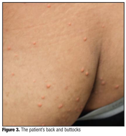
On exam in the dermatology clinic, the patient had 3 to 6mm yellowish papules with erythematous borders and central lobulation that were too numerous to count. The papules were distributed across the elbows, right knee, lower back, and buttocks (Figure 1, Figure 2, Figure 3, and Figure 4). The face, hands, flexor surfaces, and genitals were spared.
His diagnosis of molluscum contagiosum was unlikely due to the morphology and the distribution of the papules. Molluscum is usually a sexually transmitted infection in adults and therefore most commonly found in the genital area.
To secure a diagnosis, a shave biopsy of one of the papules was sent to pathology. The report from pathology found dermal histolytic inflammation with extracellular lipid deposition (Figures 5 and 6). The information from the biopsy combined with the clinical presentation and the history pointed to the dermatological diagnosis of eruptive xanthoma.
Discussion
High levels of serum triglycerides or uncontrolled diabetes mellitus generally cause eruptive xanthoma.[1–3] This patient had a history of both elevated lipoproteins and diabetes mellitus and was at high risk for development of eruptive xanthomas. The patient was sent for lab work for serum lipoprotein levels. Apolipoprotein testing was also ordered to determine if the xanthomas were a manifestation of an inherited condition.[4]
Seven days before coming to the clinic, the patient’s hemoglobin A1c was 14.7 percent and his estimated glucose was 375.5mg/dL. In 2007, the patient had a reported triglyceride level of 494mg/dL. Labs from the authors’ evaluation revealed a triglyceride level of 1478mg/dL. In addition, his total cholesterol level was 323mg/dL and his high-density lipoprotein was 36mg/dL. Due to the high triglyceride levels in the sample, his low-density lipoprotein (LDL) and very-low-density lipoprotein (VLDL) could not be calculated. An apolipoprotein evaluation could not be performed because the markedly elevated triglycerides of the sample made it unsuitable for testing. However, on follow-up electrophoresis, the patient’s triglycerides were 1718mg/dL, cholesterol was 340mg/dL, chylomicrons were moderately increased, LDL was normal, VLDL was markedly increased, and HDL was decreased. The appearance of the sample was milky and the lab interpreted the pattern as being consistent with type IV hyperlipoproteinemia.
Conclusion
The results indicate that this patient is at a great risk for both pancreatitis and early coronary artery disease.[3–5] He would benefit from early and intense treatment to lower his triglyceride levels. The treatments for eruptive xanthomas and hypertriglyceridemia are the same and involve rigorous control of the diabetes, a low-fat diet, and pharmaceutical treatment.[3,5] This patient was referred back to his endocrinologist given his poorly controlled concomitant diabetes mellitus type 2.
Recognizing eruptive xanthomas and being aware of its association with hypertriglyceridemia can help to decrease any lag between a patient being seen by a physician and treatment for a serious medical condition.
References
1. Wolff K, Johnson RA. Fitzpatrick’s Color Atlas and Synopsis of Clinical Dermatology. 6th ed. New York: McGraw Hill; 2009.
2. Fauci AS, Braunwald E, Kasper DL, Hauser SL. Harrison’s Principles of Internal Medicine. 17th ed. New York: McGraw Hill; 2008.
3. Durrington P. Dyslipidaemia. Lancet. 2003;362:717–731.
4. Sarwar N, Danesh J, Eiriksdottir G, et al. Triglycerides and the risk of coronary heart disease: 10,158 incident cases among 262,525 participants in 29 Western prospective studies. Circulation. 2007;115(4):450–458.
5. Brunzell JD. Clinical practice. Hypertriglyceridemia. N Engl J Med. 2007;357(10):1009–1017.
OX40/OX40L Costimulatory Pathway: A Potential Therapeutic Target for Allergic Contact Dermatitis?