 J Clin Aesthet Dermatol. 2023;16(12 Suppl 2):S14–S15
by Archana M. Sangha, MMS, PA-C
Ms. Sangha is a medical science liaison for Incyte in Wilmington, Delaware. Prior to that, she spent over a decade as a dermatology PA specializing in general, surgical, and cosmetic dermatology. She is a fellow of the American Academy of Physician Assistants in Alexandria, Virginia. She is also Immediate Past President of the Society of Dermatology Physician Assistants.
FUNDING: No funding was provided for this article.
DISCLOSURES: Ms. Sangha is an employee of Incyte in Wilmington, Delaware.
The worldwide prevalence of rosacea is estimated to be between 5.5 percent and 10 percent.1,2 It is a chronic, inflammatory skin disorder that is most commonly associated with fair-skinned patients. In Ireland, the incidence is estimated to be 14 percent, and in Germany, it is estimated to be 12 percent.3,4 The incidence rates in skin of color (SOC) patients are estimated to be much lower. Rosacea is thought to affect two percent of Black patients, 3.9 percent of Hispanic of Latino patients, and 2.3 percent of Asian/Pacific Islanders.5 The lower incidence rate of rosacea in SOC patients may be due to genetics and environmental factors, but it may also be due to a lower index of suspicion.9 At present time, there is no evidence to demonstrate if certain therapeutics respond better in SOC populations, thus it is recommended to follow the current management guidelines from the American Acne & Rosacea Society.10 Here, I review the nuances of diagnosing and treating rosacea in skin of color patients.
Five Pearls for Recognizing and Managing Rosacea in Skin of Color
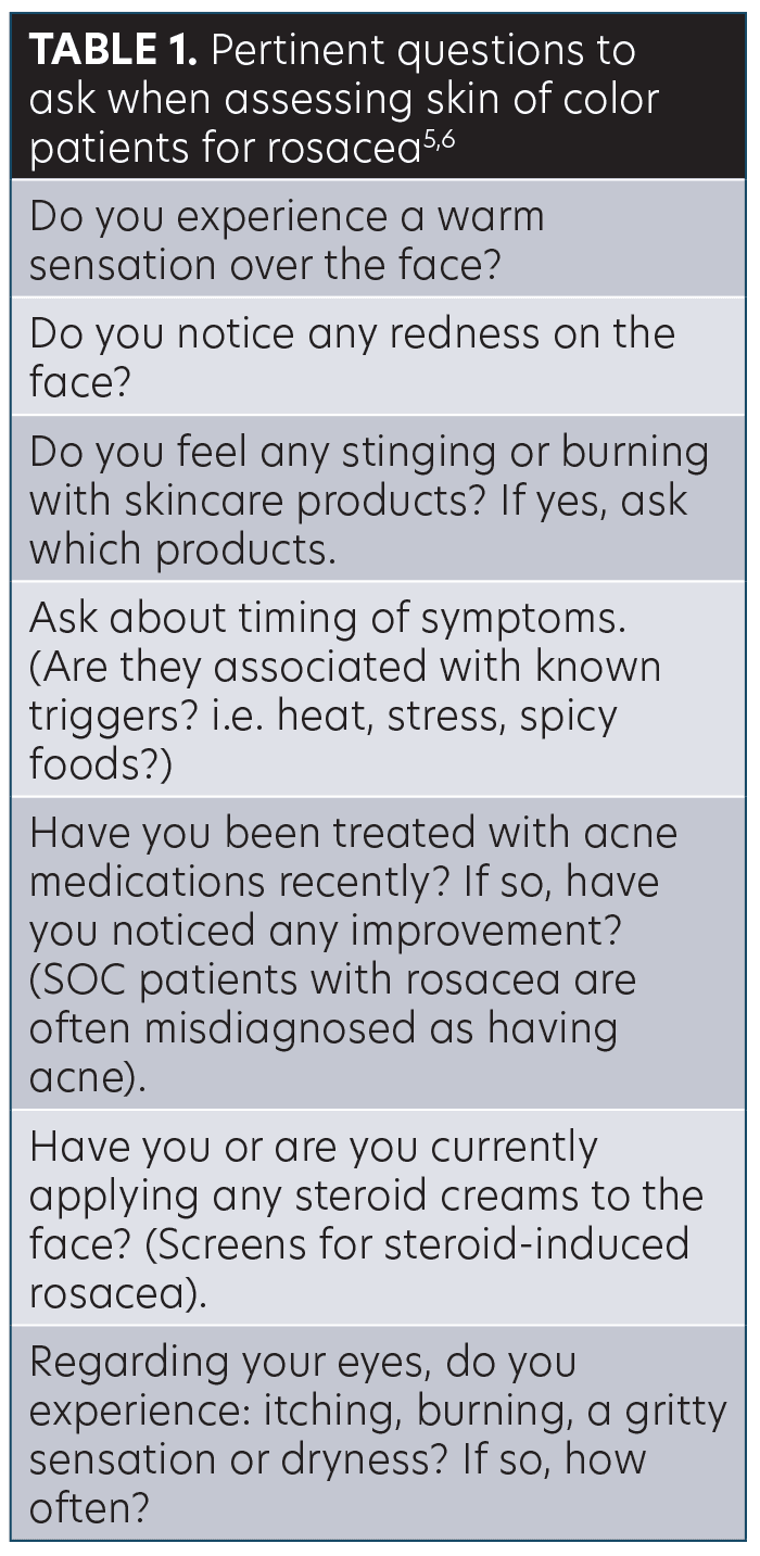
1Ask questions. The hallmark signs of rosacea include persistent facial erythema, papules, pustules, telangiectasia, and recurrent flushing. Three of these five hallmark signs (facial erythema, telangiectasia, and recurrent flushing) are more difficult to visualize in skin of color patients due to their richly pigmented skin. Thus, a thorough patient history is necessary. It’s important to not only screen for cutaneous symptoms but also ocular symptoms . One study found the incidence of ocular rosacea in female SOC patients to be 77 percent.13 See Table 1 for pertinent questions to ask.5
Look for non-classic signs. Patients with skin of color may present with dry skin, edema, and hyperpigmentation. In addition, look for skin thickening on the nose and medial cheeks as these may be early signs of phymatous changes.5 In Black patients, it may be difficult to distinguish granulomatous rosacea from sarcoidosis. Look for several firm, monomorphic papules affecting the perioral, periocular, medial, or lateral areas of the cheeks. The papules may be skin-colored, violaceous, or brown. Flushing and erythema are also less likely to occur in granulomatous rosacea.11,12
Use tools to help identify telangiectasa. A dermatoscope can be used aid in the visualization of telangiectasa. Alternatively, diascopy can be used to. Diascopy is the act of pressing a microscope slide against the skin and assessing for blanching.6 If the skin blanches, diascopy is positive for erythema from vasodilation.
Educate patients on proper skincare. Many cultures think facial dermatoses are due to improper cleansing and will often use alkaline soaps such as glycerine and black soap. However, these soaps can further impair the skin barrier due to their ability to increase the skin pH. SOC patients also tend to more frequently use astringents and toners, which can cause skin irritation.6 Counsel patients to use products that have barrier repairing properties, such as ceramides, hyaluronic acid, and niacinamide.8 Encourage use of light, water-based moisturizers and avoid gels and thin lotions.6
Recommend adherence to daily sunscreen. Often SOC patients believe their darker complexion negates the need to apply daily sunscreen. It’s important to dispel this myth and reinforce that sun protection in the setting of rosacea is particularly important, regardless of skin color. In a review by Morgado-Carrasco et al,7 ultraviolet (UV) light was found to impact skin inflammation, neoangiogenesis, telangiectasa/fibrosis, and may also initiate rosacea. A broad spectrum sunscreen with a minimum sun protection factor (SPF) of 30 should be recommended.
References
- Gether L, Overgaard LK, Egeberg A, Thyssen JP. Incidence and prevalence of rosacea: a systematic review and meta-analysis. Br J Dermatol;2018;179(2):282–289.
- Tan J, Schofer H, Araviiskaia E, et al. RISE study group. Prevalence of rosacea in the general population of Germany and Russia – The RISE study. J Eur Acad Dermatol Venereol. 2016;30:428-34.
- McAleer MA, Fitzpatrick P, Powell FC. The prevalence and pathogenesis of rosacea. Poster presentation, British Association of Dermatologists annual meeting. Liverpool, UK:1–4 Jul 2008.
- Berg M, Liden S. An epidemiological study of rosacea. Acta Dermato-Venereologica. 1989;69:419-423.
- Alexis AF, Callender VD, Baldwin HE, et al. Global epidemiology and clinical spectrum of rosacea, highlighting skin of color: review and clinical practice experience. J Am Acad Dermatol. 2019;80(6):1722–1729.
- Maliyar K, Abdulla SJ. Dermatology: how to manage rosacea in skin of colour. Drugs in Context. 2022;11.
- Morgado-Carrasco D, Granger C, Trullas C, et al. Impact of ultraviolet radiation and exposome on rosacea: key role of photoprotection in optimizing treatment. J Cosmetic Dermatol. 2021;20(11):3415–3421.
- Baldwin H, Alexis AF, Andriessen A, et al. Evidence of barrier deficiency in rosacea and the importance of integrating OTC skincare products into treatment regimens. J Drug Dermatol. 2021;20(4):384–392.
- Rainer BM, Kang S, Chien AL. Rosacea: epidemiology, pathogenesis, and treatment. Dermato-endocrinology. 2017;9(1):e1361574.
- Del Rosso J, Tanghetti E, Webster G, et al. Update on the Management of Rosacea from the American Acne & Rosacea Society (AARS). J Clin Aesthet Dermatol. 2020;13(6 Suppl):S17-S24
- Wilkin J, Dahl M, Detmar M, et al. Standard classification of rosacea: report of the National Rosacea Society Expert Committee on the Classification and Staging of Rosacea. J Am Acad Dermatol. 2002;46:584-587.
- Kelati A, Mernissi FZ. Granulomatous rosacea: a case report. J Med Case Rep. 2017;11:230.
- Al-Balbeesi AO, Almukhadeb EA, Halawani MR, et al. Manifestations of ocular rosacea in females with dark skin types. Saudi J Ophthalmol. 2019;33(2):135–141.


