 J Clin Aesthet Dermatol. 2020;13(6):46–47
by Alysha Colon, BS; Ryan Gillihan, MD; Kiran Motaparthi MD; and Maria Isabel Longo MD,PhD
Ms. Colón is with the University of Florida College of Medicine in Gainesville, Florida. Drs. Gillihan, Motaparthi, and Longo are with the Department of Dermatology at the University of Florida in Gainesville, Florida.
FUNDING: No funding was provided for this study.
DISCLOSURES: The author has no conflicts of interest relevant to the content of this article.
ABSTRACT: Desmoplastic trichilemmoma is a rare histological variant of a benign tumor of the pilosebaceous hair follicle that often clinically appears as similar in appearance to other cutaneous lesions. Here, an 81-year-old male patient with desmoplastic trichilemmoma found on the left zygoma is presented. During the dermatoscopic evaluation of the neoplasm, crown vessels with radial distribution in the periphery were displayed. Histopathologic evaluation revealed peripheral palisading lobules of tumor cells surrounded by sclerotic hyalinized stroma displacing the vessels of the tumor. This case highlights the value of using dermoscopy for improving the clinical diagnosis of desmoplastic trichilemmoma. These findings highlight a need to further investigate the diagnosis of desmoplastic trichilemmoma when crown vessels are displayed during the clinical evaluation.
Keywords: Crown vessels, desmoplastic trichilemmoma, dermoscopy
Trichilemmomas are benign adnexal tumors of the pilosebaceous hair follicle exhibiting outer root sheath differentiation and lobules of epithelial cells descending into the dermis. An uncommon histological variant, desmoplastic trichilemmoma displays these typical findings at the periphery, with a central fibroblast-mediated desmoplastic reaction distinguishing the lesion.1,2 The clinical features of these solitary neoplasms lead to a broad differential diagnosis, including basal cell carcinoma, sebaceous hyperplasia, trichilemmal carcinoma, molluscum contagiosum, and other adnexal tumors. Dermoscopy has demonstrated its utility in narrowing and improving the clinical diagnosis of cutaneous lesions.3 Dermoscopic patterns of other uncommon skin tumors have been described in the literature to provide aid in establishing the clinical diagnosis.3,4 The dermoscopic features of desmoplastic trichilemmoma specifically reveal crown vessels.5 In this case report, we attempt to further evaluate these features and correlate them with the pathology of the neoplasm.
Case Presentation
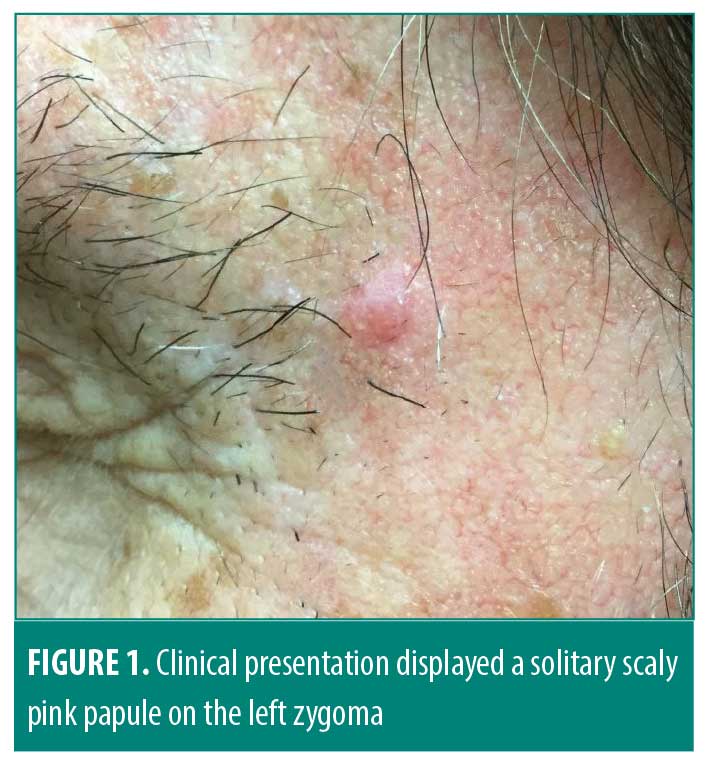
An 81-year-old male with a history of pyoderma gangrenosum and nonmelanoma skin cancer presented for evaluation of an asymptomatic pink papule on the left temple. During his physical examination, a solitary 0.5-cm pink scaly papule was observed on the left zygoma (Figure 1). A shave biopsy was performed to make the final diagnosis.

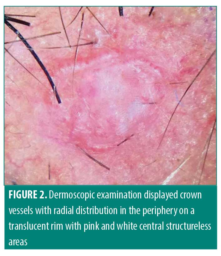
The dermatoscopic evaluation displayed crown vessels, consistent with features previously described in the literature, with radial distribution in the periphery on a translucent rim with pink and white central structureless areas (Figure 2).5

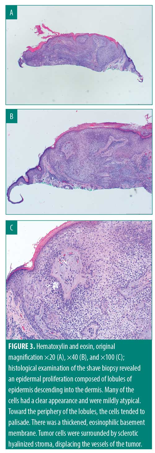
Histopathologic evaluation of the shave biopsy showed an epidermal proliferation composed of radial lobules of epidermis descending into the dermis, where many of the cells had a clear appearance due to the presence of glycogen in their cytoplasm and appeared mildly atypical. Toward the periphery of lobules, the cells tended to palisade. There was a thickened, eosinophilic basement membrane. Tumor cells appeared to be surrounded by sclerotic hyalinized stroma displacing the vessels of the tumor (Figures 3A–3C).
Discussion
Desmoplastic trichilemmoma is a variant of a benign tumor arising from the external sheath of hair follicles. Their nonspecific clinical appearance can present similarly to invasive carcinomas and lead to many differentials.6
Dermoscopy of desmoplastic trichilemmoma revealing peripheral crown vessels with pink and white central structureless areas histopathologically corresponds to a dermoplastic response involving the central infiltrating cords of sclerotic hyalinized stroma.6,7 In this context, the tumor grows downward into the dermis in a radial pattern (Figure 3C). The lobular growth surrounds the stroma, displacing the vessels of the tumor to the periphery and likely causing the appearance of the associated peripheral crown vessels described in this report.6,7

Conclusion
It is useful to describe the dermoscopic findings of desmoplastic trichilemmoma to aid in the clinical diagnosis of these lesions and to distinguish them from other cutaneous tumors with differentiating dermoscopic features. Biopsy and histologic examination are essential for making the diagnosis and determining patient care to rule out high mitotic activity, extensive mucinous stroma, and cytologic atypia suggestive of carcinoma.
References
- Hunt SJ, Kilzer B, Santa Cruz DJ. Desmoplastic trichilemmoma: histologic variant resembling invasive carcinoma. J Cutan Pathol. 1990;17(1): 45–52.
- Tellechea O., Reis JP, Baptista AP. Desmoplastic trichilemmoma. Am J Dermatopathol. 1992;14(2):107–104.
- Bryden AM, Dawe RS, Fleming C. Dermatoscopic features of benign sebaceous proliferation. Clin Exp Dermatol. 2004;29(6):676–677.
- Lallas A, Moscarella E, Argenziano G et al. Dermoscopy of uncommon skin tumours. Australas J Dermatol. 2014;55(1):53–62.
- Navarrete-Dechent C, Uribe P, González S. Desmoplastic trichilemmoma dermoscopically mimicking molluscum contagiosum. J Am Acad Dermatol. 2017;76(2S1):S22–S24.
- Kaptan MA, Kattampallil J, Rosendahl C. Trichilemmoma in continuity with pigmented basal cell carcinoma; with dermatoscopy and dermatopathology. Dermatol Pract Concept. 2015;5(2):57–59.
- Sano DT, Yang JJ, Tebcherani AJ, Bazzo LA. A rare clinical presentation of desmoplastic trichilemmoma mimicking invasive carcinoma. An Bras Dermatol. 2014;89(5):796–798.

