
Erica C. Davis, MD; Callender Skin & Laser Center, Glenn Dale, Maryland
Valerie D. Callender, MD, Callender Skin & Laser Center, Glenn Dale, Maryland and Department of Dermatology, Howard University College of Medicine, Washington, DC
Disclosure: Dr. Davis reports no relevant conflicts of interests. Dr. Callender is a consultant and speaker for Allergan, Coria, Medicis, Galderma, Sanofi-Aventis, and Stiefel. She is also a researcher for Indendis, Medicis, and Merz.
Abstract
Postinflammatory hyperpigmentation is a common sequelae of inflammatory dermatoses that tends to affect darker skinned patients with greater frequency and severity. Epidemiological studies show that dyschromias, including postinflammatory hyperpigmentation, are among the most common reasons darker racial/ethnic groups seek the care of a dermatologist. The treatment of postinflammatory hyperpigmentation should be started early to help hasten its resolution and begins with management of the initial inflammatory condition. First-line therapy typically consists of topical depigmenting agents in addition to photoprotection including a sunscreen. Topical tyrosinase inhibitors, such as hydroquinone, azelaic acid, kojic acid, arbutin, and certain licorice extracts, can effectively lighten areas of hypermelanosis. Other depigmenting agents include retinoids, mequinol, ascorbic acid, niacinamide, N-acetyl glucosamine, and soy with a number of emerging therapies on the horizon. Topical therapy is typically effective for epidermal postinflammatory hyperpigmentation; however, certain procedures, such as chemical peeling and laser therapy, may help treat recalcitrant hyperpigmentation. It is also important to use caution with all of the above treatments to prevent irritation and worsening of postinflammatory hyperpigmentation. (J Clin Aesthetic Dermatol. 2010;3(7):20–31.)
Postinflammatory hyperpigmentation (PIH) is an acquired hypermelanosis occurring after cutaneous inflammation or injury that can arise in all skin types, but more frequently affects skin-of-color patients, including African Americans, Hispanics/Latinos, Asians, Native Americans, Pacific Islanders, and those of Middle Eastern descent. PIH can have a significant psychosocial impact on skin-of-color patients (Fitzpatrick skin types IV through VI), as these pigmentary changes can occur with greater frequency and severity in these populations1 and oftentimes be more obvious in darker skin. However, there is a wide variety of safe and effective treatments for PIH in skin of color, including topical depigmenting agents, chemical peels, and laser and light therapy. Therefore, this article reviews the etiology, pathogenesis, clinical manifestations, and treatment options for PIH in skin of color.
Epidemiology
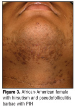
Table 1 shows the worldwide prevalence of pigmentary disorders, excluding vitiligo, in various ethnic populations. These figures typically include postinflammatory hyper- or hypopigmentation although melasma and solar lentigines may also be included. Multiple epidemiological studies have shown that PIH tends to occur more commonly among skin-of-color patients compared to Caucasian patients.[2,3] In 1983, Halder et al[2] published a study comparing the most common dermatoses seen in African American and Caucasian patients. Pigmentary disorders, other than vitiligo, were the third most common dermatoses among African-American patients (9%), but were the seventh most common among Caucasian patients (1.7%). A more recent study in 2007[3] confirmed these findings showing dyschromias to be the second most common diagnosis among African-American patients, but dyschromias failed to make it into the top 10 most common diagnoses for Caucasian patients. Of interest, in a study conducted in Singapore,[4] the authors note that PIH tended to also be more prevalent among Asians with darker skin, such as Malays and Indians, than those with lighter skin, such as the Chinese, suggesting that the degree of pigmentation rather than race/ethnicity may be more contributory to the development of PIH.
Etiology
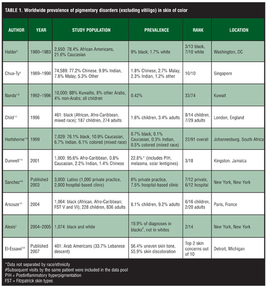
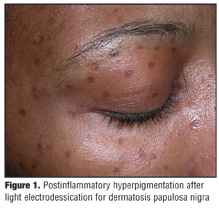
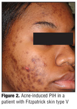
Many types of inflammatory dermatoses or cutaneous injuries can cause pigmentary changes; however, there are some diseases that show a proclivity to develop PIH rather than hypopigmentation. A wide range of etiologies for PIH exists including infections such as dermatophytoses or viral exanthems, allergic reactions such as those from insect bites or a contact dermatitis, papulosquamous diseases like psoriasis or lichen planus, medication-induced PIH from hypersensitivity reactions, or cutaneous injury from irritants, burns, or cosmetic procedures (Figure 1).[5] However, very common causes of PIH in skin of color include acne vulgaris, atopic dermatitis, and impetigo. In fact, PIH is a particularly common sequela after acne in dark-skinned patients (Figure 2). A study in 2002 evaluating acne in skin of color found that 65.3 percent of African-American (N=239), 52.7 percent of Hispanic (N=55), and 47.4 percent of Asian (N=19) patients developed acne-induced PIH.[6] Pseudofolliculitis barbae (PFB) is another common inflammatory dermatosis, particularly among African Americans, that results in PIH and is estimated to have a prevalence rate between 45 and 83 percent.[7,8] In a study by Perry et al[9] of 71 African American and Hispanic patients with PFB, 90.1 percent of patients reported hyperpigmentation; therefore, the authors suggest that PIH may be a major clinical finding in PFB (Figure 3).
Pathogenesis
PIH results from the overproduction of melanin or an irregular dispersion of pigment after cutaneous inflammation.10 When PIH is confined to the epidermis, there is an increase in the production and transfer of melanin to surrounding keratinocytes. Although the exact mechanism is unknown, this rise in melanocyte activity has been shown to be stimulated by prostanoids, cytokines, chemokines, and other inflammatory mediators as well as reactive oxygen species that are released during the inflammatory process. Multiple studies have demonstrated the melanocyte-stimulating properties of leukotrienes (LT), such as LT-C4 and LT-D4, prostaglandins E2 and D2, thromboxane-2, interleukin (IL)-1, IL-6, tumor necrosis factor (TNF)-a, epidermal growth factor, and reactive oxygen species such as nitric oxide.[1,5,11,12]
PIH within the dermis results from inflammation-induced damage to basal keratinocytes, which release large amounts of melanin. The free pigment is then phagocytosed by macrophages, now called melanophages, in the upper dermis and produces a blue-gray appearance to the skin at the site of injury.[13,14]
Clinical Manifestations
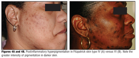
PIH typically manifests as macules or patches in the same distribution as the initial inflammatory process. The location of the excess pigment within the layers of the skin will determine its coloration. Epidermal hypermelanosis will appear tan, brown, or dark brown and may take months to years to resolve without treatment.[1] Hyperpigmentation within the dermis has a blue-gray appearance and may either be permanent or resolve over a protracted period of time if left untreated.[1,15] The intensity of PIH may also correlate with higher skin phototypes (Figure 4), although studies are needed to confirm this finding. In addition, PIH can worsen with ultraviolet (UV) irradiation or with persistent or recurrent inflammation.[16]
Treatment
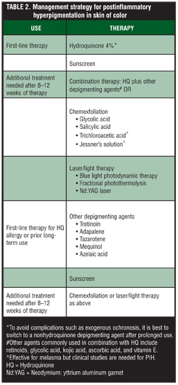
The management of PIH should begin first with addressing the underlying inflammatory dermatosis. Initiating treatment early for PIH may help hasten its resolution and prevent further darkening. However, it is important to always be mindful of the potential the treatment itself has to cause or exacerbate PIH by causing irritation.[17] There are a variety of medications and procedures in addition to photoprotection that can safely and effectively treat PIH in darker skinned patients. Topical depigmenting agents, such as hydroquinone, azelaic acid, kojic acid, licorice extract, and retinoids, can be effective alone or in combination with other agents, and procedures such as chemexfoliation and laser therapy can also be incorporated into the management strategy if needed (Table 2). Of note, topical agents are typically used to treat epidermal PIH as deeper pigmentation does not respond well to these agents.[16]
Photoprotection. An integral part in the treatment of PIH that should not be overlooked or underestimated is the importance of photoprotection to prevent the worsening of PIH. Patients should be educated on the use of daily broad-spectrum sunscreen with a sun protection factor (SPF) of 30 and sun-protective measures, such as avoidance and protective clothing. This is particularly true for those with higher skin phototypes who may not normally wear sunscreen, but also may not realize the darkening effects UV irradiation has on hyperpigmentation. In fact, a study analyzing data from the 1992 National Health Interview Survey of 1,583 African Americans regarding sun-protection behaviors found that only a minority of the respondents were very likely to use sunscreen (9 vs. 81% who were unlikely to use it), wear protective clothing (28%), or stay in the shade (45%).[18]
Although clinical studies have shown that serum vitamin D levels are reduced in sunscreen users compared to nonusers, these levels are still within normal range.[19,20] This is particularly important for dark-skinned individuals who may already be at risk for vitamin D deficiency due to inherently higher melanin concentrations in the skin.[21] The American Academy of Dermatology has released a position statement on vitamin D, which states that groups at risk for vitamin D deficiency, including dark-skinned individuals, may need a daily total dose of 1000IU of vitamin D, through diet and supplementation, according to the current United States Department of Agriculture Dietary Guidelines.[22] Therefore, proper counseling and education involves encouraging the daily use of a broad-spectrum sunscreen with an SPF of 30; sun-protective measures, such as avoidance and protective clothing; the intake of foods rich in vitamin D, such as salmon, fish liver oils, and fortified foods; and vitamin D supplementation.
Medical therapy. Hydroquinone (HQ). The mainstay of treatment for PIH remains HQ. It is a phenolic compound that blocks the conversion of dihydroxyphenylalanine (DOPA) to melanin by inhibiting tyrosinase.[10,23] Its mechanism of action may also involve inhibition of deoxyribonucleic acid (DNA) and ribonucleic acid (RNA) synthesis, selective cytotoxicity toward melanocytes, and melanosome degradation.[24,25] HQ is commonly used at concentrations from 2 to 4% but can be prescribed in strengths up to 10% and is available over the counter (OTC) at 2% in the United States.
Hydroquinone monotherapy can be effective in treating PIH, but more recently HQ has been formulated with other agents, such as retinoids, antioxidants, glycolic acid, sunscreens, and corticosteroids, to increase efficacy.10 Cook-Bolden et al[26] conducted a 12-week, open-label study of microencapsulated HQ 4% and retinol 0.15% with antioxidants in the treatment of 21 patients (81% Fitzpatrick skin types IV–VI) with PIH (n=17) and melasma (n=4). There were significant decreases in lesion size, pigmentation, and disease severity from Week 4 to the study endpoint (all P<0.032), and reflectance spectrophotometric analysis showed statistically significant reductions in melanin content as early as Week 4 as well. A similar study with a majority of skin-of-color patients treated with microentrapped HQ 4%/retinol 0.15% and sunscreen also found this agent to be safe and effective for both PIH and melasma.[27]
Irritant reactions can result from long-term daily use of 4% or higher HQ, particularly when used in combination with other agents that can be irritating, such as retinoids.[28] However, concomitant use of a topical corticosteroid can reduce irritation, thereby decreasing the risk of further hyperpigmentation.[16] An early formulation, Kligman’s formula containing 5% HQ, 0.1% tretinoin, and 0.1% dexamethasone, is one such combination that was effective yet problematic due to its use of high concentrations of tretinoin and a potent fluorinated steroid.[10] More recently, less irritative combination agents have been developed including TriLuma® (Galderma, Fort Worth, Texas), which contains 4% HQ, 0.05% tretinoin, and 0.01% fluocinolone acetonide. This triple combination agent has been shown to be both safe and effective in the treatment of melasma[29–32] and photoaging[33] in skin of color and is used successfully in clinical practice to treat PIH. However, formal clinical studies are still needed to further evaluate its use in PIH.
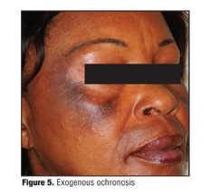
Adverse events reported with HQ use include contact dermatitis, nail discoloration, permanent leukoderma, and hypopigmentation of the surrounding normal skin that has been treated with HQ (“halo effect”).[23] Patients may also develop exogenous ochronosis (EO) (Figure 5) where homogentisic acid accumulates within the dermis causing hyperpigmentation and papules on sun-exposed areas where HQ has been applied to the skin.[10,34,35] EO is typically associated with frequent use of very high concentrations of HQ on a long-term basis although EO can still occur with short-term use of 1 to 2% HQ.[36,37] It is most commonly reported in blacks in South Africa, where the prevalence of EO is high.[6] In the United States, a low number of hydroquinone-induced EO cases have been reported.[38] However, there has been a recent rise in the illicit use of high-concentration HQ available OTC in ethnic stores in the United States.[38]
In 2006, the United States Food and Drug Administration (FDA) released a statement proposing a ban on all OTC HQ agents based on rodent studies, which suggested that oral HQ may be a carcinogen.[37] However, there have been no reports of skin cancers or internal malignancies associated with topical HQ use.[23] To date, a final ruling by the FDA is still pending.
Mequinol. A derivative and alternative to hydroquinone is 4-hydroxyanisole or mequinol. Although the two agents are related, mequinol is thought to be less irritating to the skin than HQ.[39] The drug is available by prescription in a 2% concentration and is typically formulated with 0.01% tretinoin, a retinoic acid and penetration enhancer.[40] The mechanism by which mequinol causes depigmentation may involve a competitive inhibition of tyrosinase; however, the exact pathway is unknown.[40] Several large clinical studies have shown that mequinol effectively treats solar lentigines in all patients[39,41] including ethnic populations42; however, only small clinical studies exist that evaluate its effectiveness in the treatment of PIH.[43–45] One such study[43] compared mequinol 2%/tretinoin 0.01% to HQ 4% cream in 61 skin-of-color patients with mild-to-moderate facial PIH. Patients applied each medication to contralateral sides of the face for 12 weeks. Topical mequinol/tretinoin was shown to be noninferior to 4% HQ as 81 and 85 percent of patients experienced clinical success on the mequinol-treated and the HQ-treated sides of the face, respectively.
Retinoids. Retinoids are structural and functional analogues of vitamin A, and are effective alone or in combination with other agents for the treatment of PIH in ethnic patients. Retinoids exert multiple biological effects that result in skin lightening including the modulation of cell proliferation, differentiation, and cohesiveness; induction of apoptosis; and expression of anti-inflammatory properties.[46] Topical tretinoin, all-trans-retinoic acid, is a naturally occurring metabolite of retinol and first-generation retinoid.[46] Concentrations range from 0.01 to 0.1% and tretinoin can be formulated in creams, gels, and microsphere gels, which allows for the controlled release of tretinoin leading to less irritation.[47,48] A 40-week, randomized, double-blind, vehicle-controlled clinical trial was conducted with 54 black patients to determine the safety and efficacy of 0.1% tretinoin in the treatment of PIH. Tretinoin was significantly more effective than vehicle in lightening PIH lesions when assessed by clinical (P<0.001) and colorimetric (P=0.05) analysis. However, 50 percent of patients developed retinoid dermatitis, which is the concern with using retinoids in skin of color. Starting at lower concentrations and titrating up based on treatment response and choosing more tolerable formulations, such as creams over gels, may help to decrease the risk of exacerbating PIH.[17]
Third-generation retinoids, adapalene and tazarotene, are synthetic topical agents that are also effective in the treatment of PIH. Adapalene is formulated in creams or gels in 0.1 to 0.3% concentrations; whereas, formulations of tazarotene include 0.05 and 0.1% creams or gels. Both agents have been shown in clinical studies to safely and effectively treat PIH, particularly acne-induced PIH, in darker skinned individuals.[49,50] Isotretinoin (13-cis-retinoic acid) is a naturally occurring, first-generation retinoid that is available in both oral and topical formulations. Oral isotretinoin is very effective in the treatment of severe acne; however, there has also been a case reported in the literature of significant resolution of PIH after oral isotretinoin therapy in an Asian patient.[51]
Azelaic acid. Naturally occurring as a dicarboxylic acid isolate from the organism responsible for Pityriasis versicolor, azelaic acid (AA) has been shown to be effective in the treatment of PIH.[10] AA has several mechanisms by which it depigments the skin including tyrosinase inhibition as well as selective cytotoxic and antiproliferative effects toward abnormal melanocytes through the inhibition of DNA synthesis and mitochondrial enzymes.[52,53] Available formulations include a 15% gel, typically used in the treatment of rosacea, or a 20% cream that is commonly used for acne vulgaris and melasma in addition to PIH. Lowe et al[54] conducted a randomized, double-blind, vehicle-controlled trial of 52 patients (Fitzpatrick skin types IV–VI) with facial hyperpigmentation including PIH and melasma to determine the safety and efficacy of 20% AA cream. Patients treated with AA showed greater decreases in pigmentary intensity after 24 weeks of treatment versus vehicle as measured by the investigator’s subjective scale (P=0.021) and chromometric analysis (P<0.039). Side effects were mild and transient. Multiple other studies have also shown the safety and efficacy of AA in skin of color for the treatment of melasma[55–57]; however, larger studies are needed in this patient population with PIH.
Kojic acid. Kojic acid (KA) is a fungal metabolite of certain species of Acetobacter, Aspergillus, and Penicillium.[23,52] Its depigmenting ability originates from a potent inhibition of tyrosinase by chelating copper at the active site of the enzyme.34 KA is available in 1 to 4% concentrations and can be formulated with other lightening agents, including glycolic acid and hydroquinone, to increase efficacy. Multiple studies including Caucasian and Asian patients have shown that combination therapy with 2% KA and hydroquinone improves melasma.[58,59] However, clinical studies are still needed to determine its efficacy in the treatment of PIH. In the United States and parts of Asia, KA is a frequent ingredient in cosmeceutical formulations although contact dermatitis is a common side effect with its use,[10] and multiple clinical studies have demonstrated its increased sensitizing potential.[60,61]
Arbutin. Extracted from the dried leaves of the bearberry shrub or pear, cranberry, or blueberry plants, arbutin is another derivative of HQ, but without the melanotoxic effects.[40,62] Arbutin causes depigmentation by inhibiting not only tyrosinase activity but also melanosome maturation.[23] Although its efficacy is dose-dependent, higher concentrations of arbutin can lead to a paradoxical hyperpigmentation.[63] Synthetic forms of arbutin, alpha-arbutin and deoxyarbutin, exhibit greater ability to inhibit tyrosinase than the naturally occurring compound.[64,65] A clinical study conducted by Boissy et al65 showed 3% deoxyarbutin to be effective in the treatment of solar lentigines in light-skinned patients (n=34), but there was no significant clinical response in the subset of dark-skinned patients (n=16). Arbutin is also used in a variety of cosmeceutical formulations marketed in the United States.[10,23] However, clinical studies evaluating arbutin for the treatment of PIH in higher skin phototypes is lacking.
Niacinamide. Niacinamide is the physiologically active derivative of vitamin B3 or niacin. In-vitro studies show that niacinamide significantly decreases melanosome transfer to keratinocytes without inhibiting tyrosinase activity or cell proliferation, and niacinamide may also interfere with the cell-signaling pathway between keratinocytes and melanocytes to decrease melanogenesis.[66] One of the advantages of niacinamide is its stability being unaffected by light, moisture, acids, alkalis, or oxidizers.[23] The safety and efficacy of niacinamide for PIH in darker skinned individuals has not been studied; however, topical 2 to 5% niacinamide has shown some efficacy when used alone or in combination with N-acetyl glucosamine for the treatment of melasma and UV-induced hyperpigmentation in fair-skinned patients and Asians.[66–68]
N-acetyl glucosamine. N-acetyl glucosamine (NAG) is an amino sugar that is a precursor to hyaluronic acid and is found throughout nature and human tissues.[69] Its depigmenting ability originates from the inhibition of tyrosinase glycosylation, a step necessary in the production of melanin.[69] Glucosamine itself has been reported to decrease melanogenesis; however, formulating a topical agent has been difficult due to its instability. More recently, focus has now shifted to the development of NAG-containing cosmeceuticals given its greater stability, good skin penetration, and overall tolerability.[69] NAG is typically used in 2% concentrations as monotherapy or in combination with niacinamide, which may lead to a greater clinical effect given that there are two different mechanisms of depigmentation at work.[69] Multiple double-blind, controlled clinical trials have shown the safety and efficacy of NAG alone or NAG/niacinamide combination therapy to significantly lighten hyperpigmentation secondary to solar radiation in Caucasian and Japanese patients.[68,69] NAG was generally well tolerated with mild-to-moderate skin irritation reported in a small number of patients. However, large clinical studies are still needed to determine the role of NAG in the management of PIH in all skin types.
Ascorbic acid. L-ascorbic acid (AA) or vitamin C is a naturally occurring antioxidant obtained from certain fruits and vegetables.[23] AA causes skin lightening by interacting with copper ions at the tyrosinase active site and by reducing oxidized dopaquinone, a substrate in the melanin synthetic pathway.[23,70] In addition to skin lightening, other advantages of AA include not only antioxidant effects but some studies also demonstrate anti-inflammatory and photoprotective properties.[71–76] However, early formulations of AA were unstable so esterified derivatives, such as ascorbyl-6-palmitate and magnesium ascorbyl phosphate, were created.[71] AA is typically used in 5 to 10% concentrations and can be formulated with other depigmenting agents, such as hydroquinone, which is generally well tolerated in skin of color given the good safety profile of AA.[10,40] AA and its derivatives have been shown to be safe with some efficacy in certain racial/ethnic populations including Latino and Asian patients; however, most studies involved the treatment of melasma and did not include PIH.[77,78]
Licorice. Licorice root extract (Glycyrrhiza glabra, Glycyrrhiza uralensis) is a common ingredient found in many skin-lightening cosmeceuticals,[40] and is also used in the treatment of a wide variety of diseases even outside the scope of dermatology due to its anti-inflammatory, antiviral, antimicrobial, and anticarcinogenic properties.[79] Some of the active ingredients in licorice root extract include glabridin, which inhibits tyrosinase and possesses anti-inflammatory effects,[80,81] and liquiritin, which does not inhibit tyrosinase but causes depigmentation by melanin dispersion and removal.[82] There are very few clinical trials that study the utility of licorice root extracts in the treatment of dermatological conditions. One study conducted in 20 Egyptian women showed that topical liquiritin cream (1g/day) for four weeks was both safe and effective in the treatment of melasma.[82] Side effects were minimal. Further clinical studies with racial/ethnic patients are needed to evaluate the efficacy of licorice root extract in the treatment of PIH.
Soy. The activation of protease-activated receptor 2 (PAR-2) cell receptors found on keratinocytes mediates the transfer of melanosomes from melanocytes to surrounding keratinocytes.[83] Soy proteins, such as soybean trypsin inhibitor (STI) and Bowman-Birk inhibitor (BBI), inhibit the activation of these cell receptors, and as a result, phagocytosis of melanosomes into keratinocytes is reduced leading to reversible depigmentation.[83] Soy is now being formulated alone or in combination with other agents including retinol and sunscreen into cosmeceuticals, particularly moisturizers, to help reduce the signs of photodamage as well as PIH in all skin types.[84–86] A 16-week, double-blind, placebo-controlled, clinical study of African-American, Hispanic, and Asian patients with Fitzpatrick skin types III to V and acne-induced PIH was conducted to determine the safety and efficacy of an OTC anti-acne treatment containing salicylic acid, retinol, and total soy.[87] There was a significant improvement in PIH with the soy formulation from baseline to study endpoint as well as when compared to placebo. Soy-containing products are generally well tolerated.[40] However, more large-scale clinical trials in skin of color are still needed.
Surgical therapy. Chemical peels. In 2008, chemical peeling was the fourth most common nonsurgical cosmetic procedure performed in the United States,[88] and dyschromias, such as PIH, are one of the most common indications for this procedure in skin of color.[89] For darker skinned individuals, superficial chemical peels, which penetrate into the papillary dermis,[90] are generally well tolerated with good clinical results.[10] However, care should be taken in selecting and using the specific chemical peel to avoid irritation, which can worsen PIH and lead to other complications, such as new areas of dyspigmentation, keloid formation, and hypertrophic scarring.[91] A detailed history, including other dermatological conditions, current oral and topical medications, history of herpes simplex virus (HSV) infection, past reactions to other cosmetic procedures, and a skin examination should be obtained prior to the procedure.[89,90,92]
Glycolic acid (GA), found in sugarcane, is a naturally occurring alpha-hydroxy acid (AHA) that induces epidermolysis, disperses basal layer melanin, and increases dermal collagen synthesis.[90,93] GA concentrations range from 20 to 70%, and neutralization with water or sodium bicarbonate is required to terminate the peel. Burns et al[94] conducted a 22-week clinical study of 16 African-American patients with Fitzpatrick skin types IV to VI and facial PIH. Patients were randomized to either the control group receiving 2% hydroquinone/10% GA gel and 0.05% tretinoin or the peel group receiving the same topical regimen plus six GA peels (50–68%) at three-week intervals. Significant clinical improvement from baseline was noted in the peel group by subjective measures (p<0.02), and colorimetric analysis also showed a trend of more rapid and greater improvement in the peel group. However, there were no significant differences between the two groups by either measure.
Salicylic acid (SA), derived from willow tree bark, is a beta-hydroxy acid that induces keratolysis by disrupting intercellular lipid linkages between epithelioid cells.10,90 Superficial SA peels utilize concentrations ranging from 20 to 30% without the need for neutralization. A study of 24 Korean patients with acne-induced PIH underwent 30% SA peels every two weeks for three months.[95] Colorimetric analysis showed a significant improvement in the level of lightness from baseline to the first post-peel period (p<0.02), but final levels were not significant. However, erythema was significantly decreased (p<0.0001) and improvements were also noted in greasiness, dryness, and scaliness by clinical exam. The safety and efficacy of SA peels in the treatment of PIH has also been demonstrated in even higher skin phototypes V and VI.[96]
Superficial chemical peels can also be obtained using trichloroacetic acid (TCA) or Jessner’s solution, and both agents have been efficacious in the treatment of melasma.[97,98] However, clinical evidence supportive of the use of these agents for PIH in skin of color is lacking.
Most superficial chemical peeling agents are well tolerated by Fitzpatrick skin types IV to VI. Common side effects include erythema, burning sensation, PIH, reactivation of HSV, superficial desquamation, and vesiculation.[89] Other complications include hypopigmentation, hypertrophic scarring, and keloid formation. Patients should also be educated on the importance of photoprotection to prevent or avoid worsening PIH after chemical peeling.
Laser and light-based therapies. Although topical skin-lightening agents remain the treatment of choice for PIH, lasers and light sources may be an effective adjunct to therapy or alternative for treatment failures. However, there is a paucity of literature specifically evaluating the use of devices in the treatment of PIH in all skin types. Green (510nm, 532nm), red (694nm), or near-infrared (755nm, 1064nm) lasers are pigment-specific and generate light used to selectively target intracellular melanosomes.[99] However, due to the wide absorption spectrum of melanin (250nm–1200nm), laser energy intended for deeper targets can be absorbed within the pigmented epidermis, which can lead to complications such as dyschromias, blistering, and scars.[100]
Typically, energy from short wavelength lasers is more efficiently absorbed by epidermal melanin while longer wavelengths penetrate deeper with more selective absorption by dermal targets making them safer to use for darker-skinned patients.100 The use of longer pulse durations and cooling devices can also provide a greater margin of safety while still maintaining efficacy in darker skinned individuals.[100] There have been case reports of the successful treatment of PIH with blue light photodynamic therapy, neodymium-doped yttrium aluminum garnet (Nd:YAG) laser, and fractional photothermolysis in darker skin types[101–103]; however, larger clinical studies are needed to evaluate the role of these lasers as well as other devices, such as intense pulsed light, in the treatment of PIH.
Cosmetic camouflage. Cosmetic camouflage may be useful to conceal pigmentary disorders, vascular lesions, scars, and chronic skin conditions that are not amenable to medical or surgical treatments.[104] These coverage techniques can help alleviate the patient’s distress regarding their appearance and significantly improve quality of life.[105,106] Camouflage can be particularly useful in darker skinned individuals where pigmentary changes may be more noticeable and when highly visible parts of the body are affected by the disease, such as the face, neck, and hands.[107]
The characteristics of a good cosmetic cover include a natural appearance and non-greasy feel, and the cover should also be waterproof, long lasting, and noncomedogenic with easy application.[108] There are four basic foundations: oil-based for dry skin, water-based for dry-to-normal skin, oil-free for oily skin, and water-free, which mixes oils with waxes to form thicker creams that can incorporate higher amounts of pigment to match the patient’s normal skin color.[104] Cosmetic covers can be applied for subtle coverage up to full concealment,109 and color correctors use the lesion’s color opposite to neutralize its intensity.[104]
Emerging therapies. Continued research is constantly fueled by the demand for newer, more effective depigmenting agents. Undecylenoyl phenylalanine 2% has been shown to safely and effectively treat solar lentigines in a recent randomized, double-blind, vehicle-controlled clinical trial.[110] There have also been case reports and clinical studies that have shown that topical 5% methimazole,[111] aloesin,[112] and dioic acid113 can successfully treat hyperpigmentation secondary to a variety of etiologies. Other agents that have shown some depigmenting properties but require further research include 4-(1-phenylethyl)1,3-benzenediol, paper mulberry, ellagic acid, quinolines, piperlonguminine, luteolin, calycosin, emblica, and multivitamins.[10,114,115]
Conclusion
In skin of color, postinflammatory hyperpigmentation can be a common yet troubling sequelae of cutaneous inflammation. However, there are many safe and effective treatments for this patient population including a variety of topical depigmenting agents. It is important to initiate treatment early while at the same time use caution with these agents to prevent further worsening of the hyperpigmentation. Procedures such as chemical peeling and laser therapy provide alternatives or adjuncts to topical therapy. Adding sunscreen to the treatment regimen and patient education regarding sun protection measures will also be beneficial in the management of patients with PIH.
References
1. Chang MW. Disorders of hyperpigmentation. In: Bolognia JL, Jorizzo JL, Rapini RP, eds. Dermatology. 2nd ed. Elsevier Mosby; 2009:333–389.
2. Halder RM, Grimes PE, McLaurin CI, et al. Incidence of common dermatoses in a predominately black dermatologic practice. Cutis. 1983;32:388–390.
3. Alexis AF, Sergay AB, Taylor SC. Common dermatologic disorders in skin of color: a comparative practice survey. Cutis. 2007;80:387–394.
4. Chua-Ty G, Goh CL, Koh SL. Pattern of skin diseases at the national skin centre (Singapore) from 1989–1990. Int J Dermatol. 1992;31:555–559.
5. Taylor SC, Grimes PE, Lim J, et al. Postinflammatory Hyperpigmentation. J Cutan Med Surg. 2009;13:183–191.
6. Taylor SC, Cook-Bolden F, Rahman Z, et al. Acne vulgaris in skin of color. J Am Acad Dermatol. 2002;46(2 Suppl): S98¬S106.
7. Alexander AM, Delph WI. Pseudofolliculitis barbae in the military. A medical, administrative and social problem. J Natl Med Assoc. 1974;66:459–464,479.
8. Edlich RF, Haines PC, Nichter LS, et al. Pseudofolliculitis barbae with keloids. J Emerg Med. 1986;4:283–286.
9. Perry PK, Cook-Bolden FE, Rahman Z, et al. Defining pseudofolliculitis barbae in 2001: a review of the literature and current trends. J Am Acad Dermatol. 2002;46(2 Suppl):S113–S119.
10. Grimes PE. Managment of hyperpigmentation in darker racial ethnic groups. Semin Cutan Med Surg. 2009;28: 77–85.
11. Tomita Y, Maeda K, Tagami H. Melanocyte-stimulating properties of arachidonic acid metabolites: possible role in postinflammatory pigmentation. Pigment Cell Res. 1992;5: 357–361.
12. Ortonne J. Retinoic acid and pigment cells: a review of in-vitro and in-vivo studies. Br J Dermatol. 1992;127(Suppl 41):43–47.
13. Nordlund JJ, Abdel-Malek ZA. Mechanisms for post-inflammatory hyperpigmentation and hypopigmentation. In: Bagnara JT, ed. Advances in Pigment Cell Research: Proceedings of Symposia and Lectures from the Thirteenth International Pigment Cell Conference. Tucson, AZ; October 5–9, 1986. New York, NY: Liss; 1988:219–239.
14. Masu S, Seiji M. Pigmentary incontinence in fixed drug eruptions. Histologic and electron microscopic findings. J Am Acad Dermatol. 1983;8:525–532.
15. Lacz NL, Vafaie J, Kihiczac NI, et al. Postinflammatory hyperpigmentation: a common but troubling condition. Int J Dermatol. 2004;4:362–365.
16. Ruiz-Maldonado R, Orozco-Covarrubias ML. Post-inflammatory hypopigmentation and hyperpigmentation. Semin Cutan Med Surg. 1997;16:36–43.
17. Callender VD. Acne in ethnic skin: special considerations for therapy. Dermatol Ther. 2004;17:184–195.
18. Hall HI, Rogers JD. Sun protection behaviors among African Americans. Ethn Dis. 1999;9:126–131.
19. Sollitto RB, Kraemer KH, DiGiovanna JJ. Normal vitamin D levels can be maintained despite rigorous photoprotection: six years’ experience with xeroderma pigmentosum. J Am Acad Dermatol. 1997;37:942–947.
20. Norval M, Wulf HC. Does chronic sunscreen use reduce vitamin D production to insufficient levels? Br J Dermatol. 2009;161:732–736.
21. Nesby-O’Dell S, Scanlon KS, Cogswell ME, et al. Hypovitaminosis D prevalance and determinants among African American and white women of reproductive age: third National Health and Nutrition Examination Survey, 1988–1994. Am J Clin Nutr. 2002;76:187–192.
22. Position Statement on Vitamin D. American Academy of Dermatology. November 14, 2009. http://www.aad.org/ forms/policies/Uploads/PS/PS-Vitamin%20D%2011-16-09.pdf. Accessed on March 17, 2010.
23. Badreshia-Bansal S, Draelos ZD. Insight into skin lightening cosmeceuticals for women of Color. J Drugs Dermatol. 2007;6:32–39.
24. Halder RM, Richards GM. Management of dyschromias in ethnic skin. Dermatol Ther. 2004;17:151–157.
25. Palumbo A, d’Ischia M, Misuraca G, et al. Mechanism of inhibition of melanogenesis by hydroquinone. Biochim Biophys Acta. 1991;1073:85–90.
26. Cook-Bolden FE, Hamilton SF. An open-label study of the efficacy and tolerability of microencapsulated hydroquinone 4% and retinol 0.15% with antioxidants for the treatment of hyperpigmentation. Cutis. 2008;81:365–371.
27. Grimes PE. A microsponge formulation of hydroquinone 4% and retinol 0.15% in the treatment of melasma and postinflammatory hyperpigmentation. Cutis. 2004;74: 362–368.
28. Jimbow K, Minamitsuji Y. Topical therapies for melasma and disorders of hyperpigmentation. Dermatol Ther. 2001;14: 35–45.
29. Chan R, Park KC, Lee MH, et al. A randomized controlled trial of the efficacy and safety of a fixed triple combination (fluocinolone acetonide 0.01%, hydroquinone 4%, tretinoin 0.05%) compared with hydroquinone 4% cream in Asian patients with moderate to severe melasma. Br J Dermatol. 2008;159:697–703.
30. Grimes P, Kelly AP, Torok H, et al. Community-based trial of a triple-combination agent for the treatment of facial melasma. Cutis. 2006;77:177–184.
31. Torok HM, Jones T, Rich P, et al. Hydroquinone 4%, tretinoin 0.05%, fluocinolone acetonide 0.01%: a safe and efficacious 12-month treatment for melasma. Cutis. 2005;75:57–62.
32. Cestari CF, Hassun K, Sittart A, et al. A comparison of triple combination cream and hydroquinone 4% cream for the treatment of moderate to severe facial melasma. J Cosmet Dermatol. 2007;6:36–39.
33. Hexsel D, Sidou F, Kerrouche N, et al. Combination of a triple combination cream and tretinoin cream in subjects with mottled hyperpigmentation associated with photodamage [P3210]. J Am Acad Dermatol. 2010;62(Suppl 1):AB120.
34. Ortonne JP, Passeron T. Melanin pigmentary disorders: treatment update. Dermatol Clin. 2005;23:209–226.
35. Katsambas AD. RALGA (Diacneal), a retinaldehyde and glcolyic acid association and postinflammatory hyperpigmentation in acne—a review. Dermatology. 2005;210(Suppl 1):39–45.
36. Levin CY, Maibach H. Exogenous ochronosis. An update on clinical features, causative agents and treatment options. Am J Clin Dermatol. 2001;2:213–217.
37. Levitt J. The safety of hydroquinone: a dermatologist’s response to the 2006 Federal Register. J Am Acad Dermatol. 2007;57:854–872.
38. Halder RM, Nandekar MA, Neal KW. Pigmentary disorders in pigmented skins. In: Halder RM, ed. Dermatology and Dermatological Therapy of Pigmented Skins. Boca Raton, FL: CRC/Taylor & Francis; 2006:91–114.
39. Fleischer AB, Schwartzel EH, Colby SI, et al. The combination of 2% 4-hydroxyanisole (Mequinol) and 0.01% tretinoin is effective in improving the appearance of solar lentigines and related hyperpigmented lesions in two double-blind multicenter clinical studies. J Am Acad Dermatol. 2000;42:459–467.
40. Draelos ZD. Skin lightening preparations and the hydroquinone controversy. Dermatol Ther. 2007;20:308–313.
41. Jarratt M. Mequinol 2%/tretinoin 0.01% solution: an effective and safe alternative to hydroquinone 3% in the treatment of solar lentigines. Cutis. 2004;74:319–322.
42. Draelos ZD. The combination of 2% 4-hydroxyanisole (mequinol) and 0.01% tretinoin effectively improves the appearance of solar lentigines in ethnic groups. J Cosmet Dermatol. 2006;5:239–244.
43. Taylor SC, Callender VD. A multicenter, 12-week, phase 3b trial: a combination solution of mequinol 2%/tretinoin 0.01% vs hydroquinone 4% cream in the treatment of mild to moderate postinflammatory hyperpigmentation [Abstract]. J Am Acad Dermatol. 2006;54(Suppl):AB194.
44. Callender VD. A small open-label study of a 2% 4-hydroxyanisole and 0.01% tretinoin solution for the treatment of postinflammatory hyperpigmentation [Poster]. J Am Acad Dermatol. 2004;50(Suppl):P175.
45. Piacquadio D, Farris P, Downie J, et al. Mequinol 2%/tretinoin 0.01% solution monotherapy and combination treatment of solar lentigines and postinflammatory hyperpigmentation [Poster]. J Am Acad Dermatol. 2004;52(Suppl):P145.
46. Kuenzli S, Saurat JH. Retinoids. In: Bolognia JL, Jorizzo JL, Rapini RP, eds. Dermatology. 2nd ed. Elsevier Mosby; 2009.
47. Halder RM, Brooks HL, Callender VD. Acne in ethnic skin. Dermatol Clin. 2003;21:609–615.
48. Embil K, Nacht S. The Microsponge Delivery System (MDS): a topical delivery system with reduced irritancy incorporating multiple triggering mechanisms for the release of actives. J Microencapsul. 1996;13:575–588.
49. Jacyk AK, Mpofu P. Adapalene gel 0.1% for topical treatment of acne vulgaris in African patients. Cutis. 2001;68:48–54.
50. Grimes P, Callender V. Tazarotene cream for postinflammatory hyperpigmentation and acne vulgaris in darker skin: a double-blind, randomized, vehicle-controlled study. Cutis. 2006;77:45–50.
51. Winhoven SM, Ahmed I, Owen CM, et al. Postinflammatory hyperpigmentation in an Asian patient: a dramatic response to oral isotretinoin (13-cis-retinoic acid). Br J Dermatol. 2005;152:368–369.
52. Halder RM, Richards GM. Topical agents used in the management of hyperpigmentation. Skin Therapy Lett. 2004;9:1–3.
53. Nguyen QH, Bui TP. Azelaic acid: pharmacokinetic and pharmacodynamic properties and its therapeutic role in hyperpigmentary disorders and acne. Int J Dermatol. 1995;34:75–84.
54. Lowe NJ, Rizk D, Grimes P, et al. Azelaic acid 20% cream in the treatment of facial hyperpigmentation in darker-skinned patients. Clin Ther. 1998;20:945–959.
55. Balina LM, Graupe K. The treatment of melasma: 20% azelaic acid versus 4% hydroquinone cream. Int J Dermatol. 1991;30:893–895.
56. Sarkar R, Bhalla M, Kanwar KJ. A comparative study of 20% azelaic acid cream monotherapy versus a sequential therapy in the treatment of melasma in dark-skinned patients. Dermatology. 2002;205:249–254.
57. Verallo-Rowell VM, Verallo V, Graupe K, et al. Double-blind comparison of azelaic acid and hydroquinone in the treatment of melasma. Acta Derm Venereol Suppl (Stockh). 1989;143:58–61.
58. Lim JT. Treatment of melasma using kojic acid in a gel containing hydroquinone and glycolic acid. Dermatol Surg. 1999;25:282–284.
59. Garcia A, Fulton JE. The combination of glycolic acid and hydroquinone or kojic acid for the treatment of melasma and related conditions. Dermatol Surg. 1996;22:443–447.
60. Nakagawa M, Kawai K, Kawai K. Contact allergy to kojic acid in skin care products. Contact Dermatitis. 1995;32:9–13.
61. Serra-Baldrich E, Tribo MJ, Camarasa JG. Allergic contact dermatitis from kojic acid. Contact Dermatitis. 1998;39:86.
62. Zhu W, Gao J. The use of botanical extracts as topical skin-lightening agents for the improvement of skin pigmentation disorders. J Inv Dermatol Symp Proc. 2008;13:20–24.
63. Maeda K, Fukuda M. Arbutin: mechanism of its depigmenting action in human melanocyte culture. J Pharmacol Exp Ther. 1996;276:765–769.
64. Funayama M, Arakawa H, Yamamoto R, et al. Effects of alpha- and beta-arbutin on activity of tyrosinases from mushroom and mouse melanoma. Biosci Biotechnol Biochem. 1995;59:134.
65. Boissy RE, Visscher M, deLong MA. DeoxyArbutin: a novel reversible tyrosinase inhibitor with effective in-vivo skin lightening potency. Exp Dermatol. 2005;14:601–608.
66. Hakozaki T, Minwalla L, Zhuang J, et al. The effect of niacinamide on reducing cutaneous pigmentation and suppression of melanosome transfer. Br J Dermatol. 2002;147:20–31.
67. Bissett DL, Miyamoto K, Sun P, et al. Topical niacinamide produces yellowing, wrinkling, red blotchiness, and hyperpigmented spots in aging facial skin. Int J Cosmet Sci. 2004;26:231–238.
68. Kimball AB, Kaczvinsky JR, Li J, et al. Reduction in the appearance of facial hyperpigmentation after use of moisturizers with a combination of topical niacinamide and N-acetyl glucosamine: results of a randomized, double-blind, vehicle-controlled trial. Br J Dermatol. Aug 28, 2009. [Epub ahead of print].
69. Bissett DL, Robinson LR, Raleigh PS, et al. Reduction in the appearance of facial hyperpigmentation by topical N-acetyl glucosamine. J Cosmet Dermatol. 2007;6:20–26.
70. Bolognia JL, Orlow SJ. Melanocyte Biology. In: Bolognia JL, Jorizzo JL, Rapini RP, eds. Dermatology. 2nd ed. Elsevier Mosby; 2009.
71. Farris, PK. Topical vitamin C: a useful agent for treating photoaging and other dermatologic conditions. Dermatol Surg. 2005;31:814–818.
72. Shindo Y, Witt E, Hans D, et al. Enzymic and non-enzymic antioxidants in the epidermis and dermis of human skin. J Invest Dermatol. 1994;102:122–124.
73. Lin JY, Selim MA, Shea CR, et al. UV photoprotection by combination topical antioxidants vitamin C and vitamin E. J Am Acad Dermatol. 2003;48:866–874.
74. Kobayashi S, Takehana M, Itoh S, et al. Protective effect of magnesium-L-ascorbyl-2-phosphate against skin damage induced by UVB radiation. Photochem Photobiol. 1996;64:224–228.
75. Carcamo JM, Pedraza A, Borquez-Ojeda O, et al. Vitamin C suppresses TNF alpha-induced NF kappa B activation by inhibiting I kappa B alpha phosphorylation. Biochemistry. 2002;41:12995–30002.
76. Perricone NV. The photoprotective and anti-inflammatory effects of topical ascorbyl palmitate. J Geriatr Dermatol. 1993;1:5–10.
77. Espinal-Perez LE, Moncada B, Castanedo-Cazares JP. A double-blind randomized trial of 5% ascorbic acid vs. 4% hydroquinone in melasma. Int J Dermatol. 2004;43:604–607.
78. Kameyama K, Sakai C, Kondoh S, et al. Inhibitory effect of magnesium L-ascorbyl-2-phosphate (VC-PMG) on melanogenesis in vitro and in vivo. J Am Acad Dermatol. 1996;34:29–33.
79. Asl MN, Hosseinzadeh H. Review of pharmacological effects of Glycyrrhiza sp. and its bioactive compounds. Phytother Res. 2008;22:709–724.
80. Fu B, Li H, Wang X, et al. Isolation and identification of flavonoids in licorice and a study of their inhibitory effects on tyrosinase. J Agric Food Chem. 2005;53:7408–7414.
81. Yokota T, Nishio H, Kubota Y, et al. The inhibitory effect of glabridin from licorice extracts on melanogenesis and inflammation. Pigment Cell Res. 1998;11:355–361.
82. Amer M, Metwalli M. Topical liquiritin improves melasma. Int J Dermatol. 2000;39:299–301.
83. Paine C, Sharlow E, Liebel F, et al. An alternative approach to depigmentation by soybean extracts via inhibition of the PAR-2 pathway. J Invest Dermatol. 2001;116:587–595.
84. Wallo W, Nebus J, Leyden JJ. Efficacy of a soy moisturizer in photoaging: a double-blind, vehicle-controlled, 12-week study. J Drugs Dermatol. 2007;6:917–922.
85. Hermanns JF, Petit L, Pierard-Franchimont C, et al. Assessment of topical hypopigmenting agents on solar lentigines of Asian women. Dermatology. 2002;204:281–286.
86. Finkey MB, Herndon J, Stephens T, et al. Soy moisturizer SPF 15 improves dyschromia [Poster]. J Am Acad Dermatol. 2005;52(Suppl):P170.
87. Sah A, Stephens TJ, Kurtz ES. Topical acne treatment improves postacne postinflammatory hyperpigmentation (PIH) in skin of color [Poster]. J Am Acad Dermatol. 2005;52(Suppl):P25.
88. Cosmetic Surgery National Databank Statistics. American Society for Aesthetic Plastic Surgery. http://www.surgery.org/ download/2008stats.pdf. Accessed on May 30, 2009.
89. Grimes PE, Rendon MI, Pellerano J. Superficial chemical peels. In: Grimes PE, ed. Aesthetics and Cosmetic Surgery for Darker Skin Types. Philadelphia, PA: Lippincott Williams & Wilkins; 2008:154–169.
90. Roberts WE. Chemical peeling in ethnic/dark skin. Dermatol Ther. 2004;17:196–205.
91. Sarkar R. Medium-depth chemical peels and deep chemical peels. In: Grimes PE, ed. Aesthetics and Cosmetic Surgery for Darker Skin Types. Philadelphia, PA: Lippincott Williams & Wilkins; 2008:154-169.
92. Leu D, Yoo SS. Epidermal and color improvment in ethnic skin: microdermabrasion and superficial peels. In: Alam M, Bhatia AC, Kundu RV, Yoo SS, Chan HH, eds. Cosmetic Dermatology for Skin of Color. New York, NY: McGraw Hill; 2009:29–33.
93. Song JY, Kang HA, Kim MY, et al. Damage and recovery of skin barrier function after glycolic acid chemical peeling and crystal microdermabrasion. Dermatol Surg. 2004;30: 390–394.
94. Burns RL, Prevost-Blank PL, Lawry MA, et al. Glycolic acid peels for postinflammatory hyperpigmentation in black patients: a comparative study. Dermatol Surg. 1997;23: 171–174.
95. Ahn HH, Kim IH. Whitening effect of salicyclic acid peels in Asian patients. Dermatol Surg. 2006;32:372–375.
96. Grimes PE. The safety and efficacy of salicylic acid peels in darker racial-ethnic groups. Dermatol Surg. 1999;25:18–22.
97. Ejaz A, Raza N, Iftikhar N, et al. Comparison of 30% salicylic acid with Jessner’s solution for superficial chemical peeling in epidermal melasma. J Coll Physicians Surg Pak. 2008;18:205–208.
98. Nanda S, Grover C, Reddy BS. Efficacy of hydroquinone (2%) versus tretinoin (0.025%) as adjunct topical agents for chemical peeling in patients of melasma. Dermatol Surg. 2004;30:385–388.
99. Alster TS, Tanzi EL. Laser surgery in dark skin. SKINmed. 2003;2:80–85.
100. Battle EF, Hobbs LM. Laser therapy on darker ethnic skin. Dermatol Clin. 2003;21:713–723.
101. Katz TM, Goldberg LH, Firoz BF, et al. Fractional photothermolysis for the treatment of postinflammatory hyperpigmentation. Dermatol Surg. 2009;35:1844–1848.
102. Terrell S, Aires D, Schweiger ES. Treatment of acne vulgaris using blue light photodynamic therapy in an African-American patients. J Drugs Dermatol. 2009;8:669–671.
103. Cho SB, Park SJ, Kim JS, et al. Treatment of post-inflammatory hyperpigmentation using 1064-nm Q-switched Nd:YAG laser with low fluence: report of three cases. J Eur Acad Dermatol Venereol. 2009;23:1206–1207.
104. Antoniou C, Stefanaki C. Cosmetic camouflage. J Cosmet Dermatol. 2006;5:297–301.
105. Holme SA, Beattie PE, Fleming CJ. Cosmetic camouflage advice improves quality of life. Br J Dermatol. 2002;147: 946–949.
106. Kent G. Testing a model of disfigurement: effects of a skin camouflage service on well-being and appearance anxiety. Psychol Health. 2002;17:377–386.
107. Tanioka M, Miyachi Y. Camouflage for vitiligo. Dermatolog Ther. 2009;22:90–93.
108. Roberts NG, Nordlund JJ, Wright C. The corrective cover or camouflage clinic. Ear Nose Throat J. 1989;68:480–482.
109. Rayner VL. Camouflage therapy. Dermatol Clin. 1995;13: 467–472.
110. Katoulis AC, Alevizou A, Bozi E, et al. A randomized, double-blind, vehicle-controlled study of a preparation containing undecylenoyl phenylalanine 2% in the treatment of solar lentigines. Clin Exp Dermatol. Oct 23, 2009. [Epub ahead of print].
111. Kasraee B, Handjani F, Parhizgar A, et al. Topical methimazole as a new treatment for postinflammatory hyperpigmentation: report of the first case. Dermatology. 2005;211:360–362.
112. Choi S, Park YI, Lee SK, et al. Aloesin inhibits hyperpigmentation induced by UV radiation. Clin Exp Dermatol. 2002;27:513–515.
113. Tirado-Sanchez A, Santamaria-Roman A, Ponce-Olivera RM. Efficacy of dioic acid compared with hydroquinone in the treatment of melasma. Int J Dermatol. 2009;48:893–895.
114. Choi YK, Rho YK, Yoo KH, et al. Effects of vitamin C vs. multivitamin on melanogenesis: comparative study in vitro and in vivo. Int J Dermatol. 2010;49:218–226.
115. Comparative evaluation for the efficacy and tolerance of two skin products containing either hydroquinone or emblica extract with kojic acid in female subjects with facial dyschromia [Poster]. J Am Acad Dermatol. 2009;60(Suppl 1):AB24. [[AUTHOR: Please provide authors]]
116. Nanda A, Al-Hasawi F, Alsaleh QA. A prospective survey of pediatric dermatology clinic patients in Kuwait: an analysis of 10,000 cases. Pediatr Dermatol. 1999;16:6–11.
117. Child FJ, Fuller LC, Higgins EM, et al. A study of the spectrum of skin disease occurring in a black population in southeast London. Br J Dermatol. 1999;141:512–517.
118. Hartshorne, ST. Dermatological disorders in Johannesburg, South Africa. Clin Exp Dermatol. 2003;28:661–665.
119. Dunwell P, Rose A. Study of the skin disease spectrum occurring in an Afro-Caribbean population. Int J Dermatol. 2003;42:287–289.
120. Sanchez MR. Cutaneous diseases in Latinos. Dermatol Clin. 2003;21:689–697.
121. Arsouze A, Fitoussi C, Cabotin PP, et al. Presenting skin disorders in black Afro-Caribbean patients: a multicentre study conducted in the Paris region. Ann Dermatol Venereol. 2008;135:177–182.
122. El-Essawi D, Musial JL, Hammand A, et al. A survery of skin disease and skin-related issues in Arab Americans. J Am Acad Dermatol. 2007;56:933–938.