
 J Clin Aesthet Dermatol. 2018;11(12):28–29
by Ruri D. Pamela, MD
Dr. Pamela is with the Department of Dermatology & Venereology at Dr. Suyoto Hospital in Jakarta, Indonesia.
FUNDING: This study was funded by Highside Corp. in Tokyo, Japan.
DISCLOSURES: Dr. Pamela is an employee of Highside Corp.
Abstract: Background.Chronic exposure to solar ultraviolet irradiation can result in facial fine lines and wrinkles, poor texture, and sagging skin. Growth factors are polypeptides or proteins that play a key role in the regulation of a number of physiological processes. Topical application of growth factors also reduces signs of photoaging, promotes fibroblast and keratinocyte proliferation, and induces extracellular matrix formation.
Objective. This study evaluated the efficacy of topical growth factor after microneedling treatment for the reduction of visual signs of facial photoaging in Fitzpatrick skin types III to IV.
Methods.Eight patients applied 2mL of gel containing a mixture of four different growth factors to photodamaged facial skin after microneedling procedures. The gel contained a biosynthetic mixture of epidermal growth factor, fibroblast growth factor, hepatocyte growth factor, and insulin-like growth factor. This was performed every 10 days, with three treatments total. An independent physician evaluator used the Fitzpatrick wrinkle scale to evaluate clinical photographs that were taken pre- and posttreatment (at before treatment and after four weeks of treatment). Assessment of side effects was performed after the second and third treatments. Patient questionnaires were completed in the fourth week.
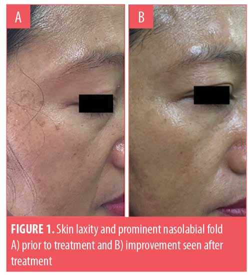
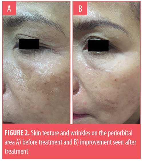
Results. Based on the independent physician’s clinical assessment, seven of the eight patients showed an improvement in texture, fine lines, and wrinkles, especially in the periorbital region. According to the patient questionnaire, four of the eight patients felt their wrinkles were improved, while all patients felt their skin texture was smoother. Five of the eight patients experienced slight redness at 1 to 2 days after treatment.
Conclusion. The application of topical growth factors after microneedling can be useful to reduce visual signs of facial photoaging by improving skin texture and minimizing the appearance of fine lines and wrinkles.
KEYWORDS: Photoaging, growth factors, microneedling
Introduction
Chronic sun exposure causes various forms of skin damage.1 Chronic exposure to solar ultraviolet irradiation can result in facial photoaging such as fine lines and wrinkles, poor texture, and sagging skin. Facial photoaging has become one of the most prevalent aesthetic concerns. New topical formulas can facilitate the skin to repair wrinkles, leading to a younger, healthier looking face and glowing skin.2 Furthermore, these topical formulas can mitigate further aging by accelerating the synthesis of collagen.2 An improved understanding of the biochemical mechanisms of skin aging has resulted in the identification of key pathways of intervention to reverse of skin aging.3
Growth factors are polypeptides or proteins that play a key role in the regulation of physiological processes. Growth factors are produced and secreted by skin cells such as fibroblasts, keratinocytes, and melanocytes. One type of these growth factors is cytokines, which are involved in regulating the immune system and repairing the skin.1,4 Many growth factors are involved in wound healing, both chronic and acute in nature. Various signals direct the cellular responses during each phase of healing, but growth factors are among the most important. For effective wound repair, the correct growth factors should be presented within the proper framework.3,5
Successful skin repair requires a balance between the function of multiple growth factors and cytokines. Within the past decade, there has been an increasing depth of knowledge about the benefits of topical growth factors for skin rejuvenation. Results from previous clinical studies demonstrate signficicantly increased production of new collagen following the topical application of physiologically balanced growth factors mixtures.3,6
The present report details a retrospective study that examined the efficacy of the application of topical growth factors (Wove Style, Tokyo, Japan) after microneedling treatment for the reduction of visual signs of facial photoaging.
Methods
Eight female patients between the ages of 35 to 60 years with Fitzpatrick skin types III to IV and mild-to-moderate photodamage, skin laxity, fine lines, and wrinkles (Fitzpatrick Wrinkle Scale: 3–6) were enrolled to receive the treatment protocol. Inclusion criteria were Fitzpatrick skin types III to IV and the presence of mild-to-moderate wrinkles (Fitzpatrick Wrinkle Scale: 3–6). Subjects who did not consent to the study and/or those with any of the following conditions were excluded: pregnant or lactating, Fitzpatrick skin types I to II and V to VI, significant skin disease in the test areas, a history of poor wound healing or keloid formation, and/or completion of previous skin rejuvenating procedures one month prior to the study.
The treatment area was thoroughly cleansed with a mild soap before each procedure. Topical anesthetic cream (lidocaine 2.5%, prilocaine 2.5%; Genero Pharmaceuticals, Bekasi, Indonesia) was applied to the treatment area for 45 minutes before treatment. Eight patients subsequently applied 2mL of gel containing a mixture of four different growth factors (Wove Style, Tokyo, Japan) to photodamaged facial skin after microneedling procedures. This was performed every 10 days, with three treatments total. The gel contained a biosynthetic mixture of epidermal growth factor, fibroblast growth factor, hepatocyte growth factor, and insulin-like growth factor. An independent physician evaluator assessed the treatment response by comparing pre- and posttreatment clinical photographs using the Fitzpatrick Wrinkle Scale (before and after four weeks of treatment). Assessment of side effects was completed after the second and third treatments. Patients were evaluated after each procedure by an independent physician evaluator using the Fitzpatrick Wrinkle Scale. Patient questionnaires were completed during the fourth week.
Results
The independent physician evaluator’s assessments of photodamage, texture, fine lines, and wrinkles were collated and analyzed using a nonparametric Wilcoxon signed-rank test. In aggregate, a significant difference was noted in the appearance of the Fitzpatrick Wrinkle Scale between the baseline and posttreatment photographs (p<0.001). Procedural complications were limited to slight erythema lasting 1 to 2 days. No instances of infection, scar, contact dermatitis, or postinflammatory pigmentary alteration were observed (Figures 1 and 2).
According to the patient questionnaires that were completed during the fourth week, four patients noted improvement in their wrinkles and fine lines and six patients noted their skin tone had become more even. One patient felt no improvement occurred in either her wrinkles or her skin tone. All patients commented that their skin texture felt smoother.
Discussion
Repetitive exposure to ultraviolet radiation accelerates skin aging, leading to the formation of peroxyl free radicals; these break down to form malondialdehyde and subsequently cross-links and polymerizes collagen, causeing the inactivation of growth factors, breakdown of matrix proteins, and membrane lipid damage.2,3 Clinical manifestations of photodamage-induced aging processes include a loss of skin elasticity and firmness, fine lines and wrinkles, and uneven skin tone.3
Growth factors are chemical messengers that travel between cells and direct them to turn on or off specific cellular activities such as cell proliferation, chemotaxis, and extracellular matrix formation.7 Topical application of growth factors also reduces signs of photoaging by promoting fibroblast and keratinocyte proliferation and inducing extracellular matrix formation.8–10
Growth factors can be derived from several sources, including epidermal cells, human foreskin, placental cells, colostrum, recombinant bacteria, yeast, and plants. Growth factors can also be produced biosynthetically.11,12
Conclusion
In this study, the application of topical growth factors after microneedling resulted in marked clinical improvement in the signs of facial photoaging as measured by an independent physician assessment of clinical photographs after three treatment sessions.
Limitations. This topical treatment is nonsterile and use should be carefully considered before introducing to open skin. In addition, prolonged use could introduce foreign materials into the skin and cause significant allergic reaction in some people. Further research over a longer period of time is needed for more thorough evaluation of efficacy and safety. Additional studies with larger patient groups and longer term follow up are required to more comprehensively assess and define the optimal benefits of topical biosynthetic growth factors.
References
- Akiba S, Shinkura R, Miyamoto K, et al. Influence of chronic sun exposure and lifestyle on facial skin photo-aging—results from a pilot study. J Epidemiol. 1999;9(6 Suppl):S126–S142.
- Mukherjee PK, Maity N, Nema NK, Sarkar BK. Bioactive compounds from natural resources against skin aging. Phytomedicine. 2011;19(1):64–73.
- Atkin DH, Trookman NS, Rizer RL, et al. Combination of physiologically balanced growth factors with antioxidants for reversal of facial photodamage. J Cosme Laser Therapy. 2010;12:14–20.
- Eming SA, Krieg T, Davidson JM. Inflammation in wound repair: molecular and cellular mechanism. J Invest Dermatol. 1007;127(3):514–525.
- Wilgus TA. Growth factor–extracellular matrix interactions regulate wound repair. Adv Wound Care (New Rochelle). 2012;1(6):249–254.
- Fitzpatrick RE, Rostan EF. Reversal of photodamage with topical growth factors: a pilot study. J Cosmetic & Laser Ther. 2003;5(1):25–34
- Babu M, Wells A. Dermal–epidermal communication in wound healing. Wounds. 2001;13(5):183–189.
- Bertaux B, Horneback W, Eisen AZ, et al. Growth stimulation of human keratinocyte by tissue inhibitor of metalloproteinases. J Invest Dermatol. 1991;97(4):679–685.
- Finch PW, Rubin JS, Miki T, et al. Human KGF is FGF related with properties of a paracrine effector of epithelial cell growth. Science. 1989;245(4919):752–755.
- Schwartz E, Cruickshank FA, Christensen CC, et al. Collagen alterations in chronically sun-damaged human skin. Photochem Photobiol. 1993;58(6):841–844.
- Bonin-Debs AL, Boche I, Gille H, et al. Development of secreted proteins as biotherapeutic agents. Exp Opin Biol Ther. 2004;4(4):551–558.
- Kiripolsky MG. Topical agents for skin health restoration. In: Obagi ZE. The Art of Skin Health Restoration and Rejuvenation. Boca Raton, FL: CRC Press/Taylor & Francis Group; 2015: 59.