
Ani Tajirian, MD; Michelle Tsui, BA
Webster Management, Inc. (Private Practice), Oakland, California
Disclosure: The authors report no relevant conflicts of interest
Abstract
Objective: Following Mohs surgery, medium-to-large defects on the central forehead can often be complicated to surgically reconstruct. In this paper, the authors discuss possible central forehead reconstructions and report their successful experience employing a simple primary vertical linear closure with a special technique to demarcate forehead rhytides rather than performing an overly complicated flap or graft. Case report: The patient was a 57-year-old man who presented with a broad superficial basal cell carcinoma that required treatment with Mohs surgery. For the resulting defect, the authors elected to perform a complex linear repair taking advantage of substantial side-to-side laxity in the supraperiosteal plane and carefully labeling and matching each forehead rhytide across the defect as the wound was sutured. Conclusion: The findings of this case demonstrate that medium-to-large wounds of the central forehead can be aesthetically repaired with a simple primary vertical linear closure. Carefully mapping and labeling horizontal forehead rhytides with a sterile surgical marking pen prior to anesthesia ensures accurate approximation during wound closure. (J Clin Aesthet Dermatol. 2016;9(8):47–49.)
Due to the prominence and broad surface of the central forehead, aesthetic reconstruction of medium to large forehead defects is imperative. Commonly utilized flaps include the island pedicle flap, rotation flaps, A-to-T flap, and O-to-T flaps (Table 1 ). The hairline as well as the horizontal rhytides along the forehead are helpful in hiding the scars and incisions necessary for complex reconstructions.[1–3]
The island pedicle flap can be considered as it is richly vascularized by the frontalis perforators and is useful when trying to avoid medial displacement of the eyebrows.[4] The island pedicle flap is a better color and texture match than a graft on the central forehead; however, the natural forehead anatomy is often disrupted and a large hypopigmented triangular scar can remain.[4]
Rotation and advancement flaps, such as the A-to-T flap and O-to-T flap can also be useful when it is difficult to achieve side-to-side movement at the immediate wound site.[5] These flaps draw from surrounding areas of increased skin laxity, but involve complex incision lines and a risk of disturbing important neurovascular structures when undermining in the subcutaneous plane.[3]
For certain defects, Z-plasties can be used to take advantage of the horizontal relaxed skin tension lines of the forehead by reorienting portions of vertical incision lines. However, a poor outcome is quite unaesthetic and when performed on longer incisions may leave the patient with a noticeable “mark of Zorro.”[6]
For medium to large wounds located in the central third of the forehead, a primary vertical linear repair that preserves natural forehead anatomy provides a much simpler way to create equally if not better aesthetic results than local flaps and Z-plasties.[5] The central forehead’s relative lack of neurovascular structures facilitates the deep plane undermining required for a vertical linear repair even for larger defects.[2] Often, advancement, rotation, and pedicle flaps can prove to be overly complicated and time-consuming to perform due to the extensive undermining required, the vascularity of the forehead, and the lack of ease of movement of the forehead.
Case Report
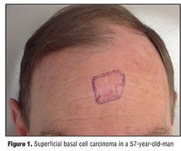
A 57-year-old man presented with a 2×2.5cm pink patch with central erosion on the central forehead (Figure 1). The skin lesion had been previously biopsied by another dermatologist, demonstrating solar keratosis and treated with three rounds of imiquimod cream. As the lesion had not resolved, a shave biopsy was performed revealing a broad superficial basal cell carcinoma. With Mohs micrographic surgery, tumor-free margins were achieved after two stages, resulting in a large 3.6×3.2cm defect to periosteum located on the central forehead (Figure 2).
At the conclusion of complete tumor extirpation, the patient was instructed to animate with the forehead, exaggerating the natural horizontal furrows. The individual forehead rhytides were carefully marked using a sterile surgical marking pen and labeled “A” through “E” on both sides of the defect (Figure 3). Anesthesia of the area was performed following the markings to prevent distortion of the patient’s natural forehead anatomy.
A vertical linear closure was designed and large Burow’s triangles were removed superiorly and inferiorly to the defect to avoid dog ears over the broad convex surface. Extensive wide undermining was then performed in the supraperiosteal plane to reduce tension and increase side-to-side laxity of the forehead skin. Following hemostasis, deep closure of the defect was achieved with 4-0 Monocryl (Ethicon), matching the labeled rhytides carefully across the wound (Figure 4). A running 6-0 polypropylene suture was then placed. The final length of the wound was 8.3cm.
The postoperative period had no complications, and at four months, full animation of the frontalis revealed no disturbance in the patient’s forehead rhytides (Figure 5). At five months, the scar was passed over with a vascular laser to treat the minor neovascularization that persisted (Figure6).
Discussion
The authors were presented with a moderate-sized wound prominently located in the central forehead. While advancement, rotation, and pedicle flaps each have their unique advantages, wounds of the central forehead can be simply and aesthetically repaired, when possible, with a primary vertical linear closure. The natural furrows that exist on the forehead necessitate careful consideration when performing wound closures. Mapping and labeling the rhytides on both sides of the defect using letters, numbers, or corresponding colors ensures an excellent aesthetic outcome.
The findings of this case demonstrate that medium-to-large wounds of the central forehead can be aesthetically repaired with a simple primary vertical linear closure. Carefully mapping and labeling horizontal forehead rhytides with a sterile surgical marking pen prior to anesthesia ensures accurate approximation during wound closure. Providers should be aware that a long thin scar may be necessary to avoid dog ears on the broad convex surface of the forehead.
References
1. Cummings C, Flint PW. Cummings Otolaryngology: Head and Neck Surgery. Philadelpha, PA: Mosby Elsevier; 2010.
2. Goldman G, Dzubow LM, Yelverton CB. Forehead. In: Goldman G, Dzubow L, eds. Facial Flaps Surgery. New York: McGraw-Hill Professional; 2012:263–281.
3. Dzubow LM, Robins P. Repair of surgical defects. I. Circular defects on the forehead. J Dermatol Surg Oncol. 1981;7:764–766.
4. Hussain W, Hafiji J, Salmon P. Frontalis-based island pedicle flaps for the single-stage repair of large defects of the forehead and frontal scalp. Br J Dermatol. 2012;166:771–774.
5. Boustany A, Ghareeb P, McClellan WT. Forehead reconstruction using a modified dual-plane A to T flap. Can J Plast Surg. 2012;20:251–254.
6. Pomaranski MR, Krull EA, Balle MR. Use of the Z-plasty technique for forehead defects. Dermatol Surg. 2005;31:1720–1723.